This website uses cookies to ensure you get the best experience on our website.


- Table of Contents
Plan GBP1 staining in paraffin sections using an IHC-validated antibody. This guide covers fixation, controls and interpretation of the cytoplasmic staining reported in a few tissues, most abundant in immune cells (HPA tissue IHC).

Expected localisation, validated protocols, controls and antibodies — the at-a-glance facts below, then the full design guide.
| Expected localisation | Cytoplasmic staining in a few tissues (HPA tissue IHC) | |
| Staining pattern | Cytoplasmic signal, most abundant in immune cells (HPA tissue IHC) | |
| Antigen retrieval | EDTA pH 8.0 HIER, heat-mediated (datasheet A03067-1) | |
| Positive control | Appendix+2 more · see all | |
| Negative control | Adrenal gland+4 more · see all |
| Fixation | Keep section fixation consistent across samples. (standard IHC practice; not target-specific) | |
| Caveat | Immune-cell staining is unannotated; RNA concordance is low (HPA tissue IHC) | |
| Regulation | Interferon inducible (UniProt) | |
| Isoform / epitope | No listed isoforms; residues 590–592 are processed (UniProt) |
Compare the catalog antibody’s IHC-P protocol (datasheet A03067-1) with three published GBP1 IHC protocols (PMC10710186; PMC11865198; PMC5978929).
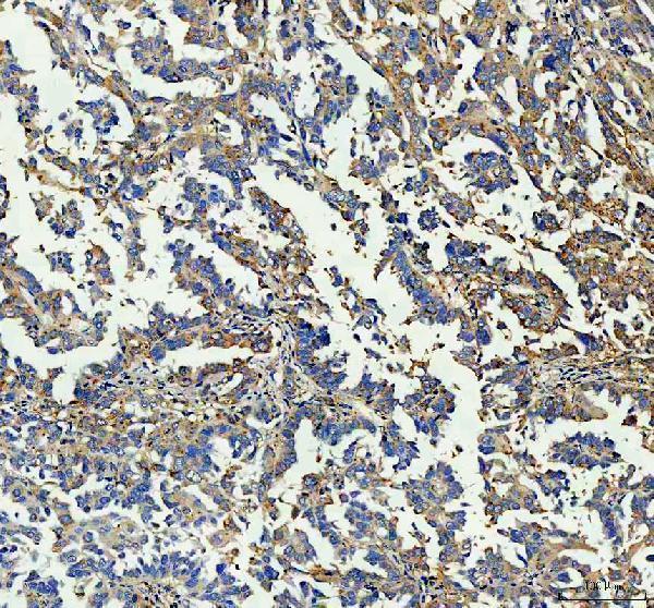
| Sample | Paraffin-embedded human ovarian cancer tissue; fixative not specified (datasheet A03067-1) |
| Fixation | Image fixative and duration unreported (datasheet A03067-1); verify before use. |
| Sectioning | 4–5 µm sections on charged slides (standard) |
| Deparaffinisation | Xylene, graded ethanol series to water (standard) |
| Antigen retrieval | Heat retrieval: EDTA pH 8.0 (datasheet A03067-1); 20 min, 95–100 °C (standard) |
| Peroxidase block | 3% H2O2, 10 min, room temperature (standard) |
| Blocking | 10% goat serum (datasheet A03067-1) |
| Primary antibody | Rabbit anti-GBP1, 2-5 μg/ml (datasheet A03067-1) |
| Primary incubation | Overnight at 4 °C (datasheet A03067-1) |
| Detection | HRP-conjugated secondary, DAB chromogen (datasheet A03067-1) |
| Counterstain | Hematoxylin, blue, dehydrate and mount (standard) |
| Expected result | GBP1-positive staining in lymphoid tissue of appendix (HPA tissue IHC: Medium). HPA tissue profile: Cytoplasmic expression in a few tissues, most abundant in immune cells. No signal in the no-primary control. |
GBP1 is chiefly cytosolic and can associate with cytoplasmic vesicle, Golgi, and cell membranes; it has no transmembrane segment (UniProt P32455 topology and subcellular location). In paraffin-section IHC, expect cytoplasmic staining in selected cells, including immune cells and some glandular cells (HPA tissue IHC). Interpret intensity cautiously: HPA rates tissue staining “Approved” but reports low consistency with RNA expression and does not annotate immune-cell staining individually (HPA tissue IHC).
| Cytoplasmic staining in appendix lymphoid tissue or small-intestinal or thyroid glandular cells, with a medium signal (HPA tissue IHC). | This matches the recorded tissue and cell patterns. Score the stained cell population and compartment together; a positive gland does not imply that every nearby cell should stain (HPA tissue IHC). |
| Predominantly nuclear staining, with little corresponding cytoplasmic signal. | Treat this as a localisation mismatch requiring investigation: UniProt lists cytosolic and membrane-associated locations, but no nuclear location (UniProt P32455 subcellular location). Review morphology, counterstain, and controls before assigning it to GBP1 (general IHC practice). |
| Strong staining in an HPA-listed negative cell population, such as adrenal glandular cells or bone-marrow hematopoietic cells (HPA tissue IHC). | Consider nonspecific binding or endogenous chromogen-detection activity, especially if a reagent-omission control also develops colour (general IHC practice). HPA's “not detected” result is cell-type specific and does not prove every cell in that tissue is negative (HPA tissue IHC). |
| Diffuse colour across cells and extracellular areas, obscuring cell boundaries. | This distribution is difficult to score as GBP1 because the documented tissue pattern is cytoplasmic in selected cells (HPA tissue IHC). Check background controls and staining development before interpreting weak cell-associated colour (general IHC practice). |
| No convincing signal in appendix lymphoid tissue or the listed positive glandular cells (HPA tissue IHC). | The expected comparator is medium staining in those specified cells, not uniform staining of the whole section (HPA tissue IHC). Assess whether those cells are present and whether the run controls worked before calling the specimen negative (general IHC practice). |
| Tissue and cell context | HPA records medium staining in appendix lymphoid tissue and small-intestinal and thyroid glandular cells; lung macrophages are low (HPA tissue IHC). Choose and score controls by the stated cell population, not by tissue name alone (general IHC practice). |
| Confidence in the tissue pattern | HPA rates the tissue IHC pattern “Approved,” reports low agreement with RNA, and leaves immune-cell staining unannotated in its summary (HPA tissue IHC). Resolve ambiguous cells against morphology and controls rather than treating the summary as a cell-by-cell map (general IHC practice). |
| GBP1 distribution | GBP1 has no transmembrane segment and is listed in cytosol and at several membranes (UniProt P32455 topology and subcellular location). Infection-associated recruitment to pathogen vacuoles or escaped bacteria is documented; do not require that specialised pattern in routine tissue (UniProt P32455 subcellular location). |
| Processing and epitope uncertainty | UniProt lists a 1–589 chain, a 590–592 propeptide, and modifications at residues 156, 589, and 590 (UniProt P32455 processing and modified residues). No antibody epitope is supplied, so these annotations alone cannot predict staining loss or a retrieval requirement. |
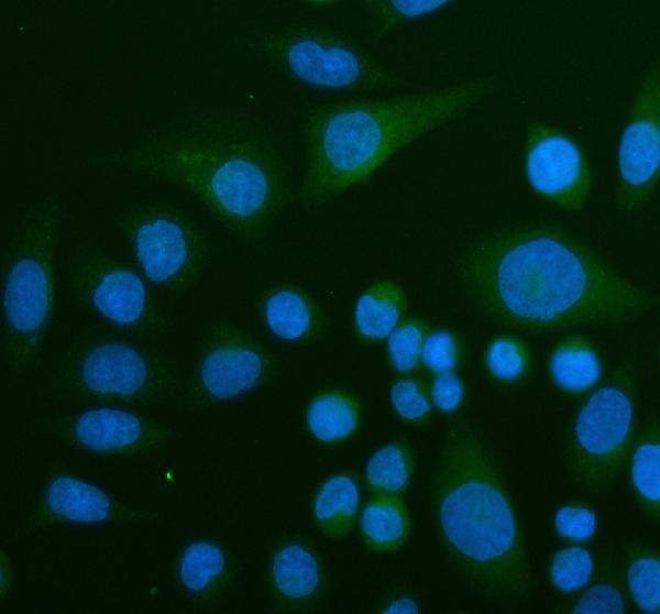
| IF/ICC Q: Can this IHC pattern be used as an IF control? | A: Use the UniProt locations as biological context, but HPA provides no ICC-IF images or main-location assignment, and the listed antibody has no ICC validation entry (UniProt P32455 subcellular location; HPA subcellular and antibody records). This page cannot establish an IF staining pattern. |
| Situation | Likely cause | Next action |
|---|---|---|
| Listed positive cells are unstained while the run control also fails. | A run-level staining or detection problem is plausible (general IHC practice). The HPA comparator is medium staining in specified appendix, small-intestinal, or thyroid cell populations (HPA tissue IHC). | Confirm the expected cells are present, then review reagent application, detection, chromogen development, and the laboratory's antigen-retrieval conditions with a working control section (general IHC practice). |
| Listed positive cells are unstained but the run control works. | Sampling or cell identification may explain the discrepancy; the HPA result applies to named cell populations and has low RNA–staining consistency (HPA tissue IHC). | Recheck morphology and the cells actually scored; compare another listed positive population before concluding that this specimen lacks GBP1 (HPA tissue IHC; general IHC practice). |
| Nuclear colour dominates the section. | The observed compartment conflicts with the listed cytosolic and membrane-associated GBP1 locations (UniProt P32455 subcellular location). Counterstain overlap or nonspecific detection can complicate interpretation (general IHC practice). | Inspect an omission control and separate chromogen from counterstain by microscopy; score GBP1 only where cell-associated signal can be assigned confidently (general IHC practice). |
| Colour appears in listed negative cells, including adrenal glandular cells (HPA tissue IHC). | Nonspecific antibody binding or endogenous detection activity is possible (general IHC practice); HPA reports those particular cells as not detected (HPA tissue IHC). | Compare a primary-antibody omission control, review blocking and detection reagents, and assess whether the signal follows identifiable cells rather than tissue deposits (general IHC practice). |
| Diffuse background hides a weak cytoplasmic pattern. | Excess detection development, inadequate washing, or nonspecific binding can raise background in chromogenic IHC (general IHC practice). | Use the omission control to locate background, review washing and chromogen-development time, and compare a listed positive cell population in the same run (general IHC practice; HPA tissue IHC). |
| Staining is present in lung macrophages but weaker than in appendix lymphoid tissue. | This can fit the reported low lung-macrophage and medium appendix-lymphoid staining levels (HPA tissue IHC). | Identify macrophages by morphology and compare like cell populations under the same scoring criteria; do not label the weaker lung signal a failed run solely from its intensity (HPA tissue IHC; general IHC practice). |


Comprehensive Human Protein Atlas IHC scoring per tissue (reliability: Approved — Low consistency between antibody staining and RNA expression data. Antibody staining in immune cells not annotated, view images.). Rows are taken directly from the HPA tissue chart — click any row's HPA link to view the source.
| Tissue | Cell type | Level | Evidence | Source |
|---|---|---|---|---|
| Appendix | Lymphoid tissue | Medium | Protein (IHC) | HPA → |
| Small intestine | Glandular cells | Medium | Protein (IHC) | HPA → |
| Thyroid gland | Glandular cells | Medium | Protein (IHC) | HPA → |
| Tissue | Cell type | Level | Evidence | Source |
|---|---|---|---|---|
| Adrenal gland | Glandular cells | Not detected | Protein (IHC) | HPA → |
| Bone marrow | Hematopoietic cells | Not detected | Protein (IHC) | HPA → |
| Breast | Adipocytes | Not detected | Protein (IHC) | HPA → |
| Bronchus | Basal cells | Not detected | Protein (IHC) | HPA → |
| Cerebellum | Cells in granular layer | Not detected | Protein (IHC) | HPA → |
Troubleshoot GBP1 staining in paraffin sections by checking retrieval, cellular localisation and controls before comparing staining across samples.
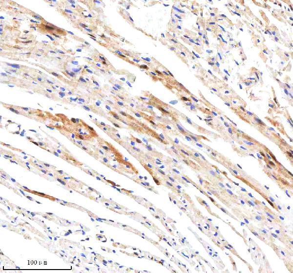
A03067-1 has IHC images from paraffin-embedded human ovarian cancer and mouse and rat heart tissue, plus IF imaging in SiHa cells (catalog image captions).
A03067-1 has pictured IHC in paraffin-embedded human ovarian cancer and mouse and rat heart tissue, and pictured IF in SiHa cells (catalog image captions). M03067-1 lists human IHC and ICC/IF applications, but has no supplied IHC or IF image captions (catalog applications; image captions).
Which to pick: Choose A03067-1 for tissue IHC in paraffin sections: its own captions show human ovarian cancer and mouse and rat heart tissue; the fixative is unreported (A03067-1 IHC image captions). Choose A03067-1 for IF/ICC when a pictured example matters: its caption shows IF in SiHa cells, while M03067-1 lists ICC/IF without a supplied image caption (catalog IF image captions; applications). For cross-species work, A03067-1 lists human, mouse and rat reactivity; M03067-1 is a rabbit monoclonal listing human reactivity only (catalog reactivity; host; clone).