This website uses cookies to ensure you get the best experience on our website.


- Table of Contents
Plan GBP5 staining in paraffin sections around cytoplasmic signal in a subset of immune cells (HPA tissue IHC). Use lung macrophages as a positive tissue reference and account for IFN-inducible expression when interpreting intensity (HPA tissue IHC; UniProt).

Expected localisation, validated protocols, controls and antibodies — the at-a-glance facts below, then the full design guide.
| Expected localisation | Cytoplasm in a subset of immune cells (HPA tissue IHC) | |
| Staining pattern | Scattered immune cells show cytoplasmic staining (HPA tissue IHC) | |
| Antigen retrieval | Citrate pH 6.0 HIER, 95–98 °C, 20 min (rule: cytoplasmic / membrane antigen) | |
| Positive control | Gallbladder+4 more · see all | |
| Negative control | Adipose tissue+4 more · see all |
| Fixation | Keep fixation consistent across sections (standard IHC practice; not target-specific) | |
| Caveat | Sparse positive cells may be missed in sampled fields (HPA tissue IHC) | |
| Regulation | IFN-inducible (UniProt) | |
| Isoform / epitope | Two isoforms; verify antibody epitope coverage (UniProt) |
The catalog antibody’s IHC-P protocol is followed by published GBP5 IHC methods for placental tissue, intestine, and NSCLC tissue microarrays (PMC13093737; PMC9201962; PMC10183185).
| Sample | Tissue sections; selected-image fixative not specified (standard IHC workflow) |
| Fixation | Image fixative and duration unreported (datasheet A07110); verify before use. |
| Sectioning | 4–5 µm sections on charged slides (standard) |
| Deparaffinisation | Xylene, graded ethanol series to water (standard) |
| Antigen retrieval | Heat-induced epitope retrieval in citrate buffer, pH 6.0, 20 min at 95–98 °C (standard rule: cytoplasmic / membrane antigen) |
| Peroxidase block | 3% H2O2, 10 min, room temperature (standard) |
| Blocking | 10% normal serum of the secondary host, 30 min, room temperature (standard) |
| Primary antibody | Rabbit anti-GBP5, 1:100–1:500 starting range (standard) |
| Primary incubation | Overnight at 4 °C (standard) |
| Detection | HRP-polymer secondary, DAB chromogen 5–10 min (standard) |
| Counterstain | Hematoxylin, blue, dehydrate and mount (standard) |
| Expected result | GBP5-positive staining in glandular cells of gallbladder (HPA tissue IHC: Medium). HPA tissue profile: Cytoplasmic expression in a subset of immune cells in several tissues. No signal in the no-primary control. |
GBP5 should appear mainly cytoplasmic, sometimes punctate or concentrated near intracellular membranes, consistent with its cytoplasmic, vesicle membrane and Golgi membrane locations and lack of a transmembrane segment (UniProt Q96PP8 topology and subcellular location). In tissue sections, expect staining in a subset of immune cells, with some other HPA-listed cell types also positive (HPA tissue IHC). HPA rates the tissue pattern Supported, with medium consistency between staining and RNA data (HPA tissue IHC).
| Cytoplasmic stain in scattered lung macrophages or cells in splenic red pulp, around the reported Medium level (HPA tissue IHC). | This fits the reported cell distribution and GBP5's cytoplasmic and intracellular membrane locations (HPA tissue IHC; UniProt Q96PP8 subcellular location). Assess the named cells rather than expecting every cell in the section to stain (HPA tissue IHC). |
| Predominantly nuclear staining, with little cytoplasmic signal. | An isolated nuclear pattern does not match the listed GBP5 locations (UniProt Q96PP8 subcellular location). Treat it as suspect and check controls and detection conditions before interpreting it as target staining (standard IHC practice). |
| Strong signal in adipocytes or adrenal gland glandular cells. | These named cell populations were Not detected by HPA tissue IHC; staining there merits a cross-reactivity or endogenous detection-activity check (HPA tissue IHC; standard IHC practice). Cell identity matters: gallbladder glandular cells were Medium, so glandular staining alone is not disqualifying (HPA tissue IHC). |
| Diffuse stain across tissue and empty areas, without a discernible cellular pattern. | That distribution cannot establish the reported cytoplasmic, cell-selective pattern (HPA tissue IHC). Review background and no-primary controls for nonspecific binding or detection background before scoring cells (standard IHC practice). |
| No signal in lung macrophages on a section expected to contain them. | HPA reports Medium staining in lung macrophages, so absent signal calls for a run check; it does not alone prove GBP5 is absent from the specimen (HPA tissue IHC). Confirm the cells are present, then review the IHC workflow and controls (standard IHC practice). |
| Cell selection for a positive comparison | Lung macrophages, splenic red-pulp cells and tonsillar non-germinal-center cells are reported Medium; choose a section containing the specified cells and score them directly (HPA tissue IHC). |
| Interpretation of epithelial staining | HPA reports Medium staining in gallbladder glandular cells and urinary-bladder urothelial cells, alongside immune-cell staining; epithelial signal needs tissue and cell context (HPA tissue IHC). |
| Location and topology | GBP5 is listed in cytoplasm and on cytoplasmic vesicle and Golgi membranes, with no transmembrane segment; intracellular granular or perinuclear accentuation can fit this distribution (UniProt Q96PP8 subcellular location and topology). |
| Strength of tissue evidence | HPA calls the tissue pattern Supported and describes medium consistency with RNA expression; this supports comparison with its images but leaves room for specimen-to-specimen differences (HPA tissue IHC). |
| Antibody evidence | The listed rabbit polyclonal antibody HPA028656 has Supported IHC status; the payload does not report Enhanced IHC validation or provide a catalog-antibody dilution (HPA antibodies). |
| Isoforms and processing | Two isoforms and a chain spanning residues 1–583 are listed, but no antibody epitope is supplied; the record cannot establish whether this stain distinguishes isoforms or processing states (UniProt Q96PP8 isoforms and processing). |
| IF/ICC Q&A: Is a GBP5 IF pattern established here? | No HPA ICC-IF image or main subcellular location is available; the UniProt locations are a biological expectation, not an image-validated IF pattern or IF protocol (HPA subcellular ICC-IF; UniProt Q96PP8 subcellular location). |
| Situation | Likely cause | Next action |
|---|---|---|
| A positive comparison section appears blank. | The selected section may lack the relevant HPA-listed cells, or the IHC run may have failed; the image alone cannot distinguish these possibilities (HPA tissue IHC; standard IHC practice). | Verify cell presence on the counterstain and compare a separate known-positive section processed in the same run; then review detection controls (HPA tissue IHC; standard IHC practice). |
| Only nuclei are colored. | A nuclear-only pattern conflicts with the recorded cytoplasmic and intracellular membrane locations and may represent nonspecific staining (UniProt Q96PP8 subcellular location; standard IHC practice). | Inspect a no-primary control and compare the staining compartment in HPA-listed positive cells before accepting the signal (standard IHC practice; HPA tissue IHC). |
| Adipocytes stain broadly. | HPA lists adipocytes in adipose tissue as Not detected; broad staining there may reflect nonspecific antibody binding or endogenous detection activity (HPA tissue IHC; standard IHC practice). | Check no-primary and detection controls, then compare staining in an HPA-listed positive cell population under the same run conditions (standard IHC practice; HPA tissue IHC). |
| Background obscures individual cells. | Diffuse background prevents assessment of the cell-selective cytoplasmic pattern HPA describes; its precise source cannot be inferred from this record (HPA tissue IHC; standard IHC practice). | Use no-primary and detection controls to localize background, then review blocking, washes and detection conditions as general IHC workflow checks (standard IHC practice). |
| Only a few cells stain in an otherwise pale section. | Sparse positive cells can fit HPA's description of cytoplasmic expression in a subset of immune cells; tissue-wide uniformity is not the reported pattern (HPA tissue IHC). | Identify the stained cells on the counterstain and compare the same cell type with HPA's tissue images before calling the run weak (HPA tissue IHC; standard IHC practice). |
| IF shows puncta but lacks a matching HPA ICC-IF image. | UniProt lists vesicle and Golgi membrane locations, while HPA supplies no ICC-IF images or main-location call for GBP5 (UniProt Q96PP8 subcellular location; HPA subcellular ICC-IF). | Describe the IF observation as provisional and assess appropriate IF controls; use this page's HPA evidence for IHC tissue interpretation, not IF image validation (HPA tissue IHC; HPA subcellular ICC-IF; standard IF practice). |


Comprehensive Human Protein Atlas IHC scoring per tissue (reliability: Supported — Medium consistency between antibody staining and RNA expression data.). Rows are taken directly from the HPA tissue chart — click any row's HPA link to view the source.
| Tissue | Cell type | Level | Evidence | Source |
|---|---|---|---|---|
| Gallbladder | Glandular cells | Medium | Protein (IHC) | HPA → |
| Lung | Macrophages | Medium | Protein (IHC) | HPA → |
| Spleen | Cells in red pulp | Medium | Protein (IHC) | HPA → |
| Tonsil | Non-germinal center cells | Medium | Protein (IHC) | HPA → |
| Urinary bladder | Urothelial cells | Medium | Protein (IHC) | HPA → |
| Tissue | Cell type | Level | Evidence | Source |
|---|---|---|---|---|
| Adipose tissue | Adipocytes | Not detected | Protein (IHC) | HPA → |
| Adrenal gland | Glandular cells | Not detected | Protein (IHC) | HPA → |
| Breast | Adipocytes | Not detected | Protein (IHC) | HPA → |
| Bronchus | Respiratory epithelial cells | Not detected | Protein (IHC) | HPA → |
| Caudate | Glial cells | Not detected | Protein (IHC) | HPA → |
Troubleshoot GBP5 staining in paraffin section chromogenic IHC by checking retrieval, cell identity, intracellular pattern, and assay controls before interpreting signal.
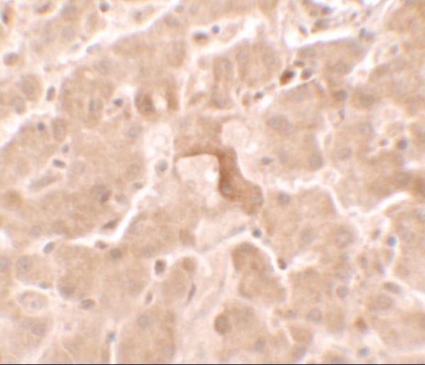
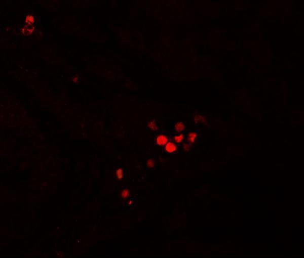
GBP5 antibody A07110 has pictured IHC and IF staining in human liver tissue (catalog: A07110 image captions). Its listed reactivity covers human, mouse, and rat (catalog: A07110 reactivity).
A07110 is the sole rendered card; its captions show IHC in human liver tissue at 2.5 μg/ml and IF in human liver tissue at 20 μg/ml (catalog: A07110 image captions). IHC-P and IF are listed applications, but the supplied staining images document human tissue only (catalog: A07110 applications and image captions).
Which to pick: For tissue IHC, pick A07110: IHC-P is listed, and its own caption shows human liver staining at 2.5 μg/ml; the fixative is unreported (catalog: A07110 applications and IHC image caption). For IF, A07110 has a human liver image; for ICC, A07110-1 lists ICC and IF at 1:50–400 but has no supplied staining image (catalog: A07110 IF image caption; A07110-1 applications, dilution data, and image captions). For cross-species work, A07110 lists human, mouse, and rat, while rabbit polyclonal A07110-1 lists human and mouse; validate staining in the chosen species because the supplied images show human tissue only (catalog: reactivity, host, clonality, and image captions).