This website uses cookies to ensure you get the best experience on our website.


- Table of Contents
Plan GCC2 paraffin IHC around its widespread cytoplasmic tissue staining (HPA tissue IHC), while treating trans-Golgi network localization as molecular context (UniProt). The catalog antibody has an IHC dilution range of 1:50–1:200 (datasheet); high-staining glandular cells and listed undetected populations offer comparison points (HPA tissue IHC).

Expected localisation, validated protocols, controls and antibodies — the at-a-glance facts below, then the full design guide.
| Expected localisation | Cytoplasmic staining in tissue sections (HPA tissue IHC) | |
| Staining pattern | Widespread cytoplasmic staining across cell types (HPA tissue IHC) | |
| Antigen retrieval | Citrate pH 6.0 HIER, 95–98 °C, 20 min (rule: cytoplasmic / membrane antigen) | |
| Positive control | Adrenal gland+4 more · see all | |
| Negative control | Oral mucosa+1 more · see all |
| Fixation | Keep fixation consistent across paraffin sections. (standard IHC practice; not target-specific) | |
| Caveat | Staining and RNA expression show medium consistency (HPA tissue IHC) | |
| Regulation | Low tissue specificity by RNA expression (HPA tissue RNA) | |
| Isoform / epitope | 2 isoforms; assess antibody epitope coverage (UniProt) |
The catalog antibody’s IHC-P protocol is followed by two published GCC2 IHC protocols: lung tissue (PMC13145344 methods) and human kidney tissue (PMC11203101 methods).
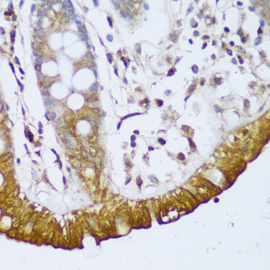
| Sample | Paraffin-embedded human colon tissue; fixative not specified (datasheet A07107) |
| Fixation | Image fixative and duration unreported (datasheet A07107); verify before use. |
| Sectioning | 4–5 µm sections on charged slides (standard) |
| Deparaffinisation | Xylene, graded ethanol series to water (standard) |
| Antigen retrieval | Heat-induced epitope retrieval in citrate buffer, pH 6.0, 20 min at 95–98 °C (standard rule: cytoplasmic / membrane antigen) |
| Peroxidase block | 3% H2O2, 10 min, room temperature (standard) |
| Blocking | 10% normal serum of the secondary host, 30 min, room temperature (standard) |
| Primary antibody | Rabbit anti-GCC2, 1:50-1:200 (datasheet A07107) |
| Primary incubation | Overnight at 4 °C (standard) |
| Detection | HRP-polymer secondary, DAB chromogen 5–10 min (standard) |
| Counterstain | Hematoxylin, blue, dehydrate and mount (standard) |
| Expected result | GCC2-positive staining in glandular cells of adrenal gland (HPA tissue IHC: High). HPA tissue profile: Ubiquitous cytoplasmic expression. No signal in the no-primary control. |
GCC2 is a cytoplasmic protein associated with the trans-Golgi network membrane despite having no transmembrane segment (UniProt Q8IWJ2: location and topology). In paraffin-section IHC, expect cytoplasmic staining across many cell types, with especially strong staining in the listed glandular and epithelial cells and cerebellar Purkinje cells (HPA: ubiquitous cytoplasmic expression; listed High cells). HPA rates the tissue IHC evidence Enhanced, while reporting medium consistency with RNA expression (HPA: tissue IHC reliability).
| Discrete cytoplasmic staining, often concentrated near the nucleus, in colon or duodenal glandular cells. | This fits GCC2’s trans-Golgi network association and the High staining reported for these cells (UniProt Q8IWJ2: location; HPA: colon and duodenum glandular cells High). Judge a compact Golgi-like focus in the context of the surrounding cytoplasmic signal; routine chromogenic IHC may not resolve the Golgi as precisely as ICC-IF (standard microscopy practice). |
| Predominantly nuclear staining, with little convincing cytoplasmic signal. | Recheck specificity and scoring because cytoplasmic expression is the tissue IHC profile and the Golgi is the main ICC-IF location (HPA: tissue IHC profile; HPA: ICC-IF main location). Do not automatically dismiss a minor nuclear component: HPA also supports additional nucleoplasmic localization in ICC-IF (HPA: ICC-IF additional location). |
| Strong staining in oral squamous epithelial cells or splenic red-pulp cells. | These cells were reported as Not detected, so strong staining is discordant with the supplied tissue observations (HPA: oral mucosa squamous cells and spleen red-pulp cells Not detected). Consider cross-reactivity or endogenous chromogen-generating activity, then compare controls and staining distribution (standard IHC practice). A negative HPA observation does not establish absolute biological absence. |
| Diffuse color across tissue, extracellular areas, and unrelated cell types. | A widespread haze without a cellular pattern is harder to assign to GCC2 than cytoplasmic staining in the expected cells (HPA: ubiquitous cytoplasmic expression; standard IHC interpretation). Assess background from nonspecific binding or the detection system with an appropriate negative control, and score cellular signal only after that comparison (standard IHC practice). |
| No staining in an adequately preserved colon or duodenum section. | Both contain glandular cells scored High by HPA, so absent signal calls the assay result into question (HPA: colon and duodenum glandular cells High). Confirm that glandular cells are present, the detection controls worked, and the section was processed as intended before calling the specimen GCC2-negative (standard IHC practice). |
| Tissue and cell selection | High staining is documented in adrenal, appendix, colon, duodenum, endometrium, and epididymis glandular cells, bronchial respiratory epithelium, and cerebellar Purkinje cells (HPA: tissue IHC High cells). Cervical glandular cells, fibroblasts in soft tissue, and adipocytes are reported Low; avoid expecting equal intensity across these cell types (HPA: tissue IHC Low cells). |
| Compartment and topology | GCC2 is associated with the trans-Golgi network membrane and has no transmembrane segment (UniProt Q8IWJ2: location and topology). Interpret a cytoplasmic or Golgi-like pattern accordingly, without treating absence of a cell-surface rim as assay failure (UniProt Q8IWJ2: location; standard IHC interpretation). |
| Antibody validation | HPA reports Enhanced IHC validation for both HPA035849 and HPA035850, and Enhanced reliability for the tissue staining profile (HPA: antibody validation; HPA: tissue IHC reliability). That supports the observed pattern, while HPA’s medium staining–RNA consistency limits how strongly any single weak or negative cell population can be interpreted (HPA: reliability description). |
| IF/ICC: what pattern should corroborate IHC? | A Golgi apparatus signal is the principal ICC-IF finding; an additional nucleoplasmic signal is supported (HPA: ICC-IF main and additional locations). This provides a compartment cross-check for IHC interpretation, while the paraffin-section decision should still be based on its own cellular pattern and controls (HPA: tissue IHC profile; standard IHC practice). |
| Isoforms and antigen retrieval | UniProt lists 2 isoforms, but the supplied record does not map the IHC antibody epitope to either one (UniProt Q8IWJ2: isoforms; supplied antibody record). Antigen retrieval may be optimized against a known-positive section as general paraffin-IHC practice; no GCC2-specific retrieval condition or fixation sensitivity is established here (standard IHC practice; supplied record). |
| Situation | Likely cause | Next action |
|---|---|---|
| The positive-control glandular cells are blank. | The expected cells may be absent from the section, or a staining step may have failed; colon and duodenum glandular cells are reported High (HPA: colon and duodenum). | Check the tissue morphology and run an appropriate detection control; then review retrieval, antibody incubation, and detection steps using the validated assay instructions (standard IHC practice). |
| Only nuclei are strongly stained. | This conflicts with the principal tissue cytoplasmic and ICC-IF Golgi patterns, although additional nucleoplasmic ICC-IF localization is supported (HPA: tissue IHC profile; HPA: ICC-IF locations). | Compare a known-positive section and negative control, and reassess whether any cytoplasmic or Golgi-like signal remains before assigning nuclear staining to GCC2 (standard IHC practice). |
| Color appears throughout the section without cell boundaries. | Nonspecific binding or detection background can obscure the expected cellular cytoplasmic pattern (standard IHC practice; HPA: tissue IHC profile). | Inspect a negative control, review blocking and washing, and adjust detection conditions according to the assay instructions (standard IHC practice). |
| Strong color appears in oral squamous cells or splenic red pulp. | Those populations are reported Not detected; cross-reactivity or endogenous detection activity is possible (HPA: oral mucosa and spleen; standard IHC practice). | Compare no-primary and appropriate detection controls, inspect whether staining follows cell boundaries, and confirm the pattern in an HPA High control (standard IHC practice; HPA: listed High cells). |
| A High control stains, but a test tissue looks weak. | Expression varies by cell type: HPA reports Low staining in cervical glandular cells, soft-tissue fibroblasts, and adipocytes (HPA: tissue IHC Low cells). | Identify the scored cell population and compare equivalent cells at the same detection settings; record weak cellular staining separately from diffuse background (standard IHC practice). |
| The Golgi-like focus is difficult to distinguish in chromogenic IHC. | The trans-Golgi network can be harder to resolve in paraffin sections than in ICC-IF images (UniProt Q8IWJ2: location; standard microscopy practice). | Score the cellular cytoplasmic pattern alongside a known-positive tissue and controls; use the HPA ICC-IF localization as a compartment reference, not as a paraffin-section protocol (HPA: ICC-IF main location; standard IHC practice). |
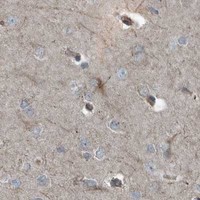

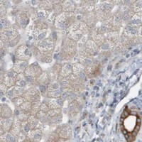


Comprehensive Human Protein Atlas IHC scoring per tissue (reliability: Enhanced — Medium consistency between antibody staining and RNA expression data.). Rows are taken directly from the HPA tissue chart — click any row's HPA link to view the source.
| Tissue | Cell type | Level | Evidence | Source |
|---|---|---|---|---|
| Adrenal gland | Glandular cells | High | Protein (IHC) | HPA → |
| Appendix | Glandular cells | High | Protein (IHC) | HPA → |
| Bronchus | Respiratory epithelial cells | High | Protein (IHC) | HPA → |
| Cerebellum | Purkinje cells | High | Protein (IHC) | HPA → |
| Colon | Glandular cells | High | Protein (IHC) | HPA → |
Troubleshoot GCC2 staining in paraffin sections by checking retrieval, cellular pattern and controls before assigning a chromogenic score.
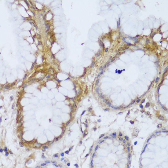
A07107 has IHC images from paraffin-embedded human colon and stomach (catalog image captions). It is listed as reactive with human and mouse samples (catalog reactivity).
A07107 will render with a human colon IHC image; the catalog also provides a human stomach IHC image, both using paraffin sections (catalog image captions). Its listed applications are IHC and WB, and its listed reactivity is human and mouse (catalog applications; catalog reactivity).
Which to pick: Choose A07107 for tissue IHC on paraffin sections; its images show human colon and stomach, while the fixative is unreported (catalog image captions). For mouse tissue, A07107 has listed reactivity but no mouse IHC image in the payload (catalog reactivity; catalog image captions). No IF/ICC application or image is listed for A07107, so this payload does not support an IF/ICC pick (catalog applications; catalog image captions).