GCLC / Glutamate--cysteine ligase catalytic subunit · IHC design guide

Design Immunohistochemistry for GCLC

Plan chromogenic GCLC IHC in paraffin sections using an IHC-validated antibody at 2–5 μg/mL (datasheet A01722). Expect variable cytoplasmic staining and interpret intensity cautiously because antibody staining has low agreement with RNA levels (HPA tissue IHC).

Evidence assembled Oct 2026 · For research use; verify linked source records and product datasheet before use
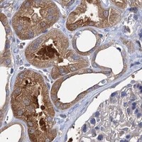
Immunohistochemistry protocol sheet for GCLC (IHC for GCLC): expected localisation Cytoplasmic staining in tissue sections (HPA tissue IHC), antibody A01722, validated IHC image, and IHC protocol steps
Printable GCLC IHC protocol sheet — expected localisation Cytoplasmic staining in tissue sections (HPA tissue IHC), antibody A01722, controls and protocol steps. Open the full GCLC IHC guide →

GCLC Immunohistochemistry Experimental Design Guide

Expected localisation, validated protocols, controls and antibodies — the at-a-glance facts below, then the full design guide.

Must know before staining
Expected localisation Cytoplasmic staining in tissue sections (HPA tissue IHC)
Staining pattern Variable cytoplasmic staining; glandular and urothelial cells high (HPA tissue IHC)
Antigen retrieval EDTA pH 8.0 HIER, heat-mediated (datasheet A01722)
Positive control ⓘ Fallopian tube+4 more · see all
Negative control ⓘ Ovary
Important caveats
Reasons your staining may differ from the expected pattern.
Fixation Keep fixation consistent across paraffin sections. (standard IHC practice; not target-specific)
Caveat Antibody staining has low agreement with RNA levels (HPA tissue IHC)
Regulation Tissue-variable protein expression (HPA tissue IHC)
Isoform / epitope No annotated isoforms or cleavage to alter the epitope map (UniProt)
Section 1

Recommended GCLC IHC & IF Protocols

The catalog antibody protocol is followed by published GCLC IHC methods for hepatocellular carcinoma tissue arrays and animal-model liver sections (PMC6603424; PMC7434920).

Recommended immunohistochemistry (IHC-P) protocol parameters
SampleParaffin-embedded human colon cancer tissue; fixative not specified (datasheet A01722)
FixationImage fixative and duration unreported (datasheet A01722); verify before use.
Sectioning4–5 µm sections on charged slides (standard)
DeparaffinisationXylene, graded ethanol series to water (standard)
Antigen retrievalHeat retrieval: EDTA pH 8.0 (datasheet A01722); 20 min, 95–100 °C (standard)
Peroxidase block3% H2O2, 10 min, room temperature (standard)
Blocking10% goat serum (datasheet A01722)
Primary antibodyRabbit anti-GCLC, 2-5 μg/ml (datasheet A01722)
Primary incubationOvernight at 4 °C (datasheet A01722)
DetectionHRP-conjugated secondary, DAB chromogen (datasheet A01722)
CounterstainHematoxylin, blue, dehydrate and mount (standard)
Expected resultGCLC-positive staining in glandular cells of fallopian tube (HPA tissue IHC: High). HPA tissue profile: Cytoplasmic expression at variable levels. No signal in the no-primary control.
💡Decision noteStart with heat-mediated EDTA retrieval at pH 8.0 for the catalog antibody (datasheet A01722); adjust using tissue controls if needed (standard IHC practice).
Section 2

What Is the Expected GCLC Staining Pattern?

In paraffin-section IHC, expect variable cytoplasmic GCLC staining, with high staining in fallopian-tube glandular cells and urinary-bladder urothelial cells (HPA tissue IHC: Approved; low consistency with RNA). GCLC has no annotated transmembrane segment or signal peptide (UniProt P48506 topology and processing). HPA ICC-IF reports mainly nucleoplasmic localization, with additional nucleolar and cytosolic signal; interpret that finding in its separate assay context (HPA subcellular ICC-IF).

What am I looking at on my slide?
Cytoplasmic staining is strong in fallopian-tube glandular cells or urinary-bladder urothelial cells.This matches the reported high-staining cell populations. Compare cells within the tissue, because HPA describes variable cytoplasmic expression overall (HPA tissue IHC).
A tissue section shows exclusively nuclear staining, without convincing cytoplasmic signal.This is discordant with the HPA tissue-IHC pattern and warrants an artefact check (HPA tissue IHC). Nuclear localization is reported separately by ICC-IF (HPA subcellular ICC-IF).
Staining predominates in cells outside the reported positive populations.Treat unexpected cell staining as unconfirmed; antibody cross-reactivity or endogenous chromogen-producing activity can mimic a positive result (standard IHC practice).
Colour spreads across cells and empty areas, obscuring cell boundaries.Diffuse background prevents a reliable cell-level localization call. Inspect the negative control and detection reagents before scoring GCLC (standard IHC practice).
Neither reported high-staining cell population shows signal.A blank result in a known-positive specimen leaves assay performance unresolved; check tissue preservation, retrieval, antibody incubation and detection (HPA tissue IHC; standard IHC practice).
💡Expected GCLC appearanceCall a positive tissue-IHC result when cytoplasmic staining is clearly visible in high-staining fallopian-tube glandular or urinary-bladder urothelial cells; isolated nuclear colour or diffuse background alone is insufficient (HPA tissue IHC; standard IHC practice).
How each factor affects the staining
Tissue and cell choiceHPA reports high staining in two specified cell populations, medium or low staining elsewhere, and no detection in ovarian stroma cells; choose and score the relevant cells (HPA tissue IHC).
Confidence in tissue patternThe HPA tissue-IHC assessment is Approved but notes low consistency between antibody staining and RNA expression. Interpret unexpected positives with that limitation in mind (HPA tissue IHC).
Antibody evidenceHPA036359, HPA036360 and CAB009569 each have an Approved IHC status; the supplied record does not assign them an Enhanced IHC status (HPA antibodies).
Topology and processingUniProt annotates no transmembrane segment or signal peptide and lists a single 1–637 chain. These annotations support checking cellular localization but do not establish retrieval conditions (UniProt P48506 topology and processing).
IF/ICC Q&A: should nuclear signal match tissue IHC?HPA ICC-IF places GCLC mainly in nucleoplasm, additionally in nucleoli and cytosol; HPA tissue IHC reports variable cytoplasmic staining. Judge each result against its assay-specific reference (HPA subcellular ICC-IF; HPA tissue IHC).
Why is my staining missing, weak or wrong?
SituationLikely causeNext action
Reported high-staining cells are weak or blank.The positive control has not demonstrated a working stain (HPA tissue IHC; standard IHC practice).Confirm the expected cells are present; review retrieval, primary-antibody incubation and detection against the assay's validated procedure (standard IHC practice).
Signal is exclusively nuclear in tissue IHC.The result conflicts with the reported cytoplasmic tissue pattern; background or misidentification remains possible (HPA tissue IHC; standard IHC practice).Check morphology and a negative control, then repeat with an IHC-validated antibody before assigning a tissue-IHC localization (HPA antibodies; standard IHC practice).
Unexpected cells stain as strongly as reported positives.Cross-reactivity or endogenous detection activity may imitate specific staining (standard IHC practice).Compare the cell distribution with HPA images; inspect an appropriate negative control and the detection-system controls (HPA tissue IHC; standard IHC practice).
Diffuse brown background obscures cellular detail.Nonspecific antibody binding or inadequately controlled endogenous detection activity may raise background (standard IHC practice).Review blocking, washes, primary-antibody concentration and the negative control; score only clearly cellular signal (standard IHC practice).
Ovarian stroma cells stain unexpectedly.HPA reports GCLC as not detected specifically in ovarian stroma cells; the result needs confirmation (HPA tissue IHC).Check cell identity, background and controls before calling these cells positive; do not generalize the reference to every cell in the ovary (HPA tissue IHC; standard IHC practice).
IF/ICC shows nuclear signal while tissue IHC is cytoplasmic.Both locations appear in HPA's separate assay records: mainly nucleoplasmic in ICC-IF and variable cytoplasmic in tissue IHC (HPA subcellular ICC-IF; HPA tissue IHC).Report the assay and compartment separately; use the tissue-IHC reference when interpreting the chromogenic paraffin section (HPA tissue IHC; HPA subcellular ICC-IF).

Sample controls for GCLC IHC & IF

🧪Run fallopian tube first and expect glandular cells to stain (HPA: High in fallopian tube glandular cells); use ovarian stroma as the negative tissue (HPA: Not detected in ovarian stroma cells). On the positive slide, assess morphologically distinct non-glandular cells for staining at background level, without assuming they are GCLC-negative (HPA: High is reported for glandular cells).
Positive control tissue: Fallopian tube (Glandular cells, HPA High)
Negative control tissue: Ovary (HPA Not detected)
ICC-IF cell lines (HPA subcellular resource): HPA ICC-IF images show GCLC in A-431, U-251MG, U2OS, with annotated localisation: Nucleoplasm (approved) (HPA subcellular).
Technical controls: Include no-primary (secondary-only) and concentration-matched rabbit IgG isotype controls, matching the primary antibody’s clonality where applicable (caption: rabbit anti-GCLC primary; standard IHC practice). A GCLC knockout is a biological negative control; block endogenous peroxidase before HRP/DAB detection in the fallopian tube section (standard IHC practice; caption: HRP/DAB detection).
⚠️Feasibility: A target-specific fixation window or fixation effect is unreported, and the selected A01722 tissue-IHC caption does not state the fixative (caption: fixative not stated). The reported paraffin-section procedure uses heat retrieval in EDTA at pH 8.0, but the evidence does not establish whether frozen sections or IF are easier (caption: EDTA heat retrieval; HPA: ICC-IF images reported). No fallopian tube-specific artefact is reported in the supplied evidence (HPA: fallopian tube staining; caption: colon cancer tissue).

HPA tissue IHC evidence for GCLC

Comprehensive Human Protein Atlas IHC scoring per tissue (reliability: Approved — Low consistency between antibody staining and RNA expression data.). Rows are taken directly from the HPA tissue chart — click any row's HPA link to view the source.

Positive expression · recommended positive controls

TissueCell typeLevelEvidenceSource
Fallopian tube Glandular cells High Protein (IHC) HPA →
Urinary bladder Urothelial cells High Protein (IHC) HPA →
Adipose tissue Adipocytes Medium Protein (IHC) HPA →
Adrenal gland Glandular cells Medium Protein (IHC) HPA →
Bone marrow Hematopoietic cells Medium Protein (IHC) HPA →

Undetected expression · recommended negative controls

TissueCell typeLevelEvidenceSource
Ovary Ovarian stroma cells Not detected Protein (IHC) HPA →
Section 3

Advanced GCLC IHC Tips

Troubleshoot GCLC staining in paraffin sections using the catalog antibody’s tissue IHC evidence and the reported cellular localisation data.

Which retrieval conditions should I start with for GCLC in paraffin sections?
Start with heat mediated antigen retrieval in EDTA at pH 8.0 (datasheet A01722). This condition preceded GCLC detection in a paraffin section of human colon cancer incubated with 2 μg/ml primary antibody overnight at 4°C (datasheet A01722). If staining is weak, adjust heating time in small increments while keeping the buffer constant, and compare sections from the same specimen (standard IHC practice). Include a section processed without primary antibody to check whether stronger heating increases background (standard IHC practice). Record retrieval time and tissue integrity alongside signal so apparent gains from damaged sections are not mistaken for improved staining (standard IHC practice).
How should I troubleshoot weak GCLC staining when fixation history is uncertain?
The selected antibody image documents a paraffin section but does not state its fixative, so target specific fixation sensitivity is unknown (datasheet A01722). Compare sections with documented fixation histories using the same EDTA pH 8.0 retrieval and primary antibody conditions (datasheet A01722; standard IHC practice). If only one block is available, inspect morphology and compare intact central tissue with edges before changing the antibody concentration (standard IHC practice). Run a no primary control on the same block to separate nonspecific chromogen deposition from candidate GCLC staining (standard IHC practice). Record fixation duration when available, since it is a potential source of variation in paraffin section staining (standard IHC practice).
Should nuclear staining be scored as GCLC in chromogenic IHC?
Assess cytoplasmic staining first because tissue IHC reports variable cytoplasmic expression, including high staining in fallopian tube glandular cells and urinary bladder urothelial cells (HPA tissue IHC). Nuclear signal also deserves review: cell imaging reports approved nucleoplasmic localisation, with additional nucleolar and cytosolic localisation (HPA subcellular). UniProt provides no subcellular annotation, so it does not independently resolve a nuclear versus cytoplasmic IHC pattern (UniProt P48506). Score nuclear and cytoplasmic compartments separately, using matched no primary controls and consistent counterstaining to check apparent nuclear signal (standard IHC practice). Do not infer a membrane pattern from this protein’s lack of a transmembrane segment (UniProt P48506 topology).
Could epitope accessibility explain discordant GCLC staining across sections?
The supplied record lists 0 isoforms and one chain spanning residues 1–637, so it supplies no isoform specific staining explanation (UniProt P48506). It lists no transmembrane segment, no glycosylation sites, and modified residues including phosphoserines at positions 5 and 8 (UniProt P48506). The catalog caption does not identify the antibody epitope, so do not attribute a staining change to those modifications without additional evidence (datasheet A01722). Compare retrieval and primary incubation on adjacent sections before assigning a molecular cause to signal loss (standard IHC practice). Keep compartment and cell type scores separate when comparing sections (HPA tissue IHC; standard IHC practice).
How can I assess GCLC by multiplex IF alongside the IHC result?
Treat IF as a separate optimisation because the selected antibody evidence describes chromogenic staining of a paraffin section (datasheet A01722). Pair GCLC with a marker for the expected cell type, such as an appropriate glandular or urothelial marker when examining those HPA positive populations (HPA tissue IHC; standard IF practice). Choose fluorophores in channels with low measured tissue autofluorescence, and include single stain and no primary controls for bleed through and background (standard IF practice). For an intracellular epitope, optimise permeabilisation to permit access to cytosolic or nuclear compartments without losing morphology (HPA subcellular; standard IF practice). Assess nuclear and cytosolic signal separately because both are reported in cell imaging (HPA subcellular).
What should I check when GCLC DAB staining appears diffuse?
First compare a no primary section with the stained section to identify background from detection reagents or tissue components (standard IHC practice). The selected workflow used a peroxidase conjugated secondary antibody and DAB, so include an endogenous peroxidase block as a general chromogenic IHC step (datasheet A01722; standard IHC practice). Its tissue section was blocked with 10% goat serum before incubation with 2 μg/ml primary antibody overnight at 4°C (datasheet A01722). If the control is clean, compare shorter development or lower primary concentration on adjacent sections while retaining EDTA pH 8.0 retrieval (datasheet A01722; standard IHC practice). Interpret diffuse colour against preserved cell boundaries and the reported cytoplasmic tissue pattern (HPA tissue IHC).
How should I quantify variable GCLC staining across tissue regions? ⚠ ANSWER MARKED FOR VERIFICATION
Define the cell population and compartment before scoring because tissue IHC reports variable cytoplasmic GCLC expression (HPA tissue IHC). For comparable regions, report the percentage of positive cells and a cytoplasmic H-score using intensity categories 0–3 applied consistently across slides (standard IHC practice). Normalise positive cell counts to the number of evaluable cells, or report positive cell density per mm² of viable tissue (standard IHC practice). Exclude necrotic and folded areas using the same rules for every section, and keep nuclear signal as a separate readout (standard IHC practice; HPA subcellular). Keep retrieval, incubation, DAB development, and image exposure consistent across the comparison set (standard IHC practice).
How can I distinguish plausible GCLC staining from section artefact?
Look for staining within intact cells and compare its compartment with the variable cytoplasmic tissue pattern reported for GCLC (HPA tissue IHC; standard IHC practice). High signal in fallopian tube glandular cells or urinary bladder urothelial cells is plausible, while ovarian stromal cells were reported as not detected (HPA tissue IHC). Nuclear staining needs separate review because nucleoplasmic, nucleolar, and cytosolic localisation comes from cell imaging rather than the tissue IHC profile (HPA subcellular; HPA tissue IHC). Discount signal concentrated at cut edges, folds, or necrotic areas, and check no primary sections for endogenous enzyme related DAB colour (standard IHC practice). Treat discordance cautiously because HPA rates tissue staining approved but reports low consistency with RNA expression (HPA tissue IHC).
Boster reagents

Best GCLC / Glutamate--cysteine ligase catalytic subunit IHC Antibodies

A01722 has human paraffin-section IHC and IF images, while PB9201 has a human paraffin-section IHC image (catalog image captions). Both list human, mouse and rat reactivity (catalog reactivity).

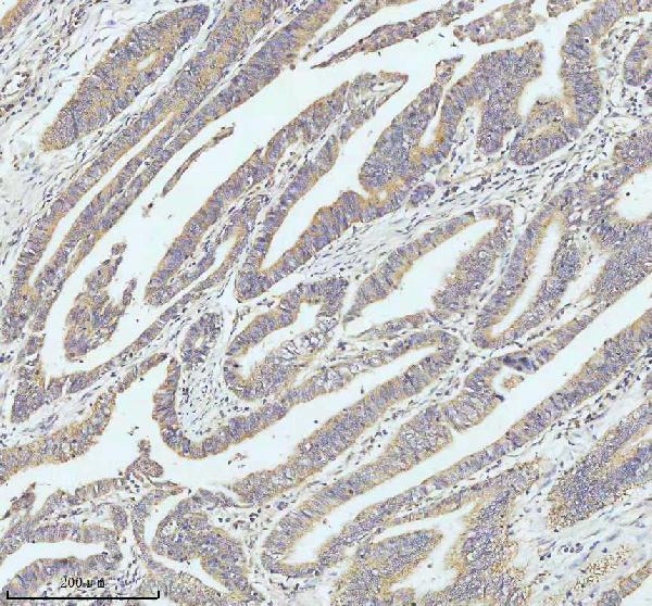
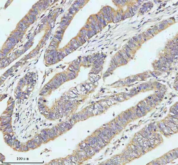
Real IHC data IHC analysis of GCLC using anti-GCLC antibody (A01722). GCLC was detected in a paraffin-embedded section of human colon cancer tissue. Heat mediated antigen retrieval was performed in EDTA buffer (pH 8.0, epitope retrieval solution). The tissue section was blocked with 10% goat serum. The tissue section was then incubated with 2 μg/ml rabbit anti-GCLC Antibody (A01722) overnight at 4°C. Peroxidase Conjugated Goat Anti-rabbit IgG was used as secondary antibody and incubated for 30 minutes at 37°C. The tissue section was developed using HRP Conjugated Rabbit IgG Super Vision Assay Kit (Catalog # SV0002) with DAB as the chromogen.
Anti-GCLC Antibody ®
Cat # A01722

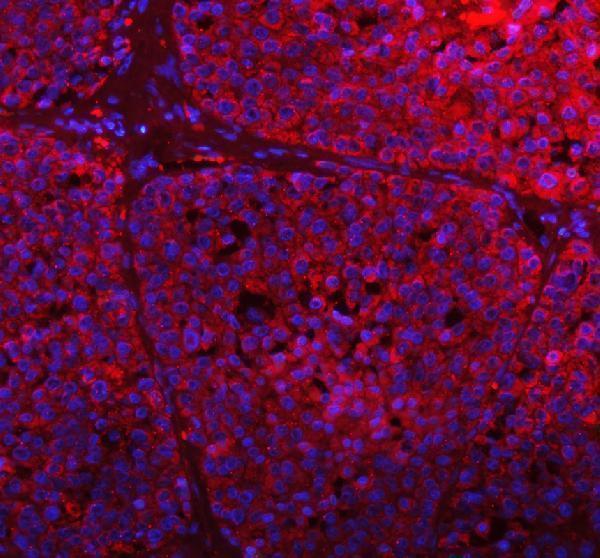
The rendered card is A01722: its IHC captions show human colon and liver cancer paraffin sections, and its IF caption shows a human liver cancer paraffin section (A01722 image captions). A01722 lists IHC and IF applications and human, mouse and rat reactivity; its supplied tissue images are human only (A01722 catalog applications/reactivity; A01722 image captions).

Which to pick: For tissue IHC, choose A01722 for its human colon and liver cancer paraffin-section examples; PB9201 has its own human colorectal cancer paraffin-section example, and neither SKU’s caption reports the fixative (A01722 IHC captions; PB9201 IHC caption). For IF, A01722 has a human liver cancer paraffin-section image; M01722-2 is a rabbit monoclonal that lists IF but supplies no IF image, and ICC validation is unreported (A01722 IF caption; M01722-2 catalog title/applications/image alts; catalog applications). For mouse or rat work, A01722 lists both IHC and IF alongside reactivity with those species, but its supplied tissue images are human (A01722 catalog applications/reactivity; A01722 image captions).

Each figure is that product's own IHC / IF validation image from its datasheet.

References

  1. UniProt Consortium. UniProt entry P48506 (GSH1_HUMAN, Glutamate--cysteine ligase catalytic subunit).
  2. Human Protein Atlas. GCLC tissue IHC expression (reliability: Approved).
  3. Human Protein Atlas. GCLC subcellular location (ICC-IF): Mainly localized to the nucleoplasm. In addition localized to the nucleoli and cytosol..
  4. Human Protein Atlas. GCLC antibody validation summary (3 antibodies).
  5. High GCLC level in tumor tissues is associated with poor prognosis of hepatocellular carcinoma after curative resection. Journal of Cancer 2019 — PMC6603424.
  6. Computational repositioning of dimethyl fumarate for treating alcoholic liver disease. Cell death & disease 2020 — PMC7434920.
  7. Neuronal glutathione loss leads to neurodegeneration involving gasdermin activation. Scientific reports 2023 — PMC9859798.
  8. Characterization of the proteome of stable and unstable carotid atherosclerotic plaques using data-independent acquisition mass spectrometry. Journal of translational medicine 2024 — PMC10921703.
  9. PubMed PMID:1350904 — UniProt-cited evidence.
  10. PubMed PMID:14574404 — UniProt-cited evidence.
  11. PubMed PMID:15489334 — UniProt-cited evidence.