GCNT2 / N-acetyllactosaminide beta-1,6-N-acetylglucosaminyl-transferase · IHC design guide

Design Immunohistochemistry for GCNT2

Plan GCNT2 chromogenic IHC around granular cytoplasmic tissue staining (HPA tissue IHC) and a Golgi membrane expectation (UniProt). The catalog antibody's human paraffin-section range is 2–5 μg/ml (datasheet A07194); score cell type and staining intensity with matched controls.

Evidence assembled Oct 2026 · For research use; verify linked source records and product datasheet before use
Immunohistochemistry protocol sheet for GCNT2 (IHC for GCNT2): expected localisation Granular cytoplasm (HPA tissue IHC); Golgi membrane (UniProt), antibody A07194, validated IHC image, and IHC protocol steps
Printable GCNT2 IHC protocol sheet — expected localisation Granular cytoplasm (HPA tissue IHC); Golgi membrane (UniProt), antibody A07194, controls and protocol steps. Open the full GCNT2 IHC guide →

GCNT2 Immunohistochemistry Experimental Design Guide

Expected localisation, validated protocols, controls and antibodies — the at-a-glance facts below, then the full design guide.

Must know before staining
Expected localisation Granular cytoplasm (HPA tissue IHC); Golgi membrane (UniProt)
Staining pattern Granular cytoplasmic staining across tissues (HPA tissue IHC)
Antigen retrieval EDTA pH 8.0 HIER, heat-mediated (datasheet A07194)
Positive control ⓘ Adrenal gland+4 more · see all
Negative control ⓘ None in HPA (detected in all 45 tissues); use no-primary + isotype controls
Important caveats
Reasons your staining may differ from the expected pattern.
Fixation Keep fixation consistent across sections (standard IHC practice; not target-specific)
Caveat Staining shows low concordance with RNA expression (HPA tissue IHC)
Regulation Prostate-enhanced RNA (HPA tissue RNA)
Isoform / epitope 3 isoforms; epitope coverage unknown (UniProt)
Section 1

Recommended GCNT2 IHC & IF Protocols

Compare the catalog antibody’s IHC-P protocol (datasheet A07194) with four published GCNT2 IHC protocols (PMC4814258; PMC6105653; PMC11940493; PMC3903410).

Recommended immunohistochemistry (IHC-P) protocol parameters
SampleParaffin-embedded human colon tissue; fixative not specified (datasheet A07194)
FixationImage fixative and duration unreported (datasheet A07194); verify before use.
Sectioning4–5 µm sections on charged slides (standard)
DeparaffinisationXylene, graded ethanol series to water (standard)
Antigen retrievalHeat retrieval: EDTA pH 8.0 (datasheet A07194); 20 min, 95–100 °C (standard)
Peroxidase block3% H2O2, 10 min, room temperature (standard)
Blocking10% goat serum (datasheet A07194)
Primary antibodyRabbit anti-GCNT2, 2-5 μg/ml (datasheet A07194)
Primary incubationOvernight at 4 °C (datasheet A07194)
DetectionHRP-conjugated secondary, DAB chromogen (datasheet A07194)
CounterstainHematoxylin, blue, dehydrate and mount (standard)
Expected resultGCNT2-positive staining in glandular cells of adrenal gland (HPA tissue IHC: Medium). HPA tissue profile: Granular cytoplasmic expression in all tissues. No signal in the no-primary control.
💡Decision noteStart with heat-mediated EDTA retrieval at pH 8.0 for the catalog antibody (datasheet A07194); the published protocols do not specify a retrieval solution (PMC4814258; PMC6105653; PMC11940493; PMC3903410).
Section 2

What Is the Expected GCNT2 Staining Pattern?

GCNT2 is a Golgi membrane enzyme with a short cytoplasmic segment and a lumenal catalytic region (UniProt Q8N0V5 topology). In paraffin IHC, expect granular cytoplasmic staining in HPA-reported glandular, hematopoietic, neuronal, and glial cells (HPA tissue IHC). HPA rates the tissue staining Approved, while reporting low consistency with RNA expression and pending external verification (HPA tissue IHC reliability).

What am I looking at on my slide?
Granular cytoplasmic signal in glandular cells of breast, appendix, adrenal gland, or bronchus.This fits the reported Medium staining in those cells (HPA tissue IHC). A compact, Golgi-like distribution is consistent with GCNT2 localization, though chromogenic IHC alone cannot establish organelle identity (UniProt Q8N0V5; HPA subcellular).
Granular staining in bone marrow hematopoietic cells or selected brain cells.HPA reports Medium staining in marrow hematopoietic cells, caudate neurons, cerebellar granular-layer cells, and cortical glia (HPA tissue IHC). Score the named cell population rather than treating the entire section as uniformly positive.
Predominantly nuclear, sharply surface-restricted, or extracellular staining.These patterns do not fit the reported granular cytoplasm or Golgi location (HPA tissue IHC; UniProt Q8N0V5). Suspect artefact or off-target signal and review the staining controls before assigning GCNT2 positivity (standard IHC practice).
Strong signal in an unexpected cell population, particularly relative to nearby reported cells.Consider cross-reactivity or endogenous chromogenic detection activity (standard IHC practice). HPA describes granular expression across tissues, so an unlisted cell type is a prompt to verify specificity, not proof that its staining is false (HPA tissue IHC).
No signal in a section containing an HPA-reported Medium cell population.A technical failure is possible, but an HPA observation is not a guaranteed positive control for every specimen (HPA tissue IHC). Check tissue preservation, primary antibody, retrieval, and detection with appropriate controls (standard IHC practice).
💡Expected GCNT2 appearanceCall a result plausible when reported cells show granular cytoplasmic, Golgi-consistent signal at roughly Medium intensity; isolated nuclear or diffuse extracellular colour warrants review (HPA tissue IHC; UniProt Q8N0V5).
How each factor affects the staining
Compartment and topologyGCNT2 occupies the Golgi membrane; residues 1–7 face cytoplasm and 24–400 face the lumen (UniProt Q8N0V5 topology). The antibody epitope is unspecified, so topology alone cannot determine antigen retrieval needs.
Tissue and cell contextHPA lists Medium staining in several glandular populations and selected marrow and brain cells, while adipocytes are Low (HPA tissue IHC). These are useful comparisons, not absolute positive or negative rules.
Evidence strengthThe HPA tissue result is Approved but has low antibody-staining/RNA consistency and awaits external verification; HPA026776 IHC is Approved, not Enhanced (HPA tissue IHC reliability; HPA antibodies). Interpret unexpected staining conservatively.
Isoforms and modificationUniProt records 3 isoforms, A, B, and C, and one glycosylation site at residue 41 (UniProt Q8N0V5). No epitope or isoform-specific staining evidence is supplied; morphology cannot identify an isoform.
Independent localization contextICC-IF places GCNT2 at the Golgi in CACO-2 and U2OS, with an Approved location (HPA subcellular; HPA antibodies). This supports a localization expectation but does not validate paraffin-section intensity or cell-type specificity.
Why is my staining missing, weak or wrong?
SituationLikely causeNext action
A reported Medium population is blank.The IHC run may have failed, or this specimen may differ from HPA examples (HPA tissue IHC; standard IHC practice).Verify a suitable positive control and inspect retrieval, primary incubation, and chromogenic detection records before interpreting the blank section (standard IHC practice).
Colour is diffuse across tissue or present in the negative control.Nonspecific binding or detection background can obscure the granular pattern (standard IHC practice; HPA tissue IHC profile).Check blocking, washing, detection reagents, and the primary-omission control; reassess only after background is controlled (standard IHC practice).
Nuclei or extracellular material dominate the signal.That distribution conflicts with Golgi membrane localization and HPA's granular cytoplasmic profile (UniProt Q8N0V5; HPA tissue IHC).Compare positive and negative controls, inspect counterstain and section morphology, and avoid scoring the misplaced colour as GCNT2 (standard IHC practice).
An unexpected cell type stains more strongly than the reported cells.Cross-reactivity or endogenous detection activity is possible; HPA's cell list is not exhaustive (standard IHC practice; HPA tissue IHC).Check a primary-omission control and cell morphology, then seek an independent specificity check before making a cell-specific claim (standard IHC practice).
Adipocytes show prominent staining.HPA reports adipocytes as Low, making strong staining a result that needs scrutiny rather than an automatic exclusion (HPA tissue IHC).Compare adjacent reported populations and controls, then document the discrepancy without treating Low as a guaranteed negative (HPA tissue IHC; standard IHC practice).
IF/ICC Q: Should signal appear at the Golgi?HPA reports Approved Golgi localization in CACO-2 and U2OS ICC-IF images (HPA subcellular; HPA antibodies).A: Use Golgi localization as a qualitative cross-check; assess IF/ICC workflow details in its separate guide (HPA subcellular).

Sample controls for GCNT2 IHC & IF

🧪Run breast first and score its glandular cells, which show medium GCNT2 staining (HPA: Breast, glandular cells, Medium). HPA lists no negative tissue and detects GCNT2 in all 45 scored tissues (HPA: no negative rows; detected in all 45 scored tissues), so use no-primary and isotype controls for the negative reference; compare any unstained nonglandular cells on the positive slide with those controls without assuming those cells are GCNT2-negative.
Positive control tissue: Adrenal gland (Glandular cells, HPA Medium)
Negative control tissue: None in HPA: GCNT2 is detected in all 45 scored tissues. Use a no-primary (secondary-only) and an isotype control instead.
ICC-IF cell lines (HPA subcellular resource): HPA ICC-IF images show GCNT2 in CACO-2, U2OS, with annotated localisation: Golgi apparatus (approved) (HPA subcellular).
Technical controls: Include a no-primary, secondary-only slide and a concentration-matched rabbit IgG isotype control for the rabbit primary (selected-SKU caption: rabbit anti-GCNT2 antibody), plus GCNT2 knockout material as a biological specificity control if available. For chromogenic detection, block endogenous peroxidase and check the breast section for tissue pigment or other background before scoring (standard IHC practice).
⚠️Feasibility: A target-specific fixation window or fixation effect is unreported, and the selected A07194 paraffin-section caption does not state the fixative (selected-SKU caption: fixative not stated). The documented paraffin workflow used heat-mediated retrieval in EDTA at pH 8.0, followed by 2 μg/ml primary antibody overnight at 4°C; this documents a working retrieval condition without establishing that retrieval is required (selected-SKU tissue-IHC caption). Frozen sections and IF are not established as easier by the supplied evidence; IF offers a Golgi localization check (HPA subcellular: Golgi apparatus), while glandular staining should be interpreted against peroxidase and tissue-background controls in chromogenic IHC (standard IHC practice).

HPA tissue IHC evidence for GCNT2

Comprehensive Human Protein Atlas IHC scoring per tissue (reliability: Approved — Low consistency between antibody staining and RNA expression data. Pending external verification.). Rows are taken directly from the HPA tissue chart — click any row's HPA link to view the source.

Positive expression · recommended positive controls

TissueCell typeLevelEvidenceSource
Adrenal gland Glandular cells Medium Protein (IHC) HPA →
Appendix Glandular cells Medium Protein (IHC) HPA →
Bone marrow Hematopoietic cells Medium Protein (IHC) HPA →
Breast Glandular cells Medium Protein (IHC) HPA →
Bronchus Respiratory epithelial cells Medium Protein (IHC) HPA →

Undetected expression · recommended negative controls

TissueCell typeLevelEvidenceSource
None in HPA: GCNT2 is detected in all 45 scored tissues. Use a no-primary (secondary-only) and an isotype control instead.
Section 3

Advanced GCNT2 IHC Tips

Use the documented paraffin section conditions as the starting point, then judge GCNT2 staining by its expected Golgi pattern and appropriate controls.

What retrieval condition should I start with for GCNT2 chromogenic IHC?
Start with heat-mediated antigen retrieval in EDTA at pH 8.0 for paraffin sections (datasheet A07194). Keep retrieval fixed while evaluating the documented 2 μg/ml antibody incubation overnight at 4°C (caption A07194). Compare adjacent sections with matched heating, cooling and DAB development to isolate retrieval effects (standard IHC practice). GCNT2 has a lumenal 24–400 region, but the antibody epitope is unspecified, so topology alone cannot predict retrieval sensitivity (UniProt Q8N0V5 topology; caption A07194). If staining remains weak, test an alternative retrieval condition on matched sections and retain the condition that improves granular cytoplasmic signal without raising background (HPA tissue IHC; standard IHC practice).
How should I handle uncertain fixation effects on GCNT2 staining?
Target-specific fixation sensitivity is unknown, and the selected paraffin section caption does not state a fixative (caption A07194). Record the fixative and fixation duration for each specimen, then compare sections processed under matched conditions (standard IHC practice). Use EDTA at pH 8.0 and the documented overnight antibody incubation at 4°C as starting conditions when assessing fixation differences (datasheet A07194; caption A07194). Include a concurrently processed control section so weak staining can be assessed against the same processing history (standard IHC practice). Do not attribute a loss of signal to fixation without comparing matched controls and checking retrieval and detection (standard IHC practice).
Which staining pattern supports GCNT2 localisation in paraffin sections?
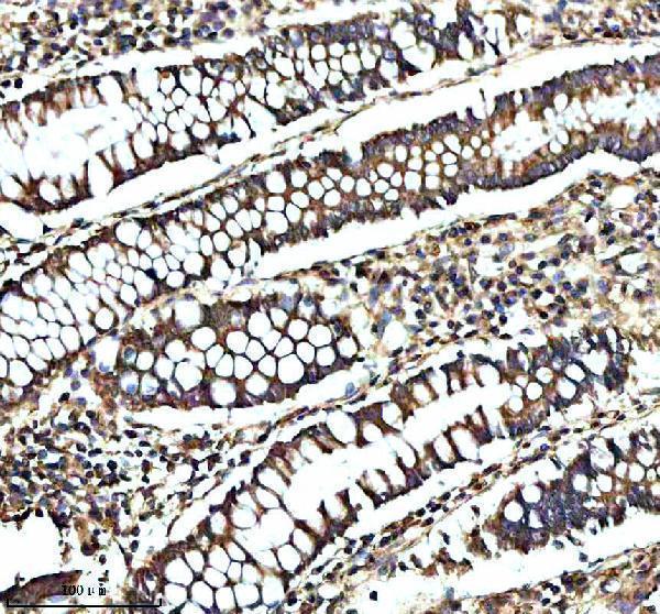
Expect predominantly granular cytoplasmic staining consistent with a Golgi membrane protein, rather than using diffuse colour alone as evidence (UniProt Q8N0V5 subcellular; HPA tissue IHC). GCNT2 has a transmembrane segment at residues 8–23 and a lumenal region at 24–400 (UniProt Q8N0V5 topology). Assess granules within intact cells using a counterstain, and compare them with staining in a no-primary control (standard IHC practice). The selected antibody produced staining in a paraffin section of human colon, although the caption does not assign it to a particular cell type (caption A07194). HPA reports granular cytoplasmic staining broadly but flags low consistency with RNA expression, so confirm localisation and cell identity independently (HPA tissue IHC).
Can this antibody distinguish GCNT2 isoforms or reveal epitope masking?
GCNT2 has 3 annotated isoforms, A, B and C, while this antibody’s epitope and isoform coverage are unspecified in the supplied evidence (UniProt Q8N0V5 isoforms; caption A07194). Therefore, do not assign isoform identity from chromogenic staining intensity or distribution alone (standard IHC interpretation). The protein has a transmembrane segment at 8–23 and an annotated glycosylation site at 41, but neither establishes whether this antibody binds nearby (UniProt Q8N0V5 topology and glycosylation). If staining differs across specimens, first repeat the documented EDTA pH 8.0 retrieval and matched detection (datasheet A07194; standard IHC practice). Establish epitope or isoform specificity with independent antibody documentation and suitable controls before interpreting those differences (standard IHC practice).
How can IF help check an ambiguous GCNT2 IHC pattern?
Use IF as a separate localisation check when granular IHC staining is difficult to assign to cells (HPA tissue IHC; standard IF practice). When examining glandular cells, multiplex GCNT2 with an epithelial cell marker and a Golgi marker to assess cell identity and compartment (HPA: appendix glandular cells; HPA subcellular; standard IF practice). Choose a far-red fluorophore and inspect an unstained specimen to distinguish signal from tissue autofluorescence (standard IF practice). Plan permeabilisation around the actual antibody epitope: GCNT2 has a short cytoplasmic 1–7 region and lumenal 24–400 region, but this epitope is unspecified (UniProt Q8N0V5 topology; caption A07194). HPA lists CACO-2 and U2OS IF images showing Golgi localisation; those observations do not establish this catalog antibody’s IF performance (HPA subcellular; caption A07194).
How do I separate GCNT2 signal from chromogenic background?
First compare the test section with no-primary and detection-only controls processed through the same DAB reaction (standard IHC practice). The documented colon staining used 10% goat serum blocking, 2 μg/ml primary antibody overnight at 4°C, and a peroxidase-conjugated secondary (caption A07194). Block endogenous peroxidase before detection, and keep DAB development matched across sections when assessing background (standard IHC practice). If staining remains widespread, titrate the primary antibody and check whether the same colour appears in control sections (standard IHC practice). Prefer discrete intracellular granules consistent with Golgi localisation over uniform deposits or staining unrelated to intact cells (HPA tissue IHC; UniProt Q8N0V5 subcellular; standard IHC interpretation).
What scoring method captures GCNT2 staining without misleading comparisons? ⚠ ANSWER MARKED FOR VERIFICATION
Define the cell population and viable tissue area before scoring, then apply the same rules to every section (standard IHC practice). An H-score combines the percentage of cells at intensity grades 0–3 into a 0–300 score; report percent positive cells alongside it (standard IHC practice). For sparse populations, positive-cell density per mm² of viable tissue can be more informative (standard IHC practice). Normalise comparisons to the same cell type or viable area, and keep retrieval, antibody concentration and DAB development consistent (standard IHC practice). Record granular cytoplasmic staining separately from diffuse colour because Golgi localisation is expected and HPA cautions that staining and RNA expression have low consistency (UniProt Q8N0V5 subcellular; HPA tissue IHC).
When should I doubt an apparent GCNT2-positive IHC result?
Give greatest weight to granular cytoplasmic staining in intact cells, consistent with GCNT2’s Golgi localisation (HPA tissue IHC; UniProt Q8N0V5 subcellular). Treat predominantly nuclear, uniform extracellular or edge-restricted staining as suspect until it survives matched controls (standard IHC interpretation). Exclude necrotic areas from scoring and check no-primary sections for endogenous peroxidase or other DAB deposits (standard IHC practice). The selected antibody has a paraffin section image of human colon, but its caption does not identify the stained cell type or fixative (caption A07194). HPA reports broad granular staining with low consistency against RNA data; corroborate unexpected positive cells with cell identity and independent evidence before assigning biological meaning (HPA tissue IHC; standard IHC practice).
Boster reagents

Best GCNT2 / N-acetyllactosaminide beta-1,6-N-acetylglucosaminyl-transferase IHC Antibodies

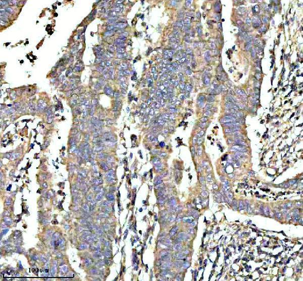
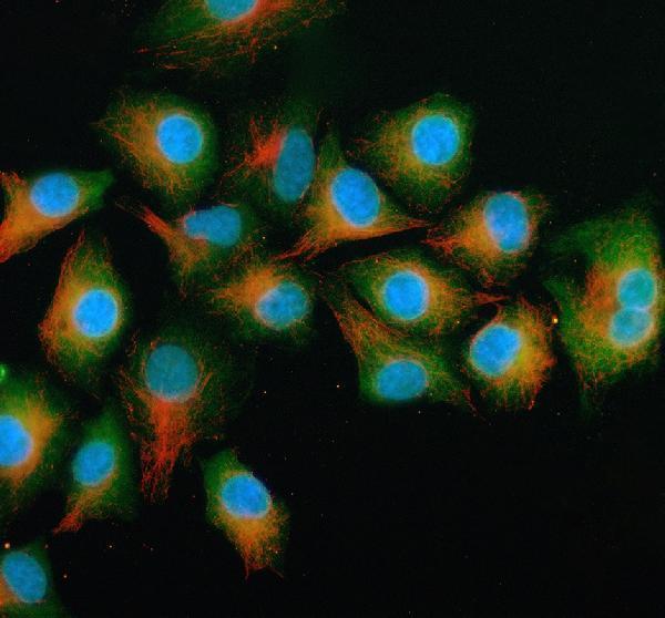
A07194 has real IHC images from human paraffin sections and IF images from A549 cells and human paraffin sections (catalog image captions).

Real IHC data IHC analysis of GCNT2 using anti-GCNT2 antibody (A07194). GCNT2 was detected in a paraffin-embedded section of human colon tissue. Heat mediated antigen retrieval was performed in EDTA buffer (pH 8.0, epitope retrieval solution). The tissue section was blocked with 10% goat serum. The tissue section was then incubated with 2 μg/ml rabbit anti-GCNT2 Antibody (A07194) overnight at 4°C. Peroxidase Conjugated Goat Anti-rabbit IgG was used as secondary antibody and incubated for 30 minutes at 37°C. The tissue section was developed using HRP Conjugated Rabbit IgG Super Vision Assay Kit (Catalog # SV0002) with DAB as the chromogen.
Anti-GCNT2 Antibody ®
Cat # A07194

A07194 is listed for IHC and IF/ICC, with IHC images from human colon, colon cancer, and prostate cancer paraffin sections (catalog applications and IHC captions). A07194 also has IF images from A549 cells and human intestine and prostate cancer paraffin sections; its listed reactivity is human, mouse, and rat (catalog IF captions and reactivity).

Which to pick: Choose A07194 for human paraffin-section chromogenic IHC: its colon caption documents EDTA retrieval at pH 8.0, a 10% goat serum block, 2 μg/ml primary antibody, and HRP/DAB detection (A07194 IHC caption). The same SKU is listed for IF/ICC, with IF images from A549 cells and human paraffin sections using 5 μg/ml primary antibody (catalog applications and A07194 IF captions). For mouse or rat samples, A07194 lists species reactivity, but the supplied IHC/IF images show human samples; clonality and the paraffin-section fixative are unreported (catalog reactivity, clone field, and image captions).

Each figure is that product's own IHC / IF validation image from its datasheet.

References

  1. UniProt Consortium. UniProt entry Q8N0V5 (GCNT2_HUMAN, N-acetyllactosaminide beta-1,6-N-acetylglucosaminyl-transferase).
  2. Human Protein Atlas. GCNT2 tissue IHC expression (reliability: Approved).
  3. Human Protein Atlas. GCNT2 subcellular location (ICC-IF): Localized to the Golgi apparatus..
  4. Human Protein Atlas. GCNT2 antibody validation summary (1 antibodies).
  5. I-branching N-acetylglucosaminyltransferase regulates prostate cancer invasiveness by enhancing α5β1 integrin signaling. Cancer science 2016 — PMC4814258.
  6. Loss of GCNT2/I-branched glycans enhances melanoma growth and survival. Nature communications 2018 — PMC6105653.
  7. Low GCNT2/I-Branching Glycan Expression Is Associated with Bladder Cancer Aggressiveness. Biomedicines 2025 — PMC11940493.
  8. Engagement of I-branching {beta}-1, 6-N-acetylglucosaminyltransferase 2 in breast cancer metastasis and TGF-{beta} signaling. Cancer research 2011 — PMC3903410.
  9. PubMed PMID:8449405 — UniProt-cited evidence.
  10. PubMed PMID:7579796 — UniProt-cited evidence.
  11. PubMed PMID:12424189 — UniProt-cited evidence.