This website uses cookies to ensure you get the best experience on our website.


- Table of Contents
This GCNT3 paraffin-section IHC guide uses the granular cytoplasmic pattern in gastrointestinal mucosa to set staining expectations (HPA tissue IHC). It highlights the Golgi membrane location (UniProt) and cautions that presumed off-target binding can complicate interpretation (HPA tissue IHC).

Expected localisation, validated protocols, controls and antibodies — the at-a-glance facts below, then the full design guide.
| Expected localisation | Granular cytoplasm in gastrointestinal mucosa (HPA tissue IHC) | |
| Staining pattern | Granular cytoplasmic staining in gastrointestinal mucosa (HPA tissue IHC) | |
| Antigen retrieval | Citrate pH 6.0 HIER, 95–98 °C, 20 min (rule: cytoplasmic / membrane antigen) | |
| Positive control | Appendix+4 more · see all | |
| Negative control | Adipose tissue+4 more · see all |
| Fixation | Matched tissue-IHC evidence does not establish the fixation claim. Validate the specimen-specific method before use. (selected-SKU IHC image A08909-1) | |
| Caveat | Presumed off-target binding may mimic GCNT3 staining (HPA tissue IHC) | |
| Regulation | Expression regulation is not annotated (UniProt) | |
| Isoform / epitope | No isoforms; lumenal vs cytoplasmic epitope affects access (UniProt) |
The catalog antibody protocol is accompanied by 3 published GCNT3 IHC protocols (PMC11651748; PMC13376619; PMC13126822).
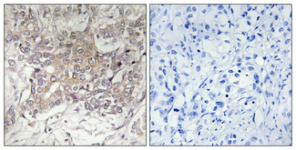
| Sample | Paraffin-embedded human lung cancer tissue; fixative not specified (datasheet A08909-1) |
| Fixation | Image fixative and duration unreported (datasheet A08909-1); verify before use. |
| Sectioning | 4–5 µm sections on charged slides (standard) |
| Deparaffinisation | Xylene, graded ethanol series to water (standard) |
| Antigen retrieval | Heat-induced epitope retrieval in citrate buffer, pH 6.0, 20 min at 95–98 °C (standard rule: cytoplasmic / membrane antigen) |
| Peroxidase block | 3% H2O2, 10 min, room temperature (standard) |
| Blocking | 10% normal serum of the secondary host, 30 min, room temperature (standard) |
| Primary antibody | Rabbit anti-GCNT3, 1:100-1:300 (datasheet A08909-1) |
| Primary incubation | Overnight at 4 °C (standard) |
| Detection | HRP-polymer secondary, DAB chromogen 5–10 min (standard) |
| Counterstain | Hematoxylin, blue, dehydrate and mount (standard) |
| Expected result | GCNT3-positive staining in enterocytes of appendix (HPA tissue IHC: High). HPA tissue profile: Granular cytoplasmic expression in gastrointestinal mucosa. No signal in the no-primary control. |
GCNT3 is a single-pass Golgi membrane protein with a short cytoplasmic tail and a luminal catalytic region (UniProt O95395 topology). In paraffin-section IHC, expect granular cytoplasmic staining chiefly in gastrointestinal mucosal cells, including appendix and rectal enterocytes (HPA: tissue IHC). HPA rates the tissue profile Enhanced, while noting medium agreement with RNA data and presumed off-target staining that it disregarded (HPA: reliability description).
| Granular cytoplasmic staining in appendix or rectal enterocytes, with a clear cell outline. | This fits the reported gastrointestinal mucosal pattern and the High staining levels in these cells (HPA: tissue IHC). A Golgi-associated target can appear as cytoplasmic granules in chromogenic sections; the stain alone does not resolve the Golgi membrane (UniProt O95395 localization; general IHC practice). |
| Predominantly nuclear staining, or a continuous outline of the cell surface. | Neither is the expected dominant compartment for GCNT3, which is assigned to the Golgi membrane and has one membrane-spanning segment (UniProt O95395 localization and topology). Treat an isolated pattern of this kind as suspect; compare it with a positive tissue control and a detection-only control before assigning it to GCNT3 (general IHC practice). |
| Strong staining in cells reported as Not detected, such as adipocytes in adipose tissue or bronchial respiratory epithelial cells. | These cells are poor matches for the HPA tissue profile (HPA: adipose tissue and bronchus IHC). Consider antibody cross-reactivity or endogenous chromogen-generating activity, and inspect detection controls (general IHC practice). HPA reports that presumed off-target binding was observed and disregarded when assessing its tissue pattern (HPA: reliability description). |
| Uniform colour across epithelium, connective tissue and empty areas, without discernible cell-associated granules. | This does not resemble the reported granular cytoplasmic expression in gastrointestinal mucosa (HPA: tissue IHC profile). Broad, structure-independent colour can arise from background in chromogenic IHC; compare a section processed without primary antibody and assess blocking, washing and chromogen development (general IHC practice). |
| No staining in appendix or rectal enterocytes while the tissue morphology remains readable. | A negative result conflicts with the High levels reported for those cells (HPA: appendix and rectum IHC). First verify that the positive-control section and detection reagents worked; then review antibody dilution and antigen-retrieval conditions as general IHC variables (general IHC practice). The supplied sources do not establish GCNT3-specific fixation sensitivity. |
| Cell and tissue selection | Appendix and rectal enterocytes are reported High; colon enterocytes and stomach glandular cells are Medium (HPA: tissue IHC). Salivary and gallbladder glandular cells are Low (HPA: tissue IHC). Interpret a weak section against its stated cell type rather than expecting equal intensity across organs. |
| Compartment and epitope orientation | GCNT3 spans the membrane at residues 7–26; residues 1–6 are cytoplasmic and 27–438 are luminal (UniProt O95395 topology). These boundaries support a Golgi-associated interpretation but do not identify the antibody epitope or establish a GCNT3-specific retrieval requirement. |
| Evidence strength | HPA calls tissue IHC Enhanced and lists HPA011154 as IHC Enhanced, but describes medium staining-to-RNA consistency and disregarded presumed off-target binding (HPA: tissue reliability; HPA: antibody validation). An unexpected positive needs control-based review even when the expected pattern is present elsewhere. |
| IF/ICC Q&A: what localization is expected? | Mainly Golgi, with additional vesicle localization; HPA lists ICC as Approved for HPA011154 and images from A-549 and Hep-G2 (HPA: subcellular ICC-IF; HPA: antibody validation). This localization check belongs to IF/ICC interpretation and supplies no paraffin-section IF protocol. |
| Situation | Likely cause | Next action |
|---|---|---|
| The expected appendix or rectum control is blank. | Possible failure of primary staining, detection or a general IHC preparation step; these enterocytes are reported High (HPA: tissue IHC). | Check a known-working positive section and reagent run, then review antibody dilution and retrieval against the antibody's validated IHC instructions (general IHC practice). Do not infer a GCNT3-specific fixation effect from this result. |
| Granules are visible, but most of the section is also coloured. | The broad signal may be detection background rather than the cell-associated granular pattern reported by HPA (HPA: tissue IHC; general IHC practice). | Compare the primary-omission control; review blocking, washes and chromogen development before scoring cell-specific staining (general IHC practice). |
| Nuclei dominate the staining. | A nuclear-dominant result disagrees with the Golgi assignment (UniProt O95395 localization; HPA: subcellular ICC-IF). | Check the positive tissue and primary-omission controls, then reassess whether cytoplasmic granules are independently discernible (general IHC practice). |
| An unexpected cell population stains strongly. | Cross-reactivity or endogenous detection activity is possible (general IHC practice); HPA notes disregarded presumed off-target binding (HPA: reliability description). | Identify the stained cell type, compare its HPA tissue entry, and inspect detection controls before reporting GCNT3 positivity (HPA: tissue IHC; general IHC practice). |
| A Low tissue stains more weakly than the positive control. | Salivary and gallbladder glandular cells are reported Low, whereas appendix and rectal enterocytes are High (HPA: tissue IHC). | Score intensity within the relevant cell population and compare with the same-run positive and detection controls; do not require equal intensity across tissues (HPA: tissue IHC; general IHC practice). |
| Kidney staining is present, but its location seems unexpected. | HPA reports High staining in Bowman's capsule; it does not describe all kidney cells as High (HPA: kidney IHC). | Locate the stained structure on the counterstained section before scoring, and distinguish Bowman's capsule staining from broad renal background (HPA: kidney IHC; general IHC practice). |


Comprehensive Human Protein Atlas IHC scoring per tissue (reliability: Enhanced — Medium consistency between antibody staining and RNA expression data. Presumed off target binding observed and disregarded.). Rows are taken directly from the HPA tissue chart — click any row's HPA link to view the source.
| Tissue | Cell type | Level | Evidence | Source |
|---|---|---|---|---|
| Appendix | Enterocytes | High | Protein (IHC) | HPA → |
| Kidney | Bowman's capsule | High | Protein (IHC) | HPA → |
| Rectum | Enterocytes | High | Protein (IHC) | HPA → |
| Colon | Enterocytes | Medium | Protein (IHC) | HPA → |
| Duodenum | Endocrine cells | Medium | Protein (IHC) | HPA → |
| Tissue | Cell type | Level | Evidence | Source |
|---|---|---|---|---|
| Adipose tissue | Adipocytes | Not detected | Protein (IHC) | HPA → |
| Adrenal gland | Glandular cells | Not detected | Protein (IHC) | HPA → |
| Bone marrow | Hematopoietic cells | Not detected | Protein (IHC) | HPA → |
| Breast | Adipocytes | Not detected | Protein (IHC) | HPA → |
| Bronchus | Respiratory epithelial cells | Not detected | Protein (IHC) | HPA → |
Troubleshoot GCNT3 staining in paraffin sections by checking retrieval, cell type and Golgi localisation before comparing signal intensity (UniProt O95395; HPA tissue IHC).
The catalog antibody has a paraffin-section IHC image from human lung cancer (image caption); IF is listed, but no IF image is supplied (catalog applications; image payload).
A08909-1 will render with an IHC image of paraffin-embedded human lung cancer at 1:100 (image caption). A08909-1 lists IHC and IF applications and Human, Mouse and Rat reactivity; no IF image is supplied (catalog applications and reactivity; image payload).
Which to pick: Choose A08909-1 for paraffin-section tissue IHC because its own image caption documents that preparation; the fixative is unreported (image caption). For IF, A08909-1 lists a 1:200–1:1000 dilution, but ICC is not separately listed and no IF image is supplied (catalog applications and dilution; image payload). A08909-1 also lists Human, Mouse and Rat reactivity, while clonality is unreported (catalog reactivity and clone field).