This website uses cookies to ensure you get the best experience on our website.


- Table of Contents
Plan chromogenic GDF15 IHC in paraffin sections with placenta as a high-staining reference and cytoplasmic trophoblast staining as the expected pattern (HPA tissue IHC). Start the catalog antibody A01583 at 1:50–1:100 (datasheet A01583), and account for GDF15 secretion when interpreting staining (UniProt).

Expected localisation, validated protocols, controls and antibodies — the at-a-glance facts below, then the full design guide.
| Expected localisation | Cytoplasmic staining in selected cells (HPA tissue IHC) | |
| Staining pattern | Trophoblast, prostate and urothelial cytoplasm; patchy GI cells (HPA tissue IHC) | |
| Antigen retrieval | Tris pH 9.0 HIER, heat-mediated (datasheet A01583) | |
| Positive control | Placenta+2 more · see all | |
| Negative control | Adipose tissue+4 more · see all |
| Fixation | Keep fixation consistent across sections (standard IHC practice; not target-specific) | |
| Caveat | Secretion can separate staining from the site of synthesis (HPA tissue IHC; UniProt) | |
| Regulation | Induced by stress (UniProt) | |
| Isoform / epitope | No annotated isoforms; cleavage yields mature chain 195–308 (UniProt) |
Use the catalog antibody’s IHC-P protocol alongside published GDF15 staining conditions for colorectal, oral, and laryngeal cancer and mouse kidney sections (PMC3101900; PMC4811520; PMC12295036; PMC12755054).
| Sample | Paraffin-embedded human placenta tissue; fixative not specified (datasheet A01583) |
| Fixation | Image fixative and duration unreported (datasheet A01583); verify before use. |
| Sectioning | 4–5 µm sections on charged slides (standard) |
| Deparaffinisation | Xylene, graded ethanol series to water (standard) |
| Antigen retrieval | Heat retrieval: Tris pH 9.0 (datasheet A01583); 20 min, 95–100 °C (standard) |
| Peroxidase block | 3% H2O2, 10 min, room temperature (standard) |
| Blocking | 10% normal serum of the secondary host, 30 min, room temperature (standard) |
| Primary antibody | Rabbit anti-GDF15, 1:50-1:100 (datasheet A01583) |
| Primary incubation | Overnight at 4 °C (standard) |
| Detection | HRP-polymer secondary, DAB chromogen 5–10 min (standard) |
| Counterstain | Hematoxylin, blue, dehydrate and mount (standard) |
| Expected result | GDF15-positive staining in syncytiotrophoblasts - cell body of placenta (HPA tissue IHC: High). HPA tissue profile: Cytoplasmic expression in placental trophoblasts, prostate, urothelium and fractions of cells in the gastrointestinal tract. No signal in the no-primary control. |
GDF15 is secreted and has no transmembrane segment (UniProt Q99988 topology). In IHC, expect cytoplasmic staining in placental syncytiotrophoblasts, with weaker staining in prostate glandular and bladder urothelial cells (HPA tissue IHC). HPA rates the tissue profile Enhanced, while noting medium agreement with RNA because a secreted protein can be found away from its production site (HPA tissue IHC).
| Strong cytoplasmic stain in placental syncytiotrophoblasts (HPA tissue IHC). | Expected positive pattern (HPA: High in syncytiotrophoblast cell bodies). |
| Predominantly nuclear stain (HPA tissue IHC comparison). | Wrong compartment; investigate artefact (HPA: cytoplasmic profile). |
| Stain in adipocytes (HPA tissue IHC comparison). | Unexpected; assess cross-reactivity or endogenous detection activity (HPA: not detected in adipocytes). |
| Diffuse stain across the section (HPA tissue IHC comparison). | Assess background before scoring; HPA describes cell-specific cytoplasmic staining (HPA tissue IHC). |
| No stain in placental syncytiotrophoblasts (HPA tissue IHC comparison). | Unexpected in a positive control; review the IHC workflow (HPA: High in syncytiotrophoblasts). |
| Secretion and processing (UniProt Q99988) | GDF15 has a signal peptide and a mature chain at residues 195–308 (UniProt Q99988). |
| Tissue control (HPA tissue IHC) | Placental syncytiotrophoblasts are High; adipocytes are not detected (HPA tissue IHC). |
| Antibody validation (HPA antibodies) | HPA011191 and CAB062554 have Enhanced IHC status (HPA antibodies). |
| IF/ICC Q&A: where is GDF15 seen? (HPA subcellular) | Golgi apparatus; this is an ICC-IF observation (HPA subcellular: enhanced). |
| Situation | Likely cause | Next action |
|---|---|---|
| No placental signal (HPA: High in syncytiotrophoblasts). | IHC workflow may have failed (general IHC practice). | Check the positive control, retrieval and detection steps (general IHC practice). |
| Nuclear-only staining (HPA: cytoplasmic profile). | Possible nonspecific signal (general IHC practice). | Check staining with an independent IHC antibody (general IHC practice). |
| Adipocyte staining (HPA: not detected in adipocytes). | Possible cross-reactivity or endogenous detection activity (general IHC practice). | Check a negative control and detection-only control (general IHC practice). |
| Diffuse section-wide staining (HPA tissue IHC comparison). | Possible nonspecific background (general IHC practice). | Review blocking, washes and antibody concentration (general IHC practice). |
| Weak placental staining (HPA: High in syncytiotrophoblasts). | Positive-control performance may be suboptimal (general IHC practice). | Review retrieval and primary-antibody conditions (general IHC practice). |
| Unexpected extracellular stain (UniProt Q99988: secreted). | Secreted GDF15 may differ in location from tissue RNA (HPA tissue IHC). | Score cell-specific stain against the placental control (HPA tissue IHC). |
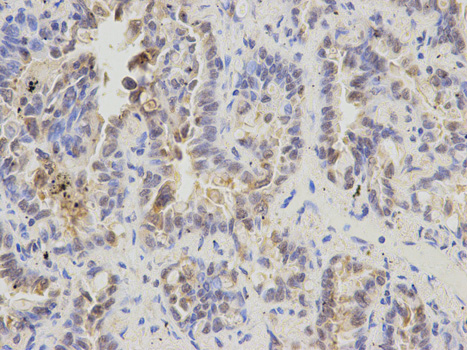
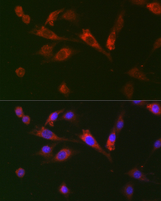


Comprehensive Human Protein Atlas IHC scoring per tissue (reliability: Enhanced — Medium consistency between antibody staining and RNA expression data. Secreted protein, tissue location of RNA and protein is expected to differ.). Rows are taken directly from the HPA tissue chart — click any row's HPA link to view the source.
| Tissue | Cell type | Level | Evidence | Source |
|---|---|---|---|---|
| Placenta | Syncytiotrophoblasts - cell body | High | Protein (IHC) | HPA → |
| Prostate | Glandular cells | Medium | Protein (IHC) | HPA → |
| Urinary bladder | Urothelial cells | Medium | Protein (IHC) | HPA → |
| Tissue | Cell type | Level | Evidence | Source |
|---|---|---|---|---|
| Adipose tissue | Adipocytes | Not detected | Protein (IHC) | HPA → |
| Adrenal gland | Glandular cells | Not detected | Protein (IHC) | HPA → |
| Bone marrow | Hematopoietic cells | Not detected | Protein (IHC) | HPA → |
| Breast | Adipocytes | Not detected | Protein (IHC) | HPA → |
| Bronchus | Respiratory epithelial cells | Not detected | Protein (IHC) | HPA → |
Troubleshoot GDF15 staining in paraffin sections by checking retrieval, compartment, tissue controls and scoring before interpreting chromogenic signal.
A01583 has pictured GDF15 IHC in paraffin-embedded human placenta and IF in BALB-3T3 cells (catalog image captions); its listed reactivity spans human, mouse and rat (catalog).
A01583 has pictured IHC in paraffin-embedded human placenta and IF in BALB-3T3 cells (catalog image captions). A01583-4 lists IHC for human and mouse, with no IHC or IF image supplied (catalog applications, reactivity and image fields).
Which to pick: For paraffin-section IHC with pictured evidence, choose A01583: its human placenta image used 1:100 and microwave retrieval in 10 mM Tris/Tris, pH 9.0; the fixative is unreported (A01583 IHC image caption). For IF/ICC, choose A01583, which lists both applications and has a pictured IF result in BALB-3T3 cells (catalog applications and IF image caption). For rat samples, A01583 is the listed option; A01583-4 lists human and mouse IHC but supplies no IHC image (catalog reactivity, applications and image fields).