This website uses cookies to ensure you get the best experience on our website.


- Table of Contents
Plan chromogenic GDF9 IHC on paraffin sections using ovarian follicles as a staining reference (HPA tissue IHC). This guide covers fixation consistency, antibody titration at 1:100–1:300 (datasheet), and interpretation of a secreted, processed protein (UniProt).

Expected localisation, validated protocols, controls and antibodies — the at-a-glance facts below, then the full design guide.
| Expected localisation | Secreted; tissue compartment unresolved (UniProt; HPA tissue IHC) | |
| Staining pattern | Low in ovarian follicle cells; compartment unspecified (HPA tissue IHC) | |
| Antigen retrieval | Citrate pH 6.0 HIER, 95–98 °C, 20 min (rule: cytoplasmic / membrane antigen) | |
| Positive control | Testis | |
| Negative control | Adipose tissue+4 more · see all |
| Fixation | Keep fixation consistent across samples (standard IHC practice; not target-specific) | |
| Caveat | Secretion may separate staining from the producing cells (UniProt) | |
| Regulation | Follicle-associated expression (HPA tissue IHC) | |
| Isoform / epitope | No isoforms; propeptide versus mature-chain epitope matters (UniProt) |
The catalog antibody’s IHC-P protocol is accompanied by 3 published ovarian IHC protocols (PMC4366263; PMC13110486; PMC4771171).
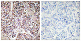
| Sample | Paraffin-embedded human lung cancer tissue; fixative not specified (datasheet A01466-1) |
| Fixation | Image fixative and duration unreported (datasheet A01466-1); verify before use. |
| Sectioning | 4–5 µm sections on charged slides (standard) |
| Deparaffinisation | Xylene, graded ethanol series to water (standard) |
| Antigen retrieval | Heat-induced epitope retrieval in citrate buffer, pH 6.0, 20 min at 95–98 °C (standard rule: cytoplasmic / membrane antigen) |
| Peroxidase block | 3% H2O2, 10 min, room temperature (standard) |
| Blocking | 10% normal serum of the secondary host, 30 min, room temperature (standard) |
| Primary antibody | Rabbit anti-GDF9, 1:100-1:300 (datasheet A01466-1) |
| Primary incubation | Overnight at 4 °C (standard) |
| Detection | HRP-polymer secondary, DAB chromogen 5–10 min (standard) |
| Counterstain | Hematoxylin, blue, dehydrate and mount (standard) |
| Expected result | GDF9-positive staining in peritubular cells of testis (HPA tissue IHC: Medium). HPA tissue profile: Expression in ovarian follicles. No signal in the no-primary control. |
GDF9 is a secreted protein with no transmembrane segment (UniProt O60383). In paraffin IHC, expect follicle-associated staining: UniProt reports protein in primary-follicle oocytes and expression in granulosa cells, while HPA reports low staining in ovarian follicle cells and medium staining in testicular peritubular cells (UniProt O60383; HPA tissue IHC). HPA rates its tissue evidence Supported and cautions that secretion complicates RNA–protein comparisons (HPA tissue IHC).
| Follicle-associated ovarian staining, potentially including oocytes and follicle cells. | This fits the reported ovarian distribution (UniProt O60383; HPA tissue IHC). HPA calls follicle-cell staining low; it gives no separate oocyte intensity, so score those cells separately (HPA tissue IHC). |
| Medium staining in testicular peritubular cells. | This matches an observed HPA IHC pattern and can serve as a tissue-level comparison (HPA tissue IHC). It does not establish that every compartment or neighboring testicular cell should stain. |
| Strong nuclear-only staining, or a sharp plasma-membrane rim without a follicle-associated pattern. | Treat this as suspect localisation and check controls. UniProt describes GDF9 as secreted, with no transmembrane segment; HPA's approved ICC-IF locations are Golgi and cytosol (UniProt O60383; HPA subcellular). Those IF locations are not a paraffin-IHC compartment standard. |
| Staining concentrated in cells HPA lists as not detected, or in unexpected cells within a section. | Investigate cross-reactivity, endogenous detection activity, and tissue identification before assigning GDF9. HPA's negative calls apply to specified cells in specified tissues, not every cell in those organs (HPA tissue IHC). |
| Diffuse brown haze, or no staining in the expected ovarian follicle or testicular peritubular compartment. | Haze alone is not a cell-specific positive result. Absence may reflect a missing target-bearing structure or a failed IHC run; ovarian follicle-cell signal is only low in HPA, so interpret a negative field against its morphology and run controls (HPA tissue IHC). |
| Secretion and processing | GDF9 has a signal peptide, propeptide, and mature chain, and is secreted (UniProt O60383). Signal need not remain confined to the producing cell; assess follicle context rather than assuming every adjacent extracellular deposit is specific. |
| Tissue and cell selection | UniProt reports primary-follicle oocytes and ovarian granulosa-cell expression; HPA reports ovarian-follicle staining and low follicle-cell signal (UniProt O60383; HPA tissue IHC). Confirm that the sampled section contains follicles before treating it as an ovarian negative. |
| Protein evidence versus RNA | HPA calls RNA tissue-enhanced in epididymis and testis but warns that secreted-protein location may differ from RNA location (HPA tissue IHC). Use the stated protein IHC observations for slide expectations, not RNA enrichment as a staining-intensity scale. |
| Antibody evidence and retrieval | HPA lists HPA069146 as IHC Supported, without an Enhanced designation (HPA antibodies). The supplied sources give no GDF9-specific retrieval condition or fixation sensitivity; use the IHC-validated antibody's protocol and optimise retrieval with run controls. |
| Situation | Likely cause | Next action |
|---|---|---|
| No signal in an ovarian section | The examined field may lack follicles, or low follicle-cell signal may be hard to distinguish from background (HPA tissue IHC). | Locate intact follicles on the counterstained section; compare an appropriate positive tissue and run controls before changing the IHC conditions. |
| No signal in a testis comparison | The expected medium HPA signal is specifically in peritubular cells; scoring other cells can give a misleading negative (HPA tissue IHC). | Identify peritubular cells morphologically. If they are present but unstained, check antibody, retrieval, detection, and control-slide performance as general IHC troubleshooting. |
| Widespread brown background | A diffuse deposit without the reported cell pattern may reflect nonspecific antibody binding or endogenous chromogenic detection activity (HPA tissue IHC for expected pattern). | Compare reagent-omission and detection controls; review blocking, washes, antibody concentration, and chromogen development as general IHC steps. |
| Strong staining in a listed negative cell type | Cross-reactivity or endogenous activity is possible; HPA reports not detected only for the named cell types and tissues (HPA tissue IHC). | Verify cell identity and control reactions, then compare the pattern with follicle or peritubular staining before calling it GDF9-specific. |
| Predominantly nuclear or sharply membranous signal | This conflicts with the secreted, non-transmembrane annotation and the approved Golgi/cytosol ICC-IF locations (UniProt O60383; HPA subcellular). | Check morphology and controls, then reassess antibody and detection conditions; do not impose the ICC-IF compartment pattern as a required IHC result. |
| What should an IF/ICC image show? | HPA reports Golgi as the main location and cytosol as an additional location in ICC-IF (HPA subcellular). | Interpret that result on the separate IF/ICC guide page. Its approved localisation supports an IF comparison, but supplies no IF protocol or paraffin-IHC intensity threshold (HPA subcellular). |


Comprehensive Human Protein Atlas IHC scoring per tissue (reliability: Supported — At least one protein variant secreted, tissue location of RNA and protein might differ and correlation is complex.). Rows are taken directly from the HPA tissue chart — click any row's HPA link to view the source.
| Tissue | Cell type | Level | Evidence | Source |
|---|---|---|---|---|
| Testis | Peritubular cells | Medium | Protein (IHC) | HPA → |
| Tissue | Cell type | Level | Evidence | Source |
|---|---|---|---|---|
| Adipose tissue | Adipocytes | Not detected | Protein (IHC) | HPA → |
| Adrenal gland | Glandular cells | Not detected | Protein (IHC) | HPA → |
| Appendix | Glandular cells | Not detected | Protein (IHC) | HPA → |
| Bone marrow | Hematopoietic cells | Not detected | Protein (IHC) | HPA → |
| Breast | Adipocytes | Not detected | Protein (IHC) | HPA → |
These questions address GDF9 staining in paraffin sections with chromogenic detection; the immunofluorescence entry covers the separate IF/ICC application.
A01466-1 has an IHC figure from paraffin-embedded human lung cancer (IHC image caption); IF is listed, but no IF figure is supplied (catalog applications; catalog IF images).
A01466-1 will render with an IHC figure from paraffin-embedded human lung cancer at 1:100 (IHC image caption). The catalog lists IHC and IF applications and Human and Mouse reactivity, but supplies no IF figure (catalog applications; catalog reactivity; catalog IF images).
Which to pick: Choose A01466-1 for paraffin-section IHC, starting within its listed 1:100–1:300 range; its own figure shows paraffin-embedded human lung cancer, with fixative unreported (datasheet: IHC 1:100–1:300; IHC image caption). For IF, A01466-1 lists 1:50, but the catalog supplies no IF figure or separate ICC validation (catalog IF dilution; catalog IF images; catalog applications). It is the catalog option for Human or Mouse samples because both are listed as reactive; the antibody is polyclonal, and the supplied IHC figure documents Human tissue only (catalog reactivity; datasheet: Polyclonal; IHC image caption).