GINS4 / DNA replication complex GINS protein SLD5 · IHC design guide

Design Immunohistochemistry for GINS4

Plan chromogenic IHC on paraffin sections using the catalog antibody at 2–5 μg/ml (datasheet A10548-1). Assess nuclear and cytoplasmic staining with appropriate controls, bearing in mind that the reported tissue pattern awaits external verification (HPA tissue IHC).

Evidence assembled Oct 2026 · For research use; verify linked source records and product datasheet before use
Immunohistochemistry protocol sheet for GINS4 (IHC for GINS4): expected localisation Nuclear and cytoplasmic staining in most tissues (HPA tissue IHC), antibody A10548-1, validated IHC image, and IHC protocol steps
Printable GINS4 IHC protocol sheet — expected localisation Nuclear and cytoplasmic staining in most tissues (HPA tissue IHC), antibody A10548-1, controls and protocol steps. Open the full GINS4 IHC guide →

GINS4 Immunohistochemistry Experimental Design Guide

Expected localisation, validated protocols, controls and antibodies — the at-a-glance facts below, then the full design guide.

Must know before staining
Expected localisation Nuclear and cytoplasmic staining in most tissues (HPA tissue IHC)
Staining pattern Variable nuclear/cytoplasmic signal; glandular cells can stain highly (HPA tissue IHC)
Antigen retrieval EDTA pH 8.0 HIER, heat-mediated (datasheet A10548-1)
Positive control ⓘ Adrenal gland+4 more · see all
Negative control ⓘ None in HPA (detected in all 45 tissues); use no-primary + isotype controls
Important caveats
Reasons your staining may differ from the expected pattern.
Fixation Matched tissue-IHC evidence does not establish the fixation claim. Validate the specimen-specific method before use. (selected-SKU IHC image A10548-1)
Caveat Tissue staining pattern awaits external verification (HPA tissue IHC)
Regulation No expression regulation annotated (UniProt)
Isoform / epitope 2 isoforms; residue-2 processing; epitope effect unknown (UniProt)
Section 1

Recommended GINS4 IHC & IF Protocols

The catalog antibody’s IHC-P protocol is accompanied by published GINS4 staining methods for colorectal cancer, lung adenocarcinoma, and glioma tissue (PMC7156840; PMC10378275; PMC8461916).

Recommended immunohistochemistry (IHC-P) protocol parameters
SampleParaffin-embedded human melanoma tissue; fixative not specified (datasheet A10548-1)
FixationImage fixative and duration unreported (datasheet A10548-1); verify before use.
Sectioning4–5 µm sections on charged slides (standard)
DeparaffinisationXylene, graded ethanol series to water (standard)
Antigen retrievalHeat retrieval: EDTA pH 8.0 (datasheet A10548-1); 20 min, 95–100 °C (standard)
Peroxidase block3% H2O2, 10 min, room temperature (standard)
Blocking10% goat serum (datasheet A10548-1)
Primary antibodyRabbit anti-GINS4, 2-5 μg/ml (datasheet A10548-1)
Primary incubationOvernight at 4 °C (datasheet A10548-1)
DetectionHRP-conjugated secondary, DAB chromogen (datasheet A10548-1)
CounterstainHematoxylin, blue, dehydrate and mount (standard)
Expected resultGINS4-positive staining in glandular cells of adrenal gland (HPA tissue IHC: High). HPA tissue profile: Nuclear and cytoplasmic expression in most tissues at variable levels. No signal in the no-primary control.
💡Decision noteStart with heat-mediated EDTA retrieval at pH 8.0 for the catalog antibody (datasheet A10548-1). The colorectal cancer study used citrate at pH 6.0 (PMC7156840).
Section 2

What Is the Expected GINS4 Staining Pattern?

GINS4 is found in the nucleus and cytoplasm and associates with chromatin (UniProt Q9BRT9). In tissue IHC, staining is reported in both compartments across most tissues, with high staining in several glandular, respiratory epithelial, and bone marrow hematopoietic cell populations (HPA tissue IHC). GINS4 has no transmembrane segment (UniProt Q9BRT9 topology). Treat the tissue pattern as provisional: HPA rates its IHC reliability Uncertain, pending external verification (HPA tissue IHC).

What am I looking at on my slide?
Nuclear staining, with some cytoplasmic staining, in glandular, respiratory epithelial, or hematopoietic cells.This fits HPA's reported distribution; selected examples include adrenal glandular cells, bronchial respiratory epithelial cells, and bone marrow hematopoietic cells, each rated High (HPA tissue IHC). Confirm cell identity on the counterstained section (general IHC practice).
Staining is confined to cell borders or extracellular material, with little nuclear or cytoplasmic signal.That compartment pattern conflicts with the reported nucleus, cytoplasm, and chromatin association and the absence of a transmembrane segment (UniProt Q9BRT9). Investigate background or antibody specificity before scoring it as GINS4 (general IHC practice).
The strongest staining is in an unexpected cell population, such as skeletal muscle myocytes.HPA lists skeletal muscle myocytes as Low, so uniformly strong staining there warrants review against a High reference tissue (HPA tissue IHC). Cross-reactivity or endogenous detection activity are possible explanations, not established causes (general IHC practice).
Diffuse colour obscures nuclei, cytoplasm, and tissue boundaries across the section.This cannot establish a GINS4 compartment pattern (HPA tissue IHC; UniProt Q9BRT9). Check the detection-only control, blocking, washing, and chromogen development to locate nonspecific signal (general IHC practice).
No staining appears in a reference section containing bone marrow hematopoietic cells.HPA rates those cells High, so absent signal calls for a technical check (HPA tissue IHC). Because the tissue IHC profile is Uncertain, a negative result alone cannot establish that GINS4 is absent (HPA tissue IHC).
💡Expected GINS4 appearanceA plausible positive has discernible nuclear and cytoplasmic staining in HPA High cell populations, while strong isolated membrane staining or widespread featureless colour should prompt specificity checks (HPA tissue IHC; UniProt Q9BRT9 topology; general IHC practice).
How each factor affects the staining
Compartment used for scoringAssess nuclear and cytoplasmic staining separately: both occur in HPA tissue IHC, and GINS4 associates with chromatin (HPA tissue IHC; UniProt Q9BRT9). A membrane-only result lacks support from these sources (UniProt Q9BRT9 topology).
Choice of reference tissueBone marrow hematopoietic cells and bronchial respiratory epithelial cells are rated High; skeletal muscle myocytes and adipocytes are listed as Low (HPA tissue IHC). Compare the named cell populations, since a whole-section average can hide their distribution (general IHC practice).
Strength of IHC evidenceHPA marks tissue IHC reliability Uncertain, pending external verification; its listed IHC antibodies HPA024663 and HPA026580 are also Uncertain (HPA tissue IHC; HPA antibodies). Interpret a matching pattern as supporting evidence, not standalone validation.
Antigen retrievalTarget-specific fixation effects are not established by the supplied assay evidence. Verify with a matched IHC source before attributing a result to fixation.
Isoforms and N-terminal processingUniProt lists 2 isoforms and full-length and N-terminally processed chains (UniProt Q9BRT9). Epitope recognition therefore depends on the antibody's stated epitope; these records do not show that processing changes tissue IHC staining (UniProt Q9BRT9).
IF/ICC Q&A: where should the signal appear?HPA reports mainly nucleoplasmic signal, with additional centrosome localisation, and marks those locations Approved in ICC-IF (HPA subcellular). That cell-image result informs localisation but does not raise the separate tissue IHC reliability rating above Uncertain (HPA tissue IHC).
Why is my staining missing, weak or wrong?
SituationLikely causeNext action
Reference tissue and study sections both lack signal.The staining run or detection chain may have failed; absence in HPA High cells is unexpected but not definitive (HPA tissue IHC; general IHC practice).Review antibody dilution, retrieval, detection reagents, and run controls; repeat with a documented High reference cell population on the same run (HPA tissue IHC; general IHC practice).
Only cell borders show strong colour.A membrane-only pattern conflicts with GINS4's reported compartments and lack of a transmembrane segment (UniProt Q9BRT9; HPA tissue IHC).Compare with a detection-only control and an HPA High reference tissue before assigning GINS4 positivity (HPA tissue IHC; general IHC practice).
Bone marrow has broad, granular colour outside identifiable cells.Endogenous detection activity or nonspecific background could contribute; the appearance alone does not identify the cause (general IHC practice).Check a detection-only control, the relevant endogenous-activity block, and whether colour remains within hematopoietic cells (HPA tissue IHC; general IHC practice).
Signal is diffuse and the counterstain no longer resolves cells.Excess background or overly developed chromogen can prevent compartment scoring (general IHC practice).Compare negative controls, review antibody concentration and washing, and shorten chromogen development if the control pattern supports that adjustment (general IHC practice).
A Low reference cell population stains more strongly than a High one.The contrast differs from HPA's listed levels, although its tissue IHC reliability is Uncertain (HPA tissue IHC).Verify cell identification and processing comparability, then repeat with controls before treating the contrast as biological (general IHC practice; HPA tissue IHC).
Nuclear staining is present, but a centrosome punctum is absent in IHC.Centrosome localisation comes from HPA ICC-IF images; it is not specified as a required tissue IHC feature (HPA subcellular; HPA tissue IHC).Score the tissue section against its nuclear and cytoplasmic pattern and appropriate controls; do not require an ICC-IF punctum for IHC positivity (HPA tissue IHC; general IHC practice).

Sample controls for GINS4 IHC & IF

🧪Run bone marrow first: hematopoietic cells should stain (HPA: High in hematopoietic cells). HPA detects GINS4 in all 45 scored tissues, so use no-primary and isotype controls for the negative comparison; putative internal negative cells should show counterstain alone, but the supplied HPA data establish no GINS4-negative cell population on this slide (HPA: no negative tissue rows).
Positive control tissue: Adrenal gland (Glandular cells, HPA High)
Negative control tissue: None in HPA: GINS4 is detected in all 45 scored tissues. Use a no-primary (secondary-only) and an isotype control instead.
ICC-IF cell lines (HPA subcellular resource): HPA ICC-IF images show GINS4 in A-431, U-251MG, U2OS, Rh30, with annotated localisation: Nucleoplasm (approved) (HPA subcellular).
Technical controls: Include a no-primary, secondary-only slide and a rabbit IgG control matched to the primary antibody’s clonality, plus GINS4-knockout material as a biological negative (catalog caption: rabbit primary antibody; standard IHC controls). Quench endogenous peroxidase in bone marrow before HRP/DAB detection (HPA: bone marrow hematopoietic cells, High; catalog caption: HRP/DAB detection; standard IHC practice).
⚠️Feasibility: No target-specific fixation window or fixation effect is reported, and the selected A10548-1 paraffin-section caption does not state a fixative (catalog caption: fixative unreported). The demonstrated paraffin-section procedure used heat retrieval in EDTA at pH 8.0; whether GINS4 staining depends on that retrieval condition, or whether frozen sections or IF are easier, is unreported (catalog caption: EDTA pH 8.0; HPA: ICC-IF images available). Endogenous peroxidase in bone marrow can complicate HRP/DAB interpretation (HPA: bone marrow hematopoietic cells, High; catalog caption: HRP/DAB detection; standard IHC practice).

HPA tissue IHC evidence for GINS4

Comprehensive Human Protein Atlas IHC scoring per tissue (reliability: Uncertain — Pending external verification.). Rows are taken directly from the HPA tissue chart — click any row's HPA link to view the source.

Positive expression · recommended positive controls

TissueCell typeLevelEvidenceSource
Adrenal gland Glandular cells High Protein (IHC) HPA →
Bone marrow Hematopoietic cells High Protein (IHC) HPA →
Bronchus Respiratory epithelial cells High Protein (IHC) HPA →
Cervix Glandular cells High Protein (IHC) HPA →
Duodenum Glandular cells High Protein (IHC) HPA →

Undetected expression · recommended negative controls

TissueCell typeLevelEvidenceSource
None in HPA: GINS4 is detected in all 45 scored tissues. Use a no-primary (secondary-only) and an isotype control instead.
Section 3

Advanced GINS4 IHC Tips

Use the selected tissue IHC conditions as a starting point, then assess GINS4 signal by compartment, cell type, and controls.

What should I change if GINS4 staining is weak after retrieval?
Start with heat-mediated retrieval in EDTA at pH 8.0 (datasheet A10548-1). The selected paraffin-section example used that retrieval before 2 μg/ml primary antibody overnight at 4°C, so check those conditions before changing the buffer (selected IHC caption: A10548-1). If staining remains weak, compare a small retrieval time series on adjacent sections while keeping antibody concentration and detection constant (standard IHC practice). Excess heating can damage morphology or increase background, making an apparent gain in signal difficult to interpret (standard IHC practice). Judge the result against nuclear and cytoplasmic staining patterns and a matched negative control (HPA tissue IHC; standard IHC practice).
Could fixation explain absent GINS4 staining in paraffin sections?
Target-specific fixation sensitivity is unknown: the selected GINS4 tissue caption identifies a paraffin section but does not state its fixative (selected IHC caption: A10548-1). Record the actual fixative and fixation duration for each specimen, then compare sections processed under documented conditions (standard IHC practice). Begin with EDTA retrieval at pH 8.0 and the reported 2 μg/ml overnight primary incubation before adjusting one variable at a time (datasheet A10548-1; selected IHC caption: A10548-1). Check preserved morphology, an appropriate positive section, and negative controls before attributing a blank slide to fixation (standard IHC practice). HPA staining patterns do not establish fixation sensitivity for this antibody (HPA tissue IHC).
How should I judge nuclear versus cytoplasmic GINS4 staining?
Assess nuclear and cytoplasmic signal separately: both are reported in tissue IHC, while GINS4 also associates with chromatin (HPA tissue IHC; UniProt Q9BRT9). Predominant nucleoplasmic localisation and an additional centrosomal location come from subcellular imaging, so a small punctum should not be scored as diffuse nuclear staining (HPA subcellular). Compare staining in intact cells with the hematoxylin counterstain and with the same compartment in a control section (standard IHC practice). Record the proportion and intensity for each compartment instead of combining them into one positive call (standard IHC practice). Treat an exclusively extracellular or luminal deposit as suspicious until controls support it (UniProt Q9BRT9 topology; standard IHC practice).
Could GINS4 isoforms or modifications explain inconsistent staining?
GINS4 has 2 annotated isoforms and an N-terminally processed chain beginning at residue 2 (UniProt Q9BRT9). Its annotated modifications include N-terminal acetylation and phosphorylation at serines 12 and 16; whether any alters this antibody's binding is unknown without its mapped epitope (UniProt Q9BRT9; selected IHC caption: A10548-1). If staining varies across preparations, compare adjacent sections under identical retrieval and detection conditions before assigning an isoform explanation (standard IHC practice). Request the antibody's immunogen or epitope information and check whether it spans a differing region if isoform discrimination matters (standard IHC practice). Report the result as GINS4 immunoreactivity unless specificity for an isoform has been demonstrated (standard IHC practice).
How can IF help investigate an ambiguous GINS4 IHC pattern?
On a separately prepared IF section, multiplex GINS4 with a marker for the expected cell population, such as hematopoietic cells in bone marrow, to identify which cells carry signal (HPA: High in bone marrow hematopoietic cells; standard IF practice). Select spectrally separated fluorophores and compare tissue autofluorescence in an unstained section, favoring a longer-wavelength channel when its background is lower (standard IF practice). GINS4 has no transmembrane segment and is reported in intracellular compartments, so use permeabilisation appropriate for access to nuclear and cytoplasmic epitopes (UniProt Q9BRT9 topology and subcellular location; standard IF practice). Compare the IF compartment pattern with the chromogenic IHC pattern, while assessing each assay's controls independently (HPA tissue IHC; standard IHC/IF practice).
What causes widespread brown signal in GINS4 IHC?
First inspect the negative control and tissue edges: widespread DAB signal there can reflect detection background or edge artefact rather than specific GINS4 staining (standard IHC practice). The selected caption used 10% goat serum block, a peroxidase-conjugated anti-rabbit secondary, and DAB development (selected IHC caption: A10548-1). Confirm the general peroxidase-blocking step, then compare a no-primary control and a shorter chromogen development under matched conditions (standard IHC practice). If background persists, titrate the primary around the reported 2 μg/ml while retaining EDTA at pH 8.0 for retrieval (selected IHC caption: A10548-1; datasheet A10548-1). Score only cell-associated staining with interpretable morphology (standard IHC practice).
How should I quantify GINS4 staining across tissue sections? ⚠ ANSWER MARKED FOR VERIFICATION
Define the cell population and compartment before scoring, because tissue IHC reports variable nuclear and cytoplasmic GINS4 expression (HPA tissue IHC). For intact cells, record the percentage positive and intensity in each compartment, or calculate an H-score from intensity categories 0–3 (standard IHC practice). For spatial analyses, report positive-cell density per mm² of viable, annotated tissue and exclude necrotic or folded areas (standard IHC practice). Normalise comparisons to the number of evaluable cells or viable tissue area, and keep retrieval, development, imaging, and scoring thresholds consistent (standard IHC practice). State the antibody and scoring rules because HPA rates its tissue IHC reliability as uncertain (HPA tissue IHC).
When is a GINS4-positive IHC result convincing?
A convincing result has cell-associated nuclear or cytoplasmic staining in intact tissue, consistent with reported localisation, and passes matched negative controls (UniProt Q9BRT9; HPA tissue IHC; standard IHC practice). Bone marrow hematopoietic cells and several glandular or respiratory epithelial populations have reported high staining, but HPA labels the tissue IHC evidence uncertain (HPA tissue IHC). Be cautious with signal confined to the wrong compartment, necrotic regions, section edges, or structures with endogenous peroxidase activity (UniProt Q9BRT9 subcellular location; standard IHC practice). Compare compartment-specific staining with morphology and controls before assigning it to GINS4, and describe uncertain patterns without claiming a validated cell state (standard IHC practice; HPA tissue IHC).
Boster reagents

Best GINS4 / DNA replication complex GINS protein SLD5 IHC Antibodies

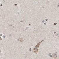
A10548-1 has real IHC data from paraffin-embedded human melanoma (A10548-1 IHC image caption). Human, mouse and rat reactivity is listed; IF/ICC data is absent (catalog reactivity/IF fields).

Real IHC data IHC analysis of SLD5/GINS4 using anti-SLD5/GINS4 antibody (A10548-1). SLD5/GINS4 was detected in a paraffin-embedded section of human melanoma tissue. Heat mediated antigen retrieval was performed in EDTA buffer (pH 8.0, epitope retrieval solution). The tissue section was blocked with 10% goat serum. The tissue section was then incubated with 2 μg/ml rabbit anti-SLD5/GINS4 Antibody (A10548-1) overnight at 4°C. Peroxidase Conjugated Goat Anti-rabbit IgG was used as secondary antibody and incubated for 30 minutes at 37°C. The tissue section was developed using HRP Conjugated Rabbit IgG Super Vision Assay Kit (Catalog # SV0002) with DAB as the chromogen.
Anti-SLD5/GINS4 Antibody ®
Cat # A10548-1

A10548-1 was demonstrated by chromogenic IHC in a paraffin-embedded human melanoma section (A10548-1 IHC image caption). It lists human, mouse and rat reactivity, but IF/ICC is absent from its applications (catalog applications/reactivity).

Which to pick: Choose A10548-1 for paraffin-section tissue IHC: its image documents EDTA retrieval at pH 8.0 and 2 μg/ml primary antibody in human melanoma; the fixative is unreported (A10548-1 IHC image caption). No listed SKU has IF/ICC validation or an IF figure (catalog applications/IF fields). For cross-species planning, A10548-1 lists human, mouse and rat reactivity, although its IHC image shows human tissue only; the antibody host is rabbit and clonality is unreported (catalog reactivity/host/clone; A10548-1 IHC image caption).

Each figure is that product's own IHC / IF validation image from its datasheet.

References

  1. UniProt Consortium. UniProt entry Q9BRT9 (SLD5_HUMAN, DNA replication complex GINS protein SLD5).
  2. Human Protein Atlas. GINS4 tissue IHC expression (reliability: Uncertain).
  3. Human Protein Atlas. GINS4 subcellular location (ICC-IF): Mainly localized to the nucleoplasm. In addition localized to the centrosome..
  4. Human Protein Atlas. GINS4 antibody validation summary (3 antibodies).
  5. GINS complex subunit 4, a prognostic biomarker and reversely mediated by Krüppel-like factor 4, promotes the growth of colorectal cancer. Cancer science 2020 — PMC7156840.
  6. SLD5/GINS4 controls dynein-dependent centrosome maturation and exposes a candidate mitotic vulnerability in cancer. bioRxiv : the preprint server for biology 2026 — PMC13192919.
  7. The Molecular Pathogenesis of Tumor-Suppressive miR-486-5p and miR-486-3p Target Genes: GINS4 Facilitates Aggressiveness in Lung Adenocarcinoma. Cells 2023 — PMC10378275.
  8. Overexpression of GINS4 is associated with poor prognosis and survival in glioma patients. Molecular medicine (Cambridge, Mass.) 2021 — PMC8461916.
  9. PubMed PMID:14702039 — UniProt-cited evidence.
  10. PubMed PMID:15489334 — UniProt-cited evidence.
  11. PubMed PMID:17170760 — UniProt-cited evidence.