This website uses cookies to ensure you get the best experience on our website.


- Table of Contents
Plan chromogenic IHC on paraffin sections using heart muscle cardiomyocytes as a high-staining reference (HPA tissue IHC). Assess cytoplasmic and membranous staining while looking for GJA1 at intercalated discs between cardiomyocytes (HPA tissue IHC; UniProt).

Expected localisation, validated protocols, controls and antibodies — the at-a-glance facts below, then the full design guide.
| Expected localisation | Membranous and cytoplasmic in tissue IHC (HPA tissue IHC) | |
| Staining pattern | Most cell types: cytoplasmic and membranous staining (HPA tissue IHC) | |
| Antigen retrieval | Citrate pH 6.0 HIER, 95–98 °C, 20 min (rule: cytoplasmic / membrane antigen) | |
| Positive control | Adrenal gland+4 more · see all | |
| Negative control | Adipose tissue+4 more · see all |
| Fixation | Keep fixation consistent across sections. (standard IHC practice; not target-specific) | |
| Caveat | Cytoplasmic signal may accompany junctional staining (HPA tissue IHC; UniProt) | |
| Regulation | Expression regulation not specified (UniProt) | |
| Isoform / epitope | 0 annotated isoforms; epitope location matters (UniProt) |
The catalog antibody’s IHC-P protocol is accompanied by published GJA1 IHC protocols for liver and goat skin sections (PMC12930822; PMC12464370).
| Sample | Tissue sections; selected-image fixative not specified (standard IHC workflow) |
| Fixation | Image fixative and duration unreported (datasheet M00599-1); verify before use. |
| Sectioning | 4–5 µm sections on charged slides (standard) |
| Deparaffinisation | Xylene, graded ethanol series to water (standard) |
| Antigen retrieval | Heat-induced epitope retrieval in citrate buffer, pH 6.0, 20 min at 95–98 °C (standard rule: cytoplasmic / membrane antigen) |
| Peroxidase block | 3% H2O2, 10 min, room temperature (standard) |
| Blocking | 10% normal serum of the secondary host, 30 min, room temperature (standard) |
| Primary antibody | Rabbit anti-GJA1, 1:200-1:1000 (datasheet M00599-1) |
| Primary incubation | Overnight at 4 °C (standard) |
| Detection | HRP-polymer secondary, DAB chromogen 5–10 min (standard) |
| Counterstain | Hematoxylin, blue, dehydrate and mount (standard) |
| Expected result | GJA1-positive staining in glandular cells of adrenal gland (HPA tissue IHC: High). HPA tissue profile: Cytoplasmic and membranous expression in most cell types. No signal in the no-primary control. |
GJA1 is a four-pass membrane protein concentrated at gap junctions; UniProt also places it in the endoplasmic reticulum and at cardiomyocyte intercalated discs (UniProt P17302 topology and subcellular location). In paraffin-section IHC, expect membranous and some cytoplasmic staining in cell populations with documented expression, especially cardiomyocytes (HPA tissue IHC: High). HPA rates the tissue staining profile Enhanced, citing agreement between antibody staining and RNA expression (HPA tissue IHC: Enhanced).
| Distinct staining marks cardiomyocyte contacts, with some cytoplasmic signal. | This fits GJA1 at intercalated discs and gap junctions (UniProt P17302 subcellular location). Cardiomyocytes stain High in HPA tissue IHC (HPA: Heart muscle, High). Assess contact-associated signal alongside the overall tissue pattern; cytoplasmic staining alone does not exclude GJA1 because HPA reports cytoplasmic and membranous expression (HPA tissue IHC). |
| Strong staining is confined to nuclei, with little contact-associated or cytoplasmic signal. | Treat a nuclear-only IHC pattern as suspect and check controls before scoring it positive: HPA describes mainly cytoplasmic and membranous tissue staining (HPA tissue IHC). HPA reports nucleoplasmic localisation only as an uncertain additional finding in ICC-IF, which does not establish nuclear-only staining as the expected paraffin-section result (HPA subcellular). |
| Adipocytes or pancreatic exocrine glandular cells stain strongly. | These particular cell populations are Not detected in the HPA tissue images (HPA: Adipocytes; pancreatic exocrine glandular cells). Consider antibody cross-reactivity or endogenous detection activity, then compare a known-positive cell population and a no-primary control (general IHC practice). Do not label the entire adipose tissue or pancreas negative based on those cell-specific entries. |
| Brown signal spreads evenly across cells, stroma and empty areas without cell-contact detail. | This distribution is less convincing than junction-associated staining with a plausible cell pattern (UniProt P17302 subcellular location; HPA tissue IHC). Excess detection background or nonspecific binding is possible (general IHC practice). Check the no-primary control and inspect whether the signal tracks tissue structures before assigning a GJA1 score. |
| There is no signal in cardiomyocytes on a heart-muscle section. | The result conflicts with HPA's High cardiomyocyte staining and UniProt's intercalated-disc localisation (HPA: Heart muscle, High; UniProt P17302 subcellular location). Treat the run as inconclusive until section quality, antibody application and chromogenic detection are checked (general IHC practice); absence here alone does not establish lack of GJA1 expression. |
| Membrane topology and antibody epitope | GJA1 has four transmembrane segments and cytoplasmic and extracellular regions (UniProt P17302 topology). If the antibody's epitope is known, use that information when interpreting accessible staining; the supplied record gives no catalog-antibody epitope, so it cannot predict a retrieval condition or staining strength. |
| Cell-specific tissue distribution | HPA reports High staining in cardiomyocytes, cervical squamous epithelial cells and placental cytotrophoblasts, but Not detected in adipocytes and pancreatic exocrine glandular cells (HPA tissue IHC). Choose and score the documented cell population, since one section can contain several cell types. |
| IHC validation scope | The HPA tissue profile is Enhanced; HPA035097 and CAB010753 each have Enhanced IHC validation, while HPA069245 has Supported IHC validation (HPA tissue IHC; HPA antibodies). Those ratings support interpretation of the reported IHC patterns, but do not establish the performance of an unspecified catalog antibody. |
| Subcellular interpretation across applications | HPA ICC-IF supports cell-junction localisation; its vesicle and nucleoplasm calls are uncertain (HPA subcellular). UniProt also lists the endoplasmic reticulum (UniProt P17302 subcellular location). These observations allow intracellular signal, but do not make every diffuse or nuclear IHC signal specific. |
| Situation | Likely cause | Next action |
|---|---|---|
| Known-positive heart muscle is blank. | A failed IHC run or inadequate detection is possible; cardiomyocytes are High in HPA tissue IHC (HPA: Heart muscle, High). | Check a known-positive section, reagent application and chromogenic detection; review retrieval against the chosen antibody's instructions (general IHC practice). No GJA1-specific retrieval condition is supplied. |
| Cardiomyocytes stain, but contact-associated detail is hard to see. | Cytoplasmic staining can coexist with membrane staining (HPA tissue IHC); a crowded or overly strong chromogenic signal can obscure fine localisation (general IHC practice). | Inspect well-preserved cell borders and intercalated-disc regions, then optimise antibody concentration or detection intensity using the positive control (general IHC practice; UniProt P17302 subcellular location). |
| Cells listed as Not detected stain strongly. | For adipocytes or pancreatic exocrine glandular cells, this conflicts with the cell-specific HPA observations; cross-reactivity or endogenous activity may contribute (HPA tissue IHC; general IHC practice). | Compare a no-primary control and a documented positive cell population before scoring; evaluate the stained cell type rather than assigning one result to the whole tissue (general IHC practice). |
| Diffuse brown haze covers the section. | Background from antibody or detection reagents is possible when staining lacks anatomical or junctional structure (general IHC practice; UniProt P17302 subcellular location). | Check the no-primary control, blocking and washes; adjust reagent concentration and detection time according to the IHC workflow (general IHC practice). Do not score uniform haze as GJA1. |
| Nuclei dominate the IHC signal. | HPA's nucleoplasmic ICC-IF call is uncertain, whereas its tissue IHC profile is cytoplasmic and membranous (HPA subcellular; HPA tissue IHC). | Check no-primary and positive controls, then re-evaluate cell borders and cytoplasm. Record nuclear-only staining as unresolved unless independently supported for this IHC assay (general IHC practice). |
| What should IF/ICC show? | HPA supports localisation at cell junctions and marks vesicles and nucleoplasm uncertain in ICC-IF (HPA subcellular). | Interpret junction-associated fluorescence as the stronger localisation evidence; treat isolated vesicular or nuclear signal cautiously (HPA subcellular). IF/ICC staining conditions belong to the separate IF/ICC guide. |
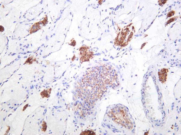
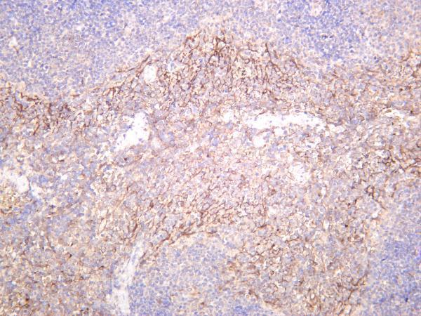


Comprehensive Human Protein Atlas IHC scoring per tissue (reliability: Enhanced — High consistency between antibody staining and RNA expression data.). Rows are taken directly from the HPA tissue chart — click any row's HPA link to view the source.
| Tissue | Cell type | Level | Evidence | Source |
|---|---|---|---|---|
| Adrenal gland | Glandular cells | High | Protein (IHC) | HPA → |
| Cerebellum | Processes in granular layer | High | Protein (IHC) | HPA → |
| Cerebral cortex | Neuropil | High | Protein (IHC) | HPA → |
| Cervix | Squamous epithelial cells | High | Protein (IHC) | HPA → |
| Endometrium | Ciliated epithelial cells | High | Protein (IHC) | HPA → |
| Tissue | Cell type | Level | Evidence | Source |
|---|---|---|---|---|
| Adipose tissue | Adipocytes | Not detected | Protein (IHC) | HPA → |
| Appendix | Glandular cells | Not detected | Protein (IHC) | HPA → |
| Bronchus | Respiratory epithelial cells | Not detected | Protein (IHC) | HPA → |
| Liver | Cholangiocytes | Not detected | Protein (IHC) | HPA → |
| Nasopharynx | Respiratory epithelial cells | Not detected | Protein (IHC) | HPA → |
Troubleshoot GJA1 chromogenic IHC by checking retrieval, staining at cell contacts, and controls before scoring expression (UniProt P17302 localisation; standard IHC practice).
The catalog lists GJA1 antibodies reactive with human, mouse, and rat samples, with paraffin-section IHC images, a rat-heart IF image, and a cell IF image (catalog; image captions).
Only M00599-1 renders as a card here (cards payload). Its IHC captions show human testis, mouse spleen, and rat spleen; IF is listed as an application, but no IF image is supplied for this SKU (M00599-1 catalog).
Which to pick: Choose M00599-1 for paraffin-section IHC across human, mouse, and rat: it is a rabbit monoclonal with IHC images for all three species (M00599-1 catalog; IHC captions). For IF in tissue, PA1026 has a rat-heart IF image; for IF/ICC in cells, A00599 has a U20S cell IF image, although IF is absent from its listed applications (PA1026 IF caption; A00599 IF caption and applications). The M00599-1 captions do not report a fixative (M00599-1 IHC captions).