This website uses cookies to ensure you get the best experience on our website.


- Table of Contents
Plan GJA3 IHC in paraffin sections using lens fiber cells as a high-staining reference (HPA tissue IHC). The guide covers fixation, detection and interpretation of the membrane and gap-junction localisation expected from UniProt, alongside HPA’s off-target staining caveat.

Expected localisation, validated protocols, controls and antibodies — the at-a-glance facts below, then the full design guide.
| Expected localisation | Cell membrane and gap junctions (UniProt) | |
| Staining pattern | High staining in lens fiber cells; subcellular pattern unspecified (HPA tissue IHC) | |
| Antigen retrieval | Citrate pH 6.0 HIER, 95–98 °C, 20 min (rule: cytoplasmic / membrane antigen) | |
| Positive control | Eye | |
| Negative control | Adipose tissue+4 more · see all |
| Fixation | Keep fixation consistent across sections. (standard IHC practice; not target-specific) | |
| Caveat | Presumed off-target staining was disregarded (HPA tissue IHC) | |
| Regulation | Expression regulation is not annotated (UniProt) | |
| Isoform / epitope | No isoforms annotated; extracellular vs cytoplasmic epitope matters (UniProt) |
The catalog antibody protocol (datasheet) is accompanied by two published GJA3 IHC protocols (PMC13543546; PMC4453443).
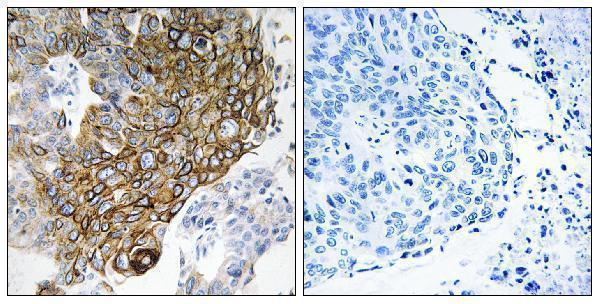
| Sample | Paraffin-embedded human lung carcinoma tissue; fixative not specified (datasheet A30696) |
| Fixation | Image fixative and duration unreported (datasheet A30696); verify before use. |
| Sectioning | 4–5 µm sections on charged slides (standard) |
| Deparaffinisation | Xylene, graded ethanol series to water (standard) |
| Antigen retrieval | Heat-induced epitope retrieval in citrate buffer, pH 6.0, 20 min at 95–98 °C (standard rule: cytoplasmic / membrane antigen) |
| Peroxidase block | 3% H2O2, 10 min, room temperature (standard) |
| Blocking | 10% normal serum of the secondary host, 30 min, room temperature (standard) |
| Primary antibody | Rabbit anti-GJA3, 1:100 (datasheet A30696) |
| Primary incubation | Overnight at 4 °C (standard) |
| Detection | HRP-polymer secondary, DAB chromogen 5–10 min (standard) |
| Counterstain | Hematoxylin, blue, dehydrate and mount (standard) |
| Expected result | GJA3-positive staining in lens fiber cells of eye (HPA tissue IHC: High). HPA tissue profile: Expression in lens. No signal in the no-primary control. |
GJA3 is a four-pass membrane protein at gap junctions (UniProt Q9Y6H8 topology and subcellular location). In paraffin-section IHC, expect staining in lens fiber cells, where HPA reports High expression (HPA: eye tissue IHC). HPA rates the tissue staining Supported, while noting presumed off-target binding that was disregarded and no internal RNA data for correlation (HPA: tissue IHC reliability).
| Strong staining outlines lens fiber cells, with signal concentrated at cell contacts. | This fits High lens fiber staining (HPA: eye tissue IHC) and membrane, gap-junction localization (UniProt Q9Y6H8). Assess the cell boundaries and tissue architecture together; a contact-enriched pattern is consistent with connexons joining adjacent cells (UniProt Q9Y6H8 function). |
| Signal is predominantly nuclear or diffusely cytoplasmic, without a convincing cell-boundary pattern. | That compartment differs from the annotated membrane and gap-junction location (UniProt Q9Y6H8). Treat it as a possible staining artefact until controls and the IHC-validated antibody's documented pattern support it; location alone cannot identify the artefact's cause. |
| Strong staining appears in unrelated cells, including adipocytes or adrenal glandular cells. | HPA reports GJA3 as Not detected in those cell types (HPA: adipose tissue and adrenal gland IHC). Investigate antibody cross-reactivity or endogenous chromogenic activity (general IHC practice); HPA also notes presumed off-target binding in its reliability assessment (HPA: tissue IHC). |
| A broad haze covers cells and surrounding tissue, obscuring boundaries. | A uniform haze does not show the membrane and junctional localization annotated for GJA3 (UniProt Q9Y6H8). Check background in a control processed without primary antibody, then review blocking, washes and detection conditions (general IHC practice). |
| Lens fiber cells show no signal despite adequate tissue morphology. | This conflicts with the High lens fiber staining reported by HPA (HPA: eye tissue IHC). First check the run's controls and the catalog antibody's IHC-P instructions; absence of signal alone cannot distinguish a technical failure from differences between specimens (general IHC practice). |
| Membrane topology | GJA3 has 4 transmembrane segments and both extracellular and cytoplasmic regions (UniProt Q9Y6H8 topology). Interpret a localized boundary signal against this anatomy; the supplied record does not identify the antibody epitope, so it cannot predict epitope-specific retrieval needs. |
| Junction assembly | Six connexins form a hemichannel; two opposing hemichannels form a gap junction (UniProt Q9Y6H8 subunit and function). Contact-enriched staining is therefore biologically plausible, but chromogenic IHC cannot by itself show that a stained junction conducts ions or small molecules. |
| Tissue and cell selection | HPA observes High staining in eye lens fiber cells and Not detected staining in several sampled cell types, including adipocytes and adrenal glandular cells (HPA: tissue IHC). Use the named cells when comparing patterns; do not extend these observations to unsampled tissues. |
| Strength of IHC evidence | The HPA tissue profile is Supported, and antibody HPA014821 is marked Supported for IHC (HPA: tissue IHC reliability; HPA: antibody validation). HPA notes presumed off-target binding and lacks internal RNA correlation, so unexpected staining needs independent control-based review. |
| RNA and protein readouts | HPA lists tissue-enhanced RNA in heart muscle, parathyroid gland and placenta, while its reported protein staining profile is expression in lens (HPA: tissue IHC and RNA specificity). The RNA list does not establish an expected positive IHC result in those tissues. |
| Processing annotations | UniProt lists no signal peptide, propeptide, annotated glycosylation sites, modified residues or isoforms for this record (UniProt Q9Y6H8 processing and annotations). These entries do not establish how fixation or antigen retrieval affects GJA3 staining; target-specific fixation sensitivity is unreported here. |
| Situation | Likely cause | Next action |
|---|---|---|
| Lens fiber cells are negative. | The result disagrees with HPA's High lens fiber staining, but the slide alone does not identify why (HPA: eye tissue IHC). | Check run controls, tissue preservation and the catalog antibody's IHC-P instructions, including its specified retrieval and dilution if supplied (general IHC practice). Do not assign a GJA3-specific fixation effect from this result. |
| Signal fills nuclei or cytoplasm more than cell borders. | The dominant compartment conflicts with membrane and gap-junction annotation (UniProt Q9Y6H8 subcellular location). | Compare with lens fiber cell boundaries, inspect a no-primary control and reassess detection background (general IHC practice). Record the discordant compartment rather than scoring it as junctional GJA3. |
| Strong staining occurs in adipocytes or adrenal glandular cells. | These cells are reported as Not detected by HPA; HPA also notes presumed off-target binding (HPA: tissue IHC and reliability). | Compare the same run's controls and the validated lens pattern. Check for antibody-dependent staining and endogenous chromogenic activity before interpreting the unexpected cells (general IHC practice). |
| Diffuse brown background obscures cell contacts. | Background can interfere with judging the annotated junctional membrane location (UniProt Q9Y6H8; general IHC practice). | Inspect a no-primary control and review blocking, washes and detection conditions (general IHC practice). Score localization only where cell boundaries remain interpretable. |
| A heart, parathyroid or placenta sample is weak or negative. | HPA's tissue-enhanced RNA category names those tissues, but its supplied IHC profile reports expression in lens (HPA: RNA specificity and tissue IHC). | Avoid treating RNA enrichment as an IHC positive-control requirement. Use the reported lens fiber pattern to assess staining performance (HPA: eye tissue IHC). |
| Can an IF/ICC image confirm the IHC pattern? | HPA summarizes the subcellular location as membrane but provides no ICC-IF image cell lines or main location here; HPA014821 has no ICC status listed (HPA: subcellular and antibody records). | Treat membrane localization as a biological expectation from UniProt and HPA, while keeping IF/ICC validation separate from this paraffin-section IHC interpretation (UniProt Q9Y6H8; HPA: subcellular record). |
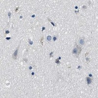

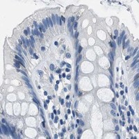
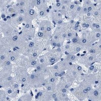
Comprehensive Human Protein Atlas IHC scoring per tissue (reliability: Supported — Presumed off target binding observed and disregarded. External characterization data supports antibody staining but no internal RNA data available for correlation.). Rows are taken directly from the HPA tissue chart — click any row's HPA link to view the source.
| Tissue | Cell type | Level | Evidence | Source |
|---|---|---|---|---|
| Eye | Lens fiber cells | High | Protein (IHC) | HPA → |
| Tissue | Cell type | Level | Evidence | Source |
|---|---|---|---|---|
| Adipose tissue | Adipocytes | Not detected | Protein (IHC) | HPA → |
| Adrenal gland | Glandular cells | Not detected | Protein (IHC) | HPA → |
| Appendix | Glandular cells | Not detected | Protein (IHC) | HPA → |
| Bone marrow | Hematopoietic cells | Not detected | Protein (IHC) | HPA → |
| Breast | Adipocytes | Not detected | Protein (IHC) | HPA → |
Troubleshoot chromogenic GJA3 IHC in paraffin sections by checking retrieval, membrane localisation, controls and cell specific scoring (UniProt Q9Y6H8; HPA tissue IHC).
A30696 has IHC image data from paraffin-embedded human lung carcinoma (A30696 IHC caption). IF/ICC are listed applications, and catalog reactivity covers human, mouse, and rat (catalog: A30696 applications/reactivity).
A30696 has an IHC image of paraffin-embedded human lung carcinoma with peptide blocking shown (A30696 IHC caption). Its listed applications include IHC, IF, and ICC, but no IF image is supplied (catalog: A30696 applications/if_image_alts).
Which to pick: Choose A30696 for paraffin-section tissue IHC based on its own human lung carcinoma image (A30696 IHC caption); the fixative is unreported (A30696 IHC caption). For IF/ICC, A30696 is listed for both at 1:100, although no IF image is supplied (catalog: A30696 applications/if_dilution/if_image_alts). For cross-species work, A30696 is a rabbit polyclonal listed as reactive with human, mouse, and rat (catalog: A30696 host/dilution_raw/reactivity).