This website uses cookies to ensure you get the best experience on our website.


- Table of Contents
GLB1 tissue IHC commonly shows granular cytoplasmic staining, with strong staining in lung macrophages (HPA tissue IHC). For paraffin sections, the catalog antibody M01829-3 has a documented 1:50 IHC workflow; assess staining by cell type with appropriate controls (datasheet: M01829-3; standard IHC practice).

Expected localisation, validated protocols, controls and antibodies — the at-a-glance facts below, then the full design guide.
| Expected localisation | Cytoplasmic staining in tissue sections (HPA tissue IHC) | |
| Staining pattern | Many cell types show granular cytoplasmic staining (HPA tissue IHC) | |
| Antigen retrieval | EDTA pH 8.0 HIER, heat-mediated (datasheet M01829-3) | |
| Positive control | Appendix+4 more · see all | |
| Negative control | Ovary+1 more · see all |
| Fixation | Keep fixation consistent across sections (standard IHC practice; not target-specific) | |
| Caveat | Lung macrophages stain strongly and may dominate the signal (HPA tissue IHC) | |
| Regulation | Low tissue specificity; no regulator established (HPA tissue IHC) | |
| Isoform / epitope | 3 isoforms; mature chain starts at residue 29, so check the epitope (UniProt) |
Compare the catalog antibody’s IHC-P protocol with published GLB1 staining of mouse bone and skeletal muscle (PMC12764366) and prostate tissue microarrays (PMC5325224).
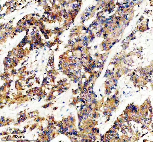
| Sample | Paraffin-embedded human colon cancer tissue; fixative not specified (datasheet M01829-3) |
| Fixation | Image fixative and duration unreported (datasheet M01829-3); verify before use. |
| Sectioning | 4–5 µm sections on charged slides (standard) |
| Deparaffinisation | Xylene, graded ethanol series to water (standard) |
| Antigen retrieval | Heat retrieval: EDTA pH 8.0 (datasheet M01829-3); 20 min, 95–100 °C (standard) |
| Peroxidase block | 3% H2O2, 10 min, room temperature (standard) |
| Blocking | 10% goat serum (datasheet M01829-3) |
| Primary antibody | Rabbit monoclonal (clone 25G11) anti-GLB1, 1:50 (datasheet M01829-3) |
| Primary incubation | Overnight at 4 °C (datasheet M01829-3) |
| Detection | HRP-conjugated secondary, DAB chromogen (datasheet M01829-3) |
| Counterstain | Hematoxylin, blue, dehydrate and mount (standard) |
| Expected result | GLB1-positive staining in glandular cells of appendix (HPA tissue IHC: High). HPA tissue profile: Ubiquitous cytoplasmic expression often with a granular pattern. No signal in the no-primary control. |
GLB1 is a lysosomal protein; UniProt also describes perinuclear cytoplasmic localization with a note that this pool is not lysosomal (UniProt P16278). In tissue IHC, expect often granular cytoplasmic staining, including strong staining in liver hepatocytes and lung macrophages (HPA tissue IHC). GLB1 has no transmembrane segment (UniProt P16278 topology). HPA rates its tissue profile Enhanced, with medium consistency between antibody staining and RNA expression (HPA tissue IHC).
| Granular cytoplasmic staining in hepatocytes or lung macrophages, with cellular outlines remaining interpretable. | This fits the reported pattern and High staining in those cells (HPA tissue IHC). Evaluate intensity within the named cell population; a dark region alone does not identify which cells stained. |
| Predominantly nuclear, surface-restricted, or uniformly extracellular chromogen. | Those compartments do not match the reported cytoplasmic pattern (HPA tissue IHC) or GLB1's lack of a transmembrane segment (UniProt P16278 topology). Check morphology and controls before interpreting the signal as GLB1. |
| Strong signal in ovarian stroma or vaginal squamous epithelium. | HPA reports GLB1 as Not detected in those cell types (HPA tissue IHC). Consider cross-reactivity or endogenous detection activity, especially if a control without primary antibody also develops color; assess the specific cells rather than the tissue as a whole. |
| Diffuse color across cells and surrounding tissue obscures granules. | The expected tissue pattern is often granular and cytoplasmic (HPA tissue IHC). Widespread haze is difficult to score and can reflect nonspecific binding or chromogen background (general IHC practice); compare sections processed with and without primary antibody. |
| No staining in hepatocytes on a liver control section. | HPA reports High hepatocyte staining (HPA tissue IHC), so an absent control signal makes a negative study section hard to interpret. Check that the hepatocytes are present, then review retrieval, antibody incubation, and detection using the established IHC workflow (general IHC practice). |
| Tissue and cell selection | High signal is reported in appendix glandular cells, bronchial ciliated cell bodies, gallbladder glandular cells, hepatocytes, lung macrophages, seminal vesicle glandular cells, and Leydig cells (HPA tissue IHC). Use the named cells when judging a positive control. |
| Low and undetected populations | HPA reports Medium adipocyte staining, Low staining in several other named cell types, and Not detected staining in ovarian stroma and vaginal squamous epithelium (HPA tissue IHC). A weak or absent result depends on which cells were examined. |
| Antibody evidence | One listed antibody has Enhanced IHC validation and another has Supported IHC validation; the ICC-supported antibody has no listed IHC status (HPA antibodies). Keep the validation status tied to the antibody used rather than transferring it across assays. |
| Processing, glycosylation, and isoforms | UniProt lists a signal peptide, propeptide, mature chain, seven glycosylation sites, and three isoforms (UniProt P16278). Without an antibody epitope or isoform-specific validation here, these annotations do not predict which forms will stain in a section. |
| Antigen retrieval | Retrieval is a general paraffin-section workflow variable (general IHC practice). No supplied GLB1 evidence identifies a preferred retrieval condition or establishes a fixation-specific change in epitope access; compare conditions against the same positive tissue control. |
| IF/ICC pattern? | HPA reports mainly vesicular localization as Supported and additional Golgi localization as Uncertain in ICC-IF (HPA subcellular). This informs compartment interpretation; it does not establish an IHC retrieval setting or make Golgi staining a required positive result. |
| Situation | Likely cause | Next action |
|---|---|---|
| Positive tissue control is blank. | A technical failure is possible because hepatocytes and lung macrophages are reported High (HPA tissue IHC); a blank section alone cannot identify which workflow step failed. | Confirm the expected cells on the section, then check the validated antibody's IHC conditions and the retrieval, detection, and counterstain steps (general IHC practice). |
| Color fills nuclei or outlines cell surfaces. | The location conflicts with the reported granular cytoplasmic pattern (HPA tissue IHC) and no transmembrane segment is annotated (UniProt P16278 topology). | Inspect cell boundaries at higher magnification and compare with the primary-omitted control; treat compartment-discordant color as unconfirmed (general IHC practice). |
| Ovarian stromal or vaginal squamous cells stain strongly. | Those cell types are reported Not detected (HPA tissue IHC); cross-reactivity or endogenous detection activity is possible. | Verify cell identity and run a control without primary antibody; if color persists, address endogenous detection activity within the chromogenic workflow (general IHC practice). |
| Granules are hidden by diffuse background. | Nonspecific binding or excessive chromogen development can reduce contrast (general IHC practice); HPA describes an often granular cytoplasmic pattern (HPA tissue IHC). | Compare the primary-omitted control, review blocking and antibody concentration, and shorten chromogen development if the established workflow permits (general IHC practice). |
| A low-expression cell population appears negative. | HPA lists Low staining in, for example, pancreatic exocrine glandular cells and cerebral cortical endothelial cells (HPA tissue IHC). Absence of obvious color there is less informative than a blank High control. | Score the identified cell type and confirm the run on High-staining cells before interpreting a negative result (HPA tissue IHC; general IHC practice). |
| IF/ICC shows vesicles plus a possible Golgi signal. | Vesicular localization is Supported, while additional Golgi localization is Uncertain (HPA subcellular). | Report the vesicular finding separately from any tentative Golgi assignment; assess IF/ICC controls in its dedicated workflow (HPA subcellular; general IF practice). |


Comprehensive Human Protein Atlas IHC scoring per tissue (reliability: Enhanced — Medium consistency between antibody staining and RNA expression data.). Rows are taken directly from the HPA tissue chart — click any row's HPA link to view the source.
| Tissue | Cell type | Level | Evidence | Source |
|---|---|---|---|---|
| Appendix | Glandular cells | High | Protein (IHC) | HPA → |
| Bronchus | Ciliated cells (cell body) | High | Protein (IHC) | HPA → |
| Gallbladder | Glandular cells | High | Protein (IHC) | HPA → |
| Liver | Hepatocytes | High | Protein (IHC) | HPA → |
| Lung | Macrophages | High | Protein (IHC) | HPA → |
Troubleshoot GLB1 staining in paraffin sections by checking retrieval, tissue preservation, cellular pattern, and controls before assigning biological meaning.
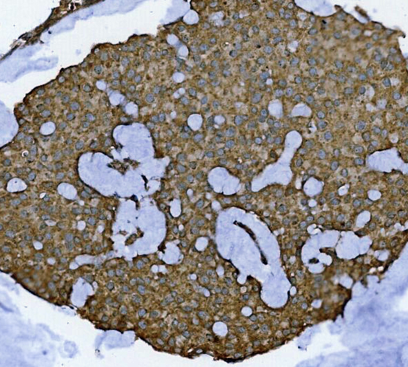
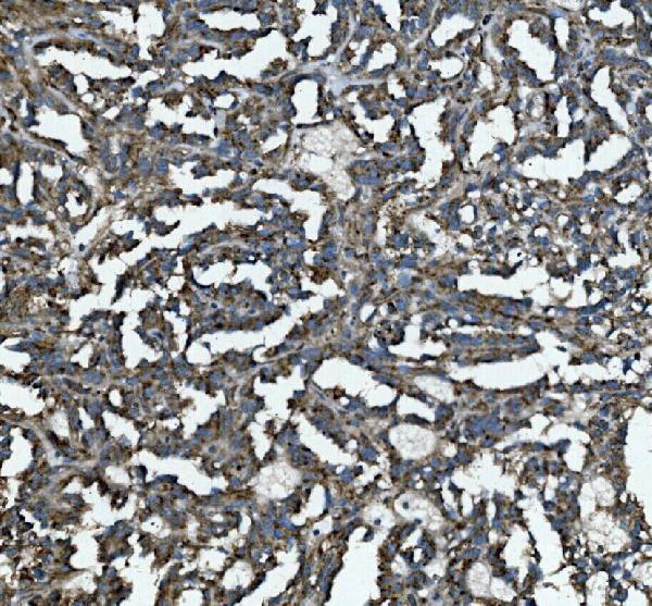
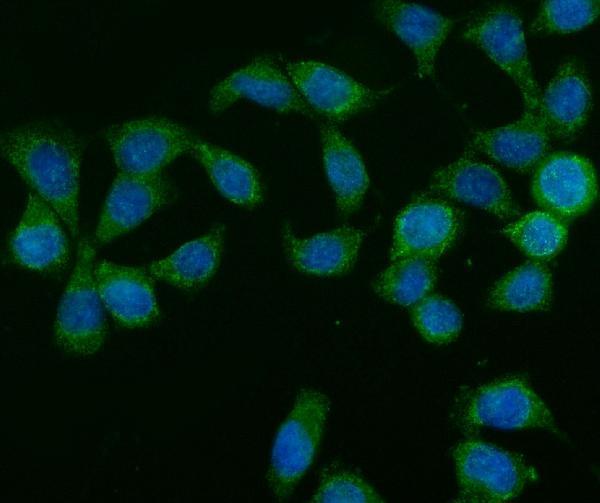
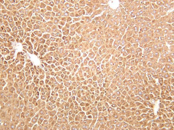
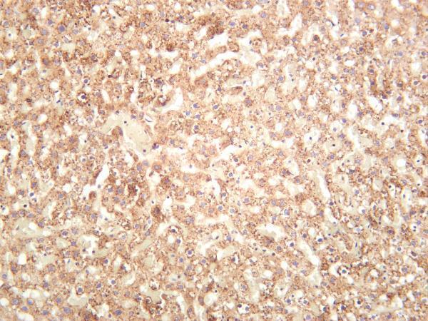
Anti-GLB1 IHC images cover human paraffin sections and mouse and rat tissues (catalog IHC captions); IF imaging covers CACO-2 cells (A01829-2 IF caption).
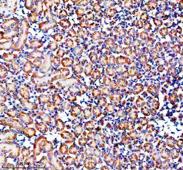
M01829-3 shows IHC staining in human colon cancer paraffin sections and mouse and rat kidney; A01829-2 shows IHC staining in human breast and lung cancer paraffin sections and IF staining in CACO-2 cells (respective image captions). M01829-4 shows IHC staining in mouse and rat liver, with human reactivity and IF also listed in its catalog entry (M01829-4 IHC captions; catalog applications and reactivity).
Which to pick: For tissue IHC across species, choose rabbit monoclonal M01829-3: its captions show human colon cancer and mouse and rat kidney paraffin sections stained at 1:50 after EDTA pH 8.0 retrieval (M01829-3 IHC captions; catalog clone). For IF/ICC, choose A01829-2: both applications are listed, and its CACO-2 IF image used 5 μg/mL (A01829-2 catalog applications; IF caption). M01829-4 is another cross-species IHC option with mouse and rat liver images and listed human reactivity; the fixative is unreported in all three antibodies’ IHC captions (M01829-4 IHC captions and catalog reactivity; A01829-2 and M01829-3 IHC captions).