Click image to see more details
-

-
-
-
-
+4
Product Info Summary
| SKU: | A01829-2 |
|---|---|
| Size: | 100 μg/vial |
| Reactive Species: | Human |
| Host: | Rabbit |
| Application: | ELISA, Flow Cytometry, IF, IHC, ICC, WB |
Customers Who Bought This Also Bought

Product info
Product Name
Anti-GLB1/Beta-galactosidase Antibody Picoband®
SKU/Catalog Number
A01829-2
Size
100 μg/vial
Form
Lyophilized
Description
Boster Bio Anti-GLB1/Beta-galactosidase Antibody Picoband® catalog # A01829-2. Tested in ELISA, Flow Cytometry, IF, IHC, ICC, WB applications. This antibody reacts with Human. The brand Picoband indicates this is a premium antibody that guarantees superior quality, high affinity, and strong signals with minimal background in Western blot applications. Only our best-performing antibodies are designated as Picoband, ensuring unmatched performance.
Storage & Handling
Store at -20˚C for one year from date of receipt. After reconstitution, at 4˚C for one month. It can also be aliquotted and stored frozen at -20˚C for six months. Avoid repeated freeze-thaw cycles.
Cite This Product
Anti-GLB1/Beta-galactosidase Antibody Picoband® (Boster Biological Technology, Pleasanton CA, USA, Catalog # A01829-2)
Host
Rabbit
Contents
Each vial contains 4 mg Trehalose, 0.9 mg NaCl and 0.2 mg Na2HPO4.
Clonality
Polyclonal
Isotype
Rabbit IgG
Immunogen
E.coli-derived human GLB1/Beta-galactosidase recombinant protein (Position: Q46-K655).
Cross-reactivity
No cross-reactivity with other proteins.
Reactive Species
A01829-2 is reactive to GLB1 in Human
Observed Molecular Weight
65-85 kDa
Calculated molecular weight
76.1 kDa
Background of GLB1
Galactosidase, beta 1, also known as GLB1, is a protein which in humans is encoded by the GLB1 gene. I t is mapped to 3p22.3. This gene encodes a member of the glycosyl hydrolase 35 family of proteins. Alternative splicing results in multiple transcript variants, at least one of which encodes a preproprotein that is proteolytically processed to generate the mature lysosomal enzyme. This enzyme catalyzes the hydrolysis of a terminal beta-linked galactose residue from ganglioside substrates and other glycoconjugates. Mutations in this gene may result in GM1-gangliosidosis and Morquio B syndrome.
Antibody Validation
Boster validates all antibodies on WB, IHC, ICC, Immunofluorescence, and ELISA with known positive control and negative samples to ensure specificity and high affinity, including thorough antibody incubations.
Application & Images
Applications
A01829-2 is guaranteed for ELISA, Flow Cytometry, IF, IHC, ICC, WB Boster Guarantee
Recommend Dilution
| Application | Dilution | Species |
|---|---|---|
| "Western blot | 0.25-0.5μg/ml | Human |
| Immunohistochemistry (Paraffin-embedded Section) | 2-5μg/ml | Human |
| Immunocytochemistry/Immunofluorescence | 5 μg/ml | Human |
| Flow Cytometry (Fixed) | 1-3μg/1x106 cells | Human |
| ELISA | 0.1-0.5μg/ml | - |
Tested application
Suggested blocking solution with 5% non-fat milk or BSA; (*)Recommended protein loading: 20-40 µg per lane
Use TE buffer pH 9.0 for antigen retrieval; (*) citrate buffer pH 6.0 is an alternative.
Validation Images & Assay Conditions
Click image to see more details
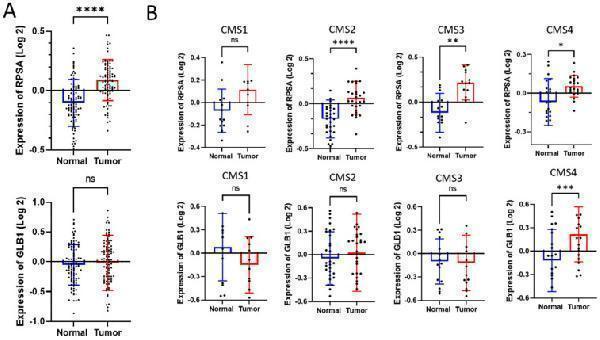
RPSA and GLB1 proteomic expression from CPTAC dataset. The proteomic expression levels of RPSA and GLB1 (β-galactosidase) were analyzed in 56 colorectal tumors (in red) and their matched normal mucosa samples (in blue) from the CPTAC dataset (Series #3, ) and expressed as the logarithmic difference in protein abundance, measured by mass spectrometry. ( A ) Unshared log 2 ratio expression of RPSA (upper panel) and GLB1 (lower panel). ( B ) Expression of RPSA (upper panels) and GLB1 (lower panels) based on transcriptomic subtypes associated with CMS1 (microsatellite instability and immune infiltration), CMS2 (WNT and MYC signaling activation), CMS3 (KRAS mutation and metabolic deregulation), CMS4 (TGFβ activation in EMT and ECM remodeling). Data are expressed as the mean ± SEM. Statistical test: paired Wilcoxon test (ns, not significant; *, p < 0.05; **, p < 0.01; ***, p < 0.001; ****, p < 0.0001).
Index in PubMed under a CC BY license. PMID: 40141206

Click image to see more details
Expression of RPSA, GLB1, and GLB1 variant 67EBP transcripts in CRC tissues. Expression levels of RPSA , GLB1, and GLB1 variant 67EBP were evaluated at the transcript levels in 25 CRC samples and corresponding resection margins from the biobank collection (Series #2, ) using quantitative RT-PCR. ( A ) Expression of RPSA . ( B ) Expression of GLB1 using primer set #1 targeting both β-galactosidase and 67EBP mRNA. ( C ) Expression of GLB1 using primer set #2 targeting exons 2–5 specific to the mRNA splice variant of 67EBP . ( D ) Expression of GLB1 using primer set #3 targeting exons 5–7 specific to the mRNA splice variant of 67EBP . RPLP0 was used as the normalizer. ( A – D , left): Expression levels were quantified using the Pfaffl method and compared to the resection margin. Results are presented as mean ± SEM. Statistical test: paired Wilcoxon test; (ns, not significant; #, p = 0.07; **, p < 0.01; ***, p < 0.001; ( A – D , right): Graphs displaying the relative amounts of RPSA and 67EBP in individual tumor tissue (T) compared to the resection margin (RM). Red lines are for a ratio of 1.0. Sample size: n = 25.
Index in PubMed under a CC BY license. PMID: 40141206

Click image to see more details
Expression of RPSA and GLB1 transcripts using a GEO DNA microarray dataset from patients with colorectal cancer. Analysis of RPSA and GLB1 transcript levels in primary tumors and their resection margins from the GEO microarray dataset (Series #4, ) using the Affymetrix Human Genome U133A Array. Only pairs whose expression was available in both the primary tumor and the corresponding resection margin were used. ( A ) RPSA expression analyzed using probe 213801_x_at. ( B ) GLB1 expression analyzed using probe 201576_s_at. Results are presented as mean ± SEM. Statistical significance was determined using the paired Wilcoxon test (ns, non-significant; ***, p < 0.001). Red lines are for a ratio of 1.0. Sample size: n = 44.
Index in PubMed under a CC BY license. PMID: 40141206

Click image to see more details
Flow Cytometry analysis of MCF-7 cells using anti-GLB1/Beta-galactosidase antibody (A01829-2).
Overlay histogram showing MCF-7 cells stained with A01829-2 (Blue line). To facilitate intracellular staining, cells were fixed with 4% paraformaldehyde and permeabilized with permeabilization buffer. The cells were blocked with 10% normal goat serum. And then incubated with rabbit anti-GLB1/Beta-galactosidase Antibody (A01829-2, 1 μg/1x106 cells) for 30 min at 20°C. DyLight®488 conjugated goat anti-rabbit IgG (BA1127, 5-10 μg/1x106 cells) was used as secondary antibody for 30 minutes at 20°C. Isotype control antibody (Green line) was rabbit IgG (1 μg/1x106) used under the same conditions. Unlabelled sample without incubation with primary antibody and secondary antibody (Red line) was used as a blank control.

Click image to see more details
Western blot analysis of GLB1/Beta-galactosidase using anti-GLB1/Beta-galactosidase antibody (A01829-2).
Electrophoresis was performed on a 5-20% SDS-PAGE gel at 70V (Stacking gel) / 90V (Resolving gel) for 2-3 hours. The sample well of each lane was loaded with 30 ug of sample under reducing conditions.
Lane 1: human Hela whole cell lysates,
Lane 2: human HepG2 whole cell lysates,
Lane 3: human SH-SY5Y whole cell lysates,
Lane 4: human MCF-7 whole cell lysates.
After electrophoresis, proteins were transferred to a nitrocellulose membrane at 150 mA for 50-90 minutes. Blocked the membrane with 5% non-fat milk/TBS for 1.5 hour at RT. The membrane was incubated with rabbit anti-GLB1/Beta-galactosidase antigen affinity purified polyclonal antibody (Catalog # A01829-2) at 0.5 μg/mL overnight at 4°C, then washed with TBS-0.1%Tween 3 times with 5 minutes each and probed with a goat anti-rabbit IgG-HRP secondary antibody at a dilution of 1:5000 for 1.5 hour at RT. The signal is developed using an Enhanced Chemiluminescent detection (ECL) kit (Catalog # EK1002) with Tanon 5200 system. A specific band was detected for GLB1/Beta-galactosidase at approximately 65-85 kDa. The expected band size for GLB1/Beta-galactosidase is at 73 kDa.

Click image to see more details
IHC analysis of GLB1/Beta-galactosidase using anti-GLB1/Beta-galactosidase antibody (A01829-2).
GLB1/Beta-galactosidase was detected in a paraffin-embedded section of human breast cancer tissue. Heat mediated antigen retrieval was performed in EDTA buffer (pH 8.0, epitope retrieval solution). The tissue section was blocked with 10% goat serum. The tissue section was then incubated with 2 μg/ml rabbit anti-GLB1/Beta-galactosidase Antibody (A01829-2) overnight at 4°C. Peroxidase Conjugated Goat Anti-rabbit IgG was used as secondary antibody and incubated for 30 minutes at 37°C. The tissue section was developed using HRP Conjugated Rabbit IgG Super Vision Assay Kit (Catalog # SV0002) with DAB as the chromogen.

Click image to see more details
IHC analysis of GLB1/Beta-galactosidase using anti-GLB1/Beta-galactosidase antibody (A01829-2).
GLB1/Beta-galactosidase was detected in a paraffin-embedded section of human lung cancer tissue. Heat mediated antigen retrieval was performed in EDTA buffer (pH 8.0, epitope retrieval solution). The tissue section was blocked with 10% goat serum. The tissue section was then incubated with 2 μg/ml rabbit anti-GLB1/Beta-galactosidase Antibody (A01829-2) overnight at 4°C. Peroxidase Conjugated Goat Anti-rabbit IgG was used as secondary antibody and incubated for 30 minutes at 37°C. The tissue section was developed using HRP Conjugated Rabbit IgG Super Vision Assay Kit (Catalog # SV0002) with DAB as the chromogen.

Click image to see more details
IF analysis of GLB1/Beta-galactosidase using anti-GLB1/Beta-galactosidase antibody (A01829-2).
GLB1/Beta-galactosidase was detected in an immunocytochemical section of CACO-2 cells. Enzyme antigen retrieval was performed using IHC enzyme antigen retrieval reagent (AR0022) for 15 mins. The cells were blocked with 10% goat serum. And then incubated with 5 μg/mL rabbit anti-GLB1/Beta-galactosidase Antibody (A01829-2) overnight at 4°C. DyLight488 Conjugated Goat Anti-Rabbit IgG (BA1127) was used as secondary antibody at 1:500 dilution and incubated for 30 minutes at 37°C. Visualize using a fluorescence microscope and filter sets appropriate for the label used.
Specific Publications For Anti-GLB1/Beta-galactosidase Antibody Picoband® (A01829-2)
Loading publications
Recommended Resources
Here are featured tools and databases that you might find useful.
- Boster's Pathways Library
- Protein Databases
- Bioscience Research Protocol Resources
- Data Processing & Analysis Software
- Photo Editing Software
- Scientific Literature Resources
- Research Paper Management Tools
- Molecular Biology Software
- Primer Design Tools
- Bioinformatics Tools
- Phylogenetic Tree Analysis
Customer Reviews
Have you used Anti-GLB1/Beta-galactosidase Antibody Picoband®?
Share your experimental results or join a short interview to earn up to $1,000 in product credits or other rewards.
0 Reviews For Anti-GLB1/Beta-galactosidase Antibody Picoband®
Customer Q&As
Have a question?
Find answers in Q&As, reviews.
Can't find your answer?
Submit your question
