This website uses cookies to ensure you get the best experience on our website.


- Table of Contents
Plan GLUD2 chromogenic IHC on paraffin sections using catalog antibody A04531 (datasheet A04531). Interpret variable cytoplasmic staining (HPA tissue IHC) alongside mitochondrial matrix localization (UniProt), with caution because the tissue staining may detect protein from more than one gene (HPA tissue IHC).

Expected localisation, validated protocols, controls and antibodies — the at-a-glance facts below, then the full design guide.
| Expected localisation | Variable cytoplasmic staining (HPA tissue IHC); mitochondrial matrix localization (UniProt) | |
| Staining pattern | Variable cytoplasmic staining in glandular and glial cells (HPA tissue IHC) | |
| Antigen retrieval | Citrate pH 6.0 HIER, heat-mediated (datasheet A04531) | |
| Positive control | Adrenal gland+4 more · see all | |
| Negative control | None in HPA (detected in all 45 tissues); use no-primary + isotype controls |
| Fixation | Keep fixation consistent across paraffin sections (standard IHC practice; not target-specific) | |
| Caveat | Staining may detect protein from more than one gene (HPA tissue IHC) | |
| Regulation | No staining-linked regulator reported (UniProt) | |
| Isoform / epitope | No annotated isoforms; mature chain spans residues 54–558 (UniProt) |
The catalog antibody’s IHC-P protocol (datasheet A04531) is paired with a published protocol for FFPE glioblastoma tissues (PMC6284416).
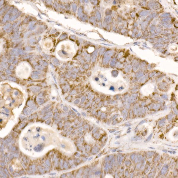
| Sample | Paraffin-embedded human colon carcinoma tissue; fixative not specified (datasheet A04531) |
| Fixation | Image fixative and duration unreported (datasheet A04531); verify before use. |
| Sectioning | 4–5 µm sections on charged slides (standard) |
| Deparaffinisation | Xylene, graded ethanol series to water (standard) |
| Antigen retrieval | Heat retrieval: Citrate pH 6.0 (datasheet A04531); 20 min, 95–100 °C (standard) |
| Peroxidase block | 3% H2O2, 10 min, room temperature (standard) |
| Blocking | 10% normal serum of the secondary host, 30 min, room temperature (standard) |
| Primary antibody | Rabbit anti-GLUD2, 1:50-1:200 (datasheet A04531) |
| Primary incubation | Overnight at 4 °C (standard) |
| Detection | HRP-polymer secondary, DAB chromogen 5–10 min (standard) |
| Counterstain | Hematoxylin, blue, dehydrate and mount (standard) |
| Expected result | GLUD2-positive staining in glandular cells of adrenal gland (HPA tissue IHC: High). HPA tissue profile: General cytoplasmic expression at variable levels. No signal in the no-primary control. |
GLUD2 is a mitochondrial matrix protein without a transmembrane segment (UniProt P49448). In IHC, assess cytoplasmic staining in the cell types HPA reports as positive, including glandular cells and caudate glial cells (HPA tissue IHC). Interpret apparent positives cautiously: HPA rates tissue IHC Uncertain because staining agrees poorly with RNA data and antibodies may detect proteins from more than one gene (HPA tissue IHC).
| Cytoplasmic staining is strongest in adrenal or appendix glandular cells, or bronchial respiratory epithelial cells (HPA: High). | This matches reported high-staining cells, but agreement alone does not establish GLUD2 specificity (HPA tissue IHC: Uncertain). Assess the distribution within cells; GLUD2 is assigned to the mitochondrial matrix (UniProt P49448). |
| Staining is mainly nuclear or extracellular, with little cytoplasmic signal. | That distribution conflicts with mitochondrial matrix localisation (UniProt P49448; HPA subcellular: mitochondria supported). Treat it as possible artefact or nonspecific detection, and review the negative control and staining distribution before scoring it positive (general IHC practice). |
| Strong staining appears in skeletal myocytes, soft-tissue fibroblasts or bone-marrow hematopoietic cells (HPA: Low). | This contrasts with HPA's low-staining examples (HPA tissue IHC). Cross-reactivity or endogenous detection activity is possible; neither can be diagnosed from location alone. Compare controls and the expected cell distribution before assigning GLUD2 positivity (general IHC practice). |
| Colour is diffuse across cells or tissue, without a discernible cellular pattern. | Diffuse background cannot reliably identify the mitochondrial matrix (UniProt P49448). Check the no-primary control for detection background, then review blocking, washing and detection conditions as general IHC variables (general IHC practice). |
| No signal appears in an adrenal or appendix section where HPA reports high glandular-cell staining (HPA: High). | First check tissue integrity, the assay control and whether the expected cells are present (general IHC practice). A negative result merits investigation, but HPA's Uncertain IHC reliability prevents treating one section as a definitive GLUD2-negative or assay-failure verdict (HPA tissue IHC). |
| Compartment and topology | GLUD2 is assigned to the mitochondrial matrix and has no transmembrane segment (UniProt P49448). Interpret cytoplasmic cellular signal in that context; nuclear or extracellular colour needs separate scrutiny (general IHC practice). |
| Protein processing | UniProt annotates the mature chain as residues 54–558 (UniProt P49448). If an antibody's epitope is known, compare it with that annotation; no epitope is supplied here, so no effect on staining can be predicted. |
| Tissue and cell context | HPA reports high staining in several glandular and epithelial populations and caudate glial cells, but low staining in skeletal myocytes, fibroblasts and bone-marrow hematopoietic cells (HPA tissue IHC). Use these as comparisons, not absolute controls. |
| IHC specificity evidence | HPA rates tissue IHC Uncertain, citing poor staining–RNA consistency and possible detection of proteins from more than one gene; its listed IHC antibodies are also rated Uncertain (HPA tissue IHC; HPA antibodies). |
| IF/ICC localisation evidence | HPA reports supported mitochondrial ICC-IF localisation, while warning that the antibodies may target proteins from multiple genes (HPA subcellular). That supports a compartment comparison; it does not validate a chromogenic IHC result or resolve GLUD2 specificity. |
| Situation | Likely cause | Next action |
|---|---|---|
| The expected glandular cells show no colour in an HPA high-staining tissue (HPA tissue IHC). | The expected cells may be absent from the section, or the IHC run may have failed (general IHC practice). HPA's uncertain tissue evidence also limits this comparison (HPA tissue IHC). | Confirm the cell population and section quality, inspect the run control, then review retrieval and antibody conditions as general IHC variables. No GLUD2-specific retrieval or fixation effect is established by these sources. |
| Every region, including low-staining comparison cells, appears strongly coloured (HPA: Low examples). | Widespread detection background, endogenous activity or cross-reactivity is possible (general IHC practice); HPA also flags possible detection of proteins from more than one gene (HPA tissue IHC). | Compare the no-primary control, inspect background outside target cells and review blocking and detection conditions (general IHC practice). Avoid scoring uniform colour as GLUD2-specific. |
| Colour concentrates in nuclei or outside cells. | The distribution conflicts with mitochondrial matrix localisation (UniProt P49448; HPA subcellular: mitochondria supported). | Review control staining and cellular boundaries; score the discordant compartment separately from cytoplasmic signal (general IHC practice). Do not infer GLUD2 localisation from the discordant colour. |
| High-staining cells vary across sections of the same tissue (HPA tissue IHC). | Different sampled cell populations or variable staining may explain the difference (general IHC practice). HPA reports variable general cytoplasmic expression and Uncertain tissue IHC reliability (HPA tissue IHC). | Compare the same cell type and tissue region across sections, document the distribution and retain the uncertainty in the interpretation (general IHC practice; HPA tissue IHC). |
| The positive signal is faint and difficult to assign to individual cells. | Weak colour or counterstain may obscure cellular boundaries (general IHC practice). A fine mitochondrial pattern may be difficult to resolve by chromogenic IHC (general IHC practice). | Review the unstained and negative-control areas, then assess counterstain and detection conditions on the IHC run (general IHC practice). Report only clearly cell-associated signal; do not require visible individual mitochondria. |
| Does mitochondrial IF/ICC staining confirm the IHC call? | HPA supports mitochondrial ICC-IF localisation but cautions that the antibodies may target proteins from multiple genes (HPA subcellular). Its tissue IHC assessment remains Uncertain (HPA tissue IHC). | Use IF/ICC as a localisation cross-check only. Judge the paraffin-section result using its own cellular pattern and controls (general IHC practice); do not transfer ICC-IF support into an IHC specificity claim. |
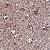


Comprehensive Human Protein Atlas IHC scoring per tissue (reliability: Uncertain — Very low consistency between antibody staining and RNA expression data. Caution, targets protein from more than one gene.). Rows are taken directly from the HPA tissue chart — click any row's HPA link to view the source.
| Tissue | Cell type | Level | Evidence | Source |
|---|---|---|---|---|
| Adrenal gland | Glandular cells | High | Protein (IHC) | HPA → |
| Appendix | Glandular cells | High | Protein (IHC) | HPA → |
| Breast | Glandular cells | High | Protein (IHC) | HPA → |
| Bronchus | Respiratory epithelial cells | High | Protein (IHC) | HPA → |
| Caudate | Glial cells | High | Protein (IHC) | HPA → |
| Tissue | Cell type | Level | Evidence | Source |
|---|---|---|---|---|
| None in HPA: GLUD2 is detected in all 45 scored tissues. Use a no-primary (secondary-only) and an isotype control instead. | ||||
Use compartment-aware controls when optimizing chromogenic GLUD2 IHC: GLUD2 is a mitochondrial matrix protein, while available tissue staining has specificity limits (UniProt P49448; HPA tissue IHC).
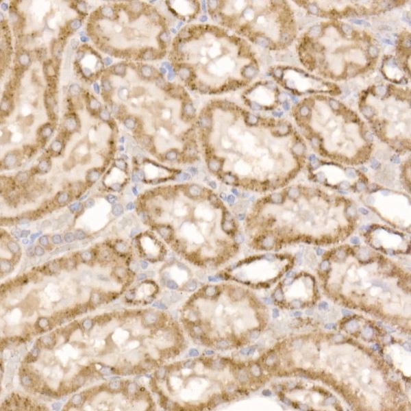
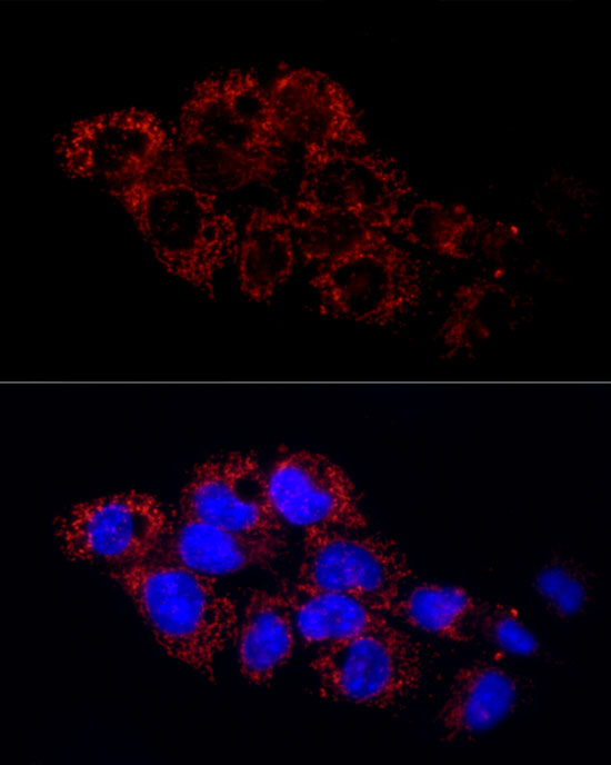
Anti-GLUD2 A04531 has IHC images from paraffin-embedded human colon carcinoma and rat kidney, plus an IF image from HepG2 cells (A04531 image captions).
A04531 is listed for IHC, IF and ICC in human, mouse and rat samples (catalog applications and reactivity). Its IHC captions show human colon carcinoma and rat kidney; its IF caption shows HepG2 cells (A04531 image captions).
Which to pick: For tissue IHC, choose A04531: its rabbit polyclonal antibody was imaged on paraffin-embedded sections at 1:50 after high-pressure retrieval in 10 mM citrate buffer, pH 6.0 (A04531 IHC captions); the fixative is unreported (A04531 IHC captions). For IF/ICC, A04531 is listed for both applications, with an IF image from HepG2 cells at 1:50 (catalog applications; A04531 IF caption). For cross-species work, A04531 lists human, mouse and rat reactivity, with IHC images for human and rat samples (catalog reactivity; A04531 IHC captions).