This website uses cookies to ensure you get the best experience on our website.


- Table of Contents
Plan paraffin-section GLUL IHC around its cytoplasmic staining in adipocytes, glia, macrophages and pericentral hepatocytes (HPA tissue IHC). This guide covers fixation, staining controls and scoring across tissue regions (standard IHC practice).

Expected localisation, validated protocols, controls and antibodies — the at-a-glance facts below, then the full design guide.
| Expected localisation | Cytoplasmic staining in tissue (HPA tissue IHC) | |
| Staining pattern | Cytoplasmic in adipocytes, glia, macrophages and pericentral hepatocytes (HPA tissue IHC) | |
| Antigen retrieval | EDTA pH 8.0 HIER, heat-mediated (datasheet A03191-3) | |
| Positive control | Adipose tissue+4 more · see all | |
| Negative control | Adrenal gland+4 more · see all |
| Fixation | Keep fixation consistent across sections. (standard IHC practice; not target-specific) | |
| Caveat | Pericentral hepatocyte staining can make liver scoring region-dependent (HPA tissue IHC) | |
| Regulation | Expression varies by cell type (HPA tissue IHC) | |
| Isoform / epitope | No annotated isoforms; mature chain begins at residue 2 (UniProt) |
The catalog antibody’s IHC-P protocol is supplemented by published GLUL staining methods for human liver and pancreas sections (PMC8814043; PMC6886125).
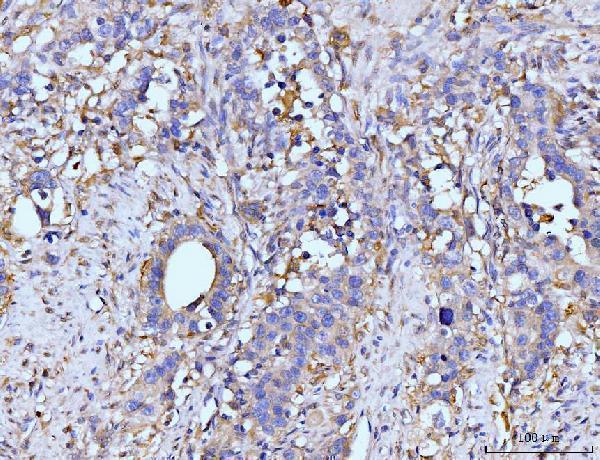
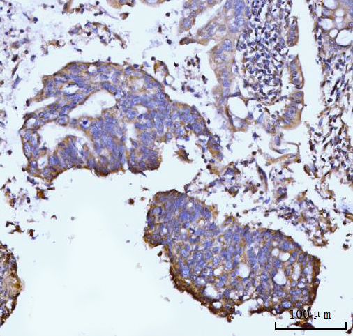
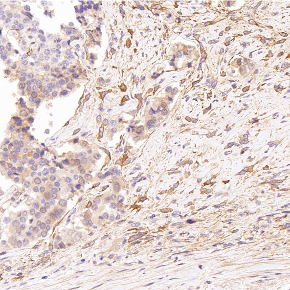
| Sample | Paraffin-embedded human appendiceal adenocarcinoma tissue; fixative not specified (datasheet A03191-3) |
| Fixation | Image fixative and duration unreported (datasheet A03191-3); verify before use. |
| Sectioning | 4–5 µm sections on charged slides (standard) |
| Deparaffinisation | Xylene, graded ethanol series to water (standard) |
| Antigen retrieval | Heat retrieval: EDTA pH 8.0 (datasheet A03191-3); 20 min, 95–100 °C (standard) |
| Peroxidase block | 3% H2O2, 10 min, room temperature (standard) |
| Blocking | 10% goat serum (datasheet A03191-3) |
| Primary antibody | Rabbit anti-GLUL, 2-5 μg/ml (datasheet A03191-3) |
| Primary incubation | Overnight at 4 °C (datasheet A03191-3) |
| Detection | Streptavidin-biotin complex (SABC), DAB chromogen (datasheet A03191-3) |
| Counterstain | Hematoxylin, blue, dehydrate and mount (standard) |
| Expected result | GLUL-positive staining in adipocytes of adipose tissue (HPA tissue IHC: High). HPA tissue profile: Cytoplasmic expression in several different cell types. Distinct expression in adipocytes, macrophages, glial cells and in hepatocytes around the central vein. No signal in the no-primary control. |
Expect predominantly cytoplasmic GLUL staining in adipocytes, glial cells, macrophages, and hepatocytes around the central vein (HPA: tissue IHC). GLUL mainly resides in the cytosol, although a fraction associates with the cell membrane; it has no transmembrane segment (UniProt P15104: localization and topology). HPA rates the tissue profile Approved, with medium consistency between antibody staining and RNA expression (HPA: tissue IHC reliability).
| Distinct cytoplasmic staining in adipocytes or glial cells. | This fits the reported pattern; HPA grades adipocytes and glial cells in caudate, cerebral cortex, and hippocampus High (HPA: tissue IHC). Compare cells within the section: an expected compartment and cell type together make a stronger interpretation than stain intensity alone (general IHC practice). |
| Strong staining confined to nuclei, with little cytoplasmic signal. | Predominantly nuclear staining conflicts with GLUL's mainly cytosolic localization (UniProt P15104: localization; HPA: subcellular). Check antibody and detection controls before calling it GLUL; a membrane-associated fraction can occur, so limited peripheral staining alone is not a wrong-compartment result (UniProt P15104: localization). |
| Prominent staining in a cell population HPA lists as not detected. | For example, HPA reports Not detected in lymph-node germinal-center cells and kidney glomerular cells; that finding does not describe every cell in either tissue (HPA: tissue IHC). Verify the cell identity and inspect controls for cross-reactivity or endogenous detection activity before assigning target expression (general IHC practice). |
| Weak, diffuse color covers cells and surrounding tissue. | This is difficult to score as GLUL when cytoplasmic cell boundaries are obscured (HPA: tissue IHC profile; general IHC practice). Compare a no-primary control, inspect endogenous detection activity, and assess blocking, washing, and chromogen development as general IHC workflow checks (general IHC practice). |
| No signal in adipocytes or glial cells expected to stain strongly. | An absent signal in these known-positive cell populations argues for a technical check before a biological absence call (HPA: High in adipocytes and glial cells). Verify that the relevant cells are present, then review antibody use, detection reagents, retrieval, and slide quality (general IHC practice). |
| Cell and tissue context | HPA reports High staining in adipocytes, glial cells, placental trophoblasts, testicular Leydig cells, and bladder urothelial cells; it also describes hepatocytes around the central vein (HPA: tissue IHC). Judge the identified cell population, since HPA's not-detected entries are cell-specific rather than whole-organ negatives (HPA: tissue IHC). |
| Compartment and topology | GLUL is mainly cytosolic, with a membrane-associated fraction and no transmembrane segment (UniProt P15104: localization and topology). HPA also approves cytosol and mitochondria as main ICC-IF locations (HPA: subcellular). Thus, predominantly cytoplasmic IHC staining fits the evidence; avoid requiring a crisp membrane outline (UniProt P15104: localization). |
| Interpretive confidence | HPA's tissue profile is Approved but has medium staining-versus-RNA consistency; paired antibodies show high similarity (HPA: tissue IHC reliability). Its listed antibodies HPA007316, HPA007571, and CAB008636 each have Approved IHC status (HPA: antibodies). Treat this as support for the reported pattern, while checking unusual results against controls (general IHC practice). |
| Antigen retrieval | Use the validated IHC-P conditions for the chosen antibody when available, and optimize retrieval with a known-positive control if needed (general IHC practice). The supplied UniProt and HPA records give no GLUL-specific retrieval condition or fixation-sensitivity result; neither tissue staining nor topology establishes one (UniProt P15104; HPA: tissue IHC). |
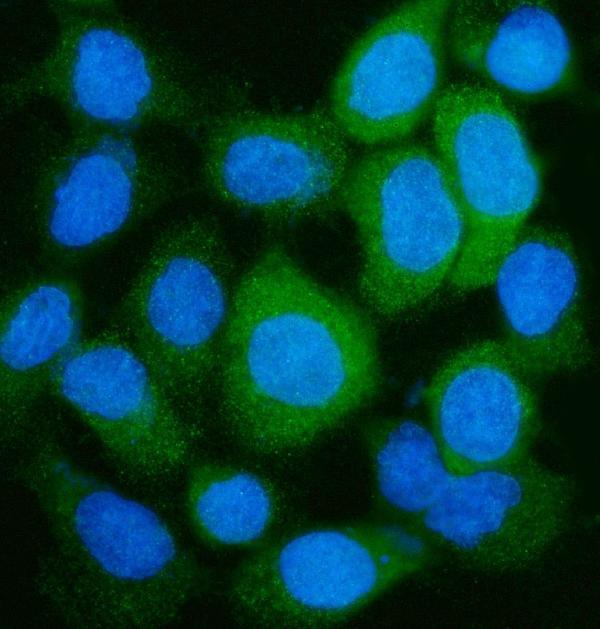
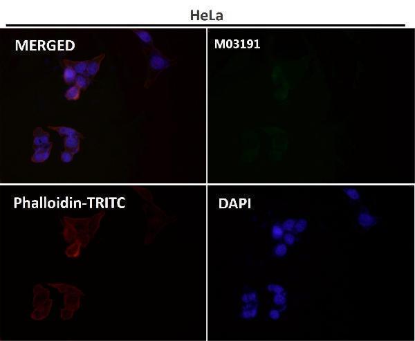
| IF/ICC: what localization should I expect? | HPA approves cytosol and mitochondria as main ICC-IF locations, with plasma membrane and connecting piece as additional locations (HPA: subcellular). UniProt describes mainly cytosolic GLUL with a membrane-associated fraction (UniProt P15104: localization). Use the separate IF/ICC guide for that application. |
| Situation | Likely cause | Next action |
|---|---|---|
| Positive control shows no staining. | The expected cells may be absent from the section, or an IHC workflow step may have failed (HPA: tissue IHC; general IHC practice). | Confirm adipocytes or glial cells are present; then check antibody use, retrieval, detection reagents, and slide quality with the chosen antibody's IHC-P instructions (HPA: High in those cells; general IHC practice). |
| Signal is mainly nuclear. | That distribution conflicts with the reported predominantly cytoplasmic pattern (HPA: tissue IHC; UniProt P15104: localization). | Review counterstain and no-primary controls, confirm which color is chromogen, and reassess whether cytoplasmic signal is present (general IHC practice). Do not score nuclear color alone as convincing GLUL. |
| Many unrelated cell types stain uniformly. | Diffuse binding or endogenous detection activity can obscure the cell-specific pattern (general IHC practice; HPA: tissue IHC profile). | Check a no-primary control and the detection system; review blocking, washing, and chromogen development (general IHC practice). Compare the result with expected cytoplasmic staining in identified cells (HPA: tissue IHC). |
| A supposed negative tissue contains positive cells. | HPA's negative labels apply to named cells, such as glomerular cells in kidney, not necessarily to every cell in that organ (HPA: tissue IHC). | Identify the stained population before calling the control failed. Compare like cells across sections, and use a no-primary control if the staining remains unexpected (HPA: tissue IHC; general IHC practice). |
| Peripheral staining appears alongside cytoplasmic signal. | A fraction of GLUL can associate with the cell membrane, despite lacking a transmembrane segment (UniProt P15104: localization and topology). | Assess whether cytoplasmic staining remains the dominant pattern and whether the stained cells fit HPA tissue observations (UniProt P15104: localization; HPA: tissue IHC). Check detection controls if the peripheral signal is widespread (general IHC practice). |
| Positive cells stain much less than expected. | Apparent intensity depends on both the sampled cell population and assay performance; HPA levels describe observed cells, not a guaranteed intensity in every run (HPA: tissue IHC; general IHC practice). | Verify cell identity against HPA's named populations, compare an appropriate positive control, and review the chosen antibody's IHC-P instructions and detection development (HPA: tissue IHC; general IHC practice). |
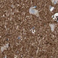
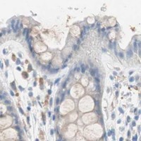
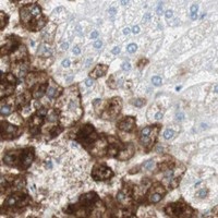


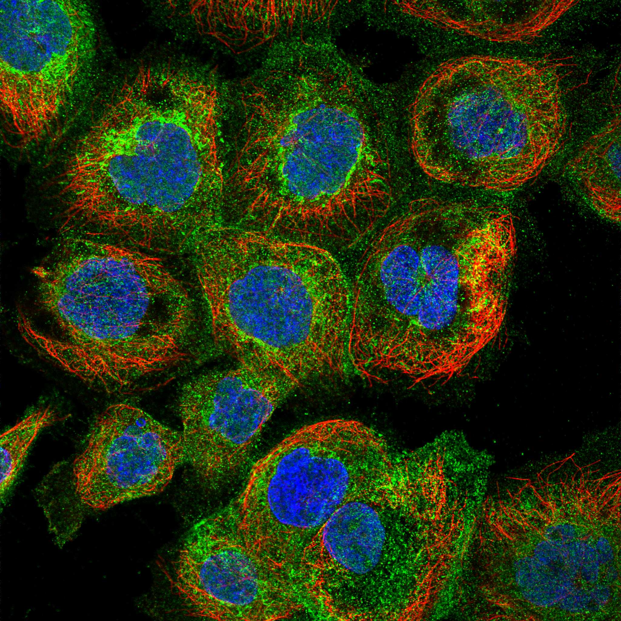
Comprehensive Human Protein Atlas IHC scoring per tissue (reliability: Approved — Medium consistency between antibody staining and RNA expression data. Paired antibodies with high similarity supports the protein expression profile.). Rows are taken directly from the HPA tissue chart — click any row's HPA link to view the source.
| Tissue | Cell type | Level | Evidence | Source |
|---|---|---|---|---|
| Adipose tissue | Adipocytes | High | Protein (IHC) | HPA → |
| Caudate | Glial cells | High | Protein (IHC) | HPA → |
| Cerebral cortex | Glial cells | High | Protein (IHC) | HPA → |
| Epididymis | Glandular cells | High | Protein (IHC) | HPA → |
| Hippocampus | Glial cells | High | Protein (IHC) | HPA → |
| Tissue | Cell type | Level | Evidence | Source |
|---|---|---|---|---|
| Adrenal gland | Glandular cells | Not detected | Protein (IHC) | HPA → |
| Appendix | Glandular cells | Not detected | Protein (IHC) | HPA → |
| Endometrium | Cells in endometrial stroma | Not detected | Protein (IHC) | HPA → |
| Heart muscle | Cardiomyocytes | Not detected | Protein (IHC) | HPA → |
| Kidney | Cells in glomeruli | Not detected | Protein (IHC) | HPA → |
Troubleshoot GLUL chromogenic IHC in paraffin sections by checking retrieval, staining controls, compartment, cell identity, and scoring against documented expression patterns.
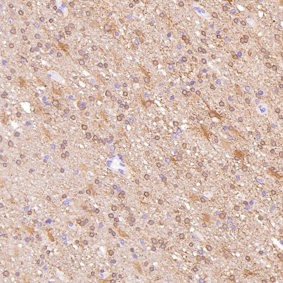
Anti-GLUL antibodies have IHC images from human, mouse and rat tissues (catalog IHC image captions), with an IF/ICC image from A431 cells (A03191-3 IF image caption).
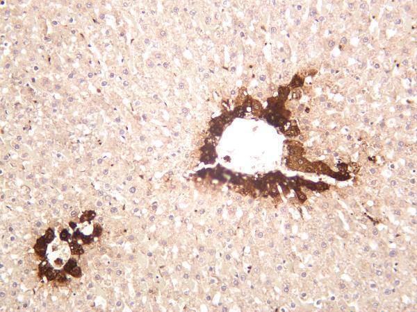
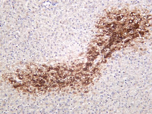
A03191-3 has IHC images from human appendiceal, gastric, liver and lung tumors (A03191-3 IHC image captions); M03191 has paraffin-section images from human glioblastoma and prostate cancer, rat kidney and mouse ovary (M03191 IHC image captions). M03191-2 has IHC images from rat and mouse liver and human hepatocellular carcinoma (M03191-2 IHC image captions).
Which to pick: For paraffin-section IHC, choose A03191-3 if its documented EDTA retrieval at pH 8.0 and 2 μg/ml staining example suit your tissue; the fixative is unreported (A03191-3 IHC image caption). For IF/ICC, choose A03191-3: its A431 cell image documents 5 μg/ml primary antibody and a fluorescent secondary (A03191-3 IF image caption). For IHC across human, mouse and rat, consider the rabbit monoclonal M03191-2, which has an IHC application listing and images from all three species; its image captions do not report section processing or fixative (M03191-2 catalog applications, title and IHC image captions).