GMNN / Geminin · IHC design guide

Design Immunohistochemistry for GMNN

Plan chromogenic GMNN IHC in paraffin sections using the nuclear staining seen in highly proliferating cells as a reference (HPA tissue IHC). The catalog antibody A06060-3 has an IHC working range of 2–5 μg/ml (datasheet); account for GMNN degradation during mitosis when interpreting staining (UniProt).

Evidence assembled Oct 2026 · For research use; verify linked source records and product datasheet before use
Immunohistochemistry protocol sheet for GMNN (IHC for GMNN): expected localisation Nuclear in highly proliferating cells (HPA tissue IHC), antibody A06060-3, validated IHC image, and IHC protocol steps
Printable GMNN IHC protocol sheet — expected localisation Nuclear in highly proliferating cells (HPA tissue IHC), antibody A06060-3, controls and protocol steps. Open the full GMNN IHC guide →

GMNN Immunohistochemistry Experimental Design Guide

Expected localisation, validated protocols, controls and antibodies — the at-a-glance facts below, then the full design guide.

Must know before staining
Expected localisation Nuclear in highly proliferating cells (HPA tissue IHC)
Staining pattern Nuclear staining in highly proliferating cells (HPA tissue IHC)
Antigen retrieval EDTA pH 8.0 HIER, heat-mediated (datasheet A06060-3)
Positive control ⓘ Appendix+4 more · see all
Negative control ⓘ Adipose tissue+4 more · see all
Important caveats
Reasons your staining may differ from the expected pattern.
Fixation Keep fixation consistent across paraffin sections. (standard IHC practice; not target-specific)
Caveat Mitotic degradation may reduce staining (UniProt)
Regulation Degraded during mitosis (UniProt)
Isoform / epitope No annotated isoforms; one 1–209 chain (UniProt)
Section 1

Recommended GMNN IHC & IF Protocols

The catalog antibody’s IHC-P protocol (datasheet A06060-3) is accompanied by three published GMNN IHC workflows (PMC9764478; PMC9814937; PMC7260192).

Recommended immunohistochemistry (IHC-P) protocol parameters
SampleParaffin-embedded human gall bladder adenosquamous carcinoma tissue; fixative not specified (datasheet A06060-3)
FixationImage fixative and duration unreported (datasheet A06060-3); verify before use.
Sectioning4–5 µm sections on charged slides (standard)
DeparaffinisationXylene, graded ethanol series to water (standard)
Antigen retrievalHeat retrieval: EDTA pH 8.0 (datasheet A06060-3); 20 min, 95–100 °C (standard)
Peroxidase block3% H2O2, 10 min, room temperature (standard)
Blocking10% goat serum (datasheet A06060-3)
Primary antibodyRabbit anti-GMNN, 2-5 μg/ml (datasheet A06060-3)
Primary incubationOvernight at 4 °C (datasheet A06060-3)
DetectionStreptavidin-biotin complex (SABC), DAB chromogen (datasheet A06060-3)
CounterstainHematoxylin, blue, dehydrate and mount (standard)
Expected resultGMNN-positive staining in endocrine cells of appendix (HPA tissue IHC: High). HPA tissue profile: Nuclear expression in highly proliferating cells. No signal in the no-primary control.
💡Decision noteStart with heat-mediated EDTA, pH 8.0 (datasheet A06060-3). If optimizing retrieval, a published ACC workflow used citrate, pH 6.0 (PMC9764478).
Section 2

What Is the Expected GMNN Staining Pattern?

GMNN should show predominantly nuclear IHC staining in highly proliferating cells (HPA tissue IHC: Enhanced reliability; nuclear profile). High staining is reported in oral mucosal squamous epithelial cells, intestinal endocrine cells and testicular pachytene spermatocytes (HPA tissue IHC). Interpret cytoplasmic signal with care: GMNN can occur in the cytoplasm and relocate to the nucleus (UniProt O75496: subcellular location); it has no transmembrane segment (UniProt O75496: topology).

What am I looking at on my slide?
Discrete nuclear staining in the expected cells, with little staining outside them.This matches the reported IHC profile. Assess the fraction of positive nuclei within the relevant cell population; a whole-section impression can obscure selective staining (HPA tissue IHC: nuclear expression in highly proliferating cells).
Predominantly diffuse cytoplasmic staining, with no convincing nuclei in a positive-control section.This is discordant with the tissue IHC pattern (HPA tissue IHC: nuclear profile). Cytoplasmic GMNN remains biologically possible (UniProt O75496: subcellular location), so check the control and detection background before calling the compartment artefactual.
Strong staining in adipocytes while the expected positive cells lack nuclear staining.Treat this as possible cross-reactivity or endogenous detection activity, then review controls (HPA tissue IHC: adipocytes not detected). A reference negative is a comparison, not proof that every specimen must be negative.
Uniform color over nuclei, cytoplasm and surrounding tissue.This obscures compartment and cell-type assignment. Consider nonspecific background from the IHC workflow; the reported pattern is selective nuclear expression (HPA tissue IHC: profile), so do not score the diffuse color as GMNN-positive nuclei.
No convincing nuclear signal in an oral mucosa or testis positive-control section.Check the staining run before interpreting the study sample: squamous epithelial cells in oral mucosa and pachytene spermatocytes in testis have high reported staining (HPA tissue IHC). Their absence does not identify a GMNN-specific fixation defect.
💡Expected GMNN appearanceCall a section positive when distinct nuclei stain in the relevant cells, including high staining in oral mucosal squamous cells or pachytene spermatocytes (HPA tissue IHC: High); widespread diffuse color or isolated unexpected-cell staining warrants a background or specificity check (HPA tissue IHC: nuclear profile).
How each factor affects the staining
Cell population and proliferationGMNN staining is reported as nuclear in highly proliferating cells (HPA tissue IHC: profile); compare the named cell population, since signal need not be uniform across a tissue section.
Cell-cycle timingGMNN inhibits replication licensing and is degraded during mitosis (UniProt O75496: function). Variable nuclear positivity can therefore reflect cell state; IHC alone cannot assign a specific cell-cycle phase.
Compartment and topologyUniProt lists both cytoplasm and nucleus and no transmembrane segment (UniProt O75496: location; topology). For tissue IHC, use the HPA nuclear profile as the primary pattern; investigate exclusively cytoplasmic chromogen.
Antibody validationSeveral listed antibodies have Enhanced IHC validation (HPA antibodies: HPA049977, CAB011458, CAB047327). That supports the reported pattern but does not validate a different reagent or every specimen.
IF/ICC: where should signal appear?Mainly in the nucleoplasm (HPA subcellular ICC-IF: supported); cytosol and basal body are additional uncertain locations (HPA subcellular ICC-IF). This answers the IF/ICC pattern question, without defining an IF protocol.
Why is my staining missing, weak or wrong?
SituationLikely causeNext action
Positive-control nuclei are blank.The run may have failed, or staining conditions may be unsuitable; the result alone cannot establish target-specific fixation sensitivity.Confirm the chosen control contains the expected cells (HPA tissue IHC: oral mucosa squamous cells High; testis pachytene spermatocytes High). Check reagent application, retrieval, dilution and detection as general IHC workflow steps.
Cytoplasmic color dominates a tissue section.Cytoplasmic GMNN is possible (UniProt O75496: location), but a cytoplasm-only result differs from the reported nuclear tissue profile (HPA tissue IHC).Inspect nuclei in an HPA-supported positive cell population (HPA tissue IHC); compare with negative controls and review background before assigning a specific cause.
Adipocytes or other unexpected cells stain strongly.The pattern may reflect nonspecific binding or endogenous detection activity; adipocytes are reported as not detected (HPA tissue IHC).Compare a no-primary control and, where relevant, an endogenous-activity control (general IHC practice). Check whether expected positive nuclei also stain before interpreting the unexpected cells.
Color covers most of the section.High background can prevent recognition of the selective nuclear pattern (HPA tissue IHC: profile); the appearance alone does not identify its source.Review blocking, washes, primary-antibody dilution and detection exposure as general IHC steps. Reassess compartment and cell-type selectivity after background is reduced.
Only a small subset of nuclei is positive.Selective staining may be consistent with expression in highly proliferating cells and GMNN cell-cycle regulation (HPA tissue IHC: profile; UniProt O75496: function).Identify and score the relevant cell population separately; compare it with an HPA-supported positive control (HPA tissue IHC). Avoid treating the percentage alone as a failed run.
Two antibodies give different cell or compartment patterns.Validation belongs to the tested antibody; several listed reagents have Enhanced IHC status, but this does not transfer to another reagent (HPA antibodies: IHC status).Confirm each reagent's IHC validation, compare the same control tissue and review negative controls. Prefer the reproducible selective nuclear pattern reported for tissue IHC (HPA tissue IHC).

Sample controls for GMNN IHC & IF

🧪Run oral mucosa first and look for staining in squamous epithelial cells (HPA: High in oral mucosa squamous epithelial cells). Use adipose tissue as the negative comparator, focusing on adipocytes (HPA: Not detected in adipocytes); on the oral mucosa slide, assess morphologically distinct stromal cells for background staining rather than assuming every stromal cell is GMNN-negative.
Positive control tissue: Appendix (Endocrine cells, HPA High)
Negative control tissue: Adipose tissue (HPA Not detected)
ICC-IF cell lines (HPA subcellular resource): HPA ICC-IF images show GMNN in U-251MG, U2OS, A-431, ASC52telo, RPTEC/TERT1, hTERT-RPE1 (serum starved), with annotated localisation: Nucleoplasm (supported) (HPA subcellular).
Technical controls: Include a no-primary (secondary-only) control, a host-species- and class-matched nonimmune rabbit IgG isotype control, and GMNN knockout material if available as a biological negative (caption: rabbit primary and anti-rabbit IgG secondary; standard IHC practice). Quench endogenous peroxidase and check for endogenous biotin background in the oral mucosa section when using the caption’s SABC/DAB detection (caption: SABC/DAB; standard IHC practice).
⚠️Feasibility: No target-specific fixation window or fixation effect is reported, and the fixative for the selected A06060-3 paraffin-section caption is unreported (caption: paraffin section; fixative not stated). The caption uses heat retrieval in EDTA at pH 8.0, so that is a documented starting condition, but retrieval dependence has not been established (caption: EDTA retrieval, pH 8.0). The supplied evidence does not establish whether frozen sections or IF are easier; for IF localisation assessment, nucleoplasmic signal has support, while cytosolic and basal-body locations are uncertain (HPA: subcellular localisation).

HPA tissue IHC evidence for GMNN

Comprehensive Human Protein Atlas IHC scoring per tissue (reliability: Enhanced — Medium consistency between antibody staining and RNA expression data.). Rows are taken directly from the HPA tissue chart — click any row's HPA link to view the source.

Positive expression · recommended positive controls

TissueCell typeLevelEvidenceSource
Appendix Endocrine cells High Protein (IHC) HPA →
Colon Endocrine cells High Protein (IHC) HPA →
Duodenum Endocrine cells High Protein (IHC) HPA →
Oral mucosa Squamous epithelial cells High Protein (IHC) HPA →
Rectum Endocrine cells High Protein (IHC) HPA →

Undetected expression · recommended negative controls

TissueCell typeLevelEvidenceSource
Adipose tissue Adipocytes Not detected Protein (IHC) HPA →
Adrenal gland Glandular cells Not detected Protein (IHC) HPA →
Bone marrow Hematopoietic cells Not detected Protein (IHC) HPA →
Caudate Glial cells Not detected Protein (IHC) HPA →
Cerebral cortex Endothelial cells Not detected Protein (IHC) HPA →
Section 3

Advanced GMNN IHC Tips

Use the catalog antibody’s paraffin section example as the starting point, then judge staining against GMNN localisation and proliferating cell patterns.

What retrieval should I use if nuclear GMNN staining is weak?
Start with heat mediated retrieval in EDTA at pH 8.0 for paraffin sections (datasheet A06060-3). The selected antibody’s tissue example used that buffer before 2 μg/ml primary antibody overnight at 4°C, so keep those conditions consistent while checking retrieval (datasheet A06060-3). If staining remains weak, compare a small retrieval time series on matched sections, watching for tissue damage or rising background (standard IHC practice). Include a section with expected nuclear staining in proliferating cells to distinguish failed retrieval from a specimen with little detectable GMNN (HPA: nuclear expression in highly proliferating cells).
Could fixation explain loss of GMNN staining in my paraffin sections?
The selected paraffin section caption does not state a fixative, so target specific GMNN fixation sensitivity is unknown (datasheet A06060-3: fixative not stated). Record the specimen’s fixative, fixation duration and processing history before attributing a weak result to antigen masking (standard IHC practice). Compare matched sections processed under documented conditions while holding EDTA retrieval at pH 8.0 and antibody concentration at 2 μg/ml constant (datasheet A06060-3). Assess morphology alongside staining, and use a positive control section in the same run to identify a processing or detection problem (standard IHC practice).
How should I assess nuclear versus cytoplasmic GMNN staining?
Score nuclear and cytoplasmic signals separately: tissue IHC reports nuclear expression in highly proliferating cells, while the subcellular dataset supports nucleoplasmic localisation (HPA: tissue IHC; HPA: nucleoplasm supported). Cytoplasmic signal is biologically possible because GMNN can occur in the cytoplasm and relocalise to the nucleus, but diffuse staining alone needs validation (UniProt O75496: subcellular location). Compare cells of the same type within the section, preserving the nuclear boundary with a counterstain and recording intensity by compartment (standard IHC practice). If a proposed positive population stains only in the cytoplasm, inspect negative controls and repeat detection before calling it GMNN (standard IHC practice; HPA: nuclear tissue profile).
Can isoforms or epitope masking account for discordant staining?
The supplied record lists 0 isoforms and a single 1–209 GMNN chain, so it provides no annotated isoform explanation for discordant paraffin staining (UniProt O75496: isoforms and processing). It lists no transmembrane segment or glycosylation sites, but does list modified residues including acetyllysine 27 and phosphoserines 34, 36, 49 and 63 (UniProt O75496: topology and modifications). Without a mapped antibody epitope, those modifications cannot establish whether this antibody’s binding changes (datasheet A06060-3: epitope not specified). Recheck retrieval and compare an independently validated antibody where available, using matched sections and controls (standard IHC practice).
How can IF help resolve an ambiguous GMNN IHC pattern?
Use IF as a separate localisation check, pairing GMNN with a validated marker for the cell population under study; tissue IHC places GMNN in highly proliferating cells (HPA: tissue IHC profile). Choose a fluorophore channel with low specimen autofluorescence, often a far red channel, and inspect single label controls before interpreting overlap (standard IF practice). Because GMNN is intracellular and has no transmembrane segment, permeabilise cells for intracellular access, but do not assume a mapped epitope or membrane side (UniProt O75496: location and topology; datasheet A06060-3: epitope not specified). Compare nuclear signal with a nuclear counterstain, recognising that nucleoplasm is the supported IF location (HPA: nucleoplasm supported).
What should I check when DAB appears broadly positive?
First inspect a no primary control and the section’s morphology to separate antibody dependent staining from detection background (standard IHC practice). The selected paraffin example used a biotinylated secondary, a streptavidin biotin complex and DAB, so evaluate each detection step when broad pigment appears (datasheet A06060-3). Apply a peroxidase block and check for endogenous biotin interference where relevant to this detection chemistry (standard chromogenic IHC practice). Reduce nonspecific signal by checking blocking and primary concentration against the documented 10% goat serum block and 2 μg/ml primary starting conditions, while retaining a positive control (datasheet A06060-3; standard IHC practice).
How should I quantify GMNN across paraffin sections? ⚠ ANSWER MARKED FOR VERIFICATION
Define cell populations and regions before scoring, then report the percentage of positive nuclei and either staining intensity or an H-score (standard IHC scoring practice; HPA: nuclear expression in highly proliferating cells). An H-score can combine intensity categories 0–3 with the percentage of cells in each category, using the same threshold across matched sections (standard IHC scoring practice). Normalise positive nuclei to all evaluable nuclei in the defined cell population; if measuring density, report positive nuclei per mm² of viable tissue (standard IHC quantification practice). Exclude necrosis and damaged edges consistently, and report cytoplasmic staining separately because the expected tissue profile is nuclear (standard IHC practice; HPA: tissue IHC profile).
Which staining patterns support a true GMNN positive result?
A convincing paraffin section result shows interpretable nuclear staining in the relevant proliferating cells, consistent with the tissue IHC profile and supported nucleoplasmic localisation (HPA: tissue IHC profile; HPA: nucleoplasm supported). Check the cell type: HPA reports high staining in oral mucosa squamous epithelial cells and testis pachytene spermatocytes, but no detection in adipocytes of adipose tissue (HPA: tissue IHC). Treat staining confined to cut edges, necrotic areas or the no primary control as suspect, and assess endogenous peroxidase contribution to DAB deposits (standard IHC practice). Cytoplasmic staining can occur, but an exclusively cytoplasmic pattern requires independent support before interpretation as GMNN (UniProt O75496: subcellular location; HPA: nuclear tissue profile).
Boster reagents

Best GMNN / Geminin IHC Antibodies

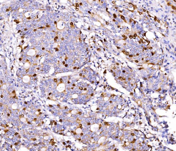
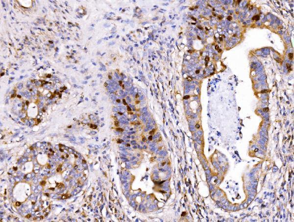
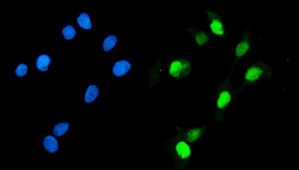
A06060-3 has human paraffin-section IHC and Caco-2 ICC/IF images (catalog image captions); human and mouse reactivity is listed (catalog reactivity).

Real IHC data IHC analysis of Geminin/GMNN using anti-Geminin/GMNN antibody (A06060-3). Geminin/GMNN was detected in a paraffin-embedded section of human gall bladder adenosquamous carcinoma tissue. Heat mediated antigen retrieval was performed in EDTA buffer (pH 8.0, epitope retrieval solution). The tissue section was blocked with 10% goat serum. The tissue section was then incubated with 2 μg/ml rabbit anti-Geminin/GMNN Antibody (A06060-3) overnight at 4°C. Biotinylated goat anti-rabbit IgG was used as secondary antibody and incubated for 30 minutes at 37°C. The tissue section was developed using Strepavidin-Biotin-Complex (SABC) (Catalog # SA1022) with DAB as the chromogen.
Anti-Geminin/GMNN Antibody ®
Cat # A06060-3

A06060-3 will render with its human gall bladder adenosquamous carcinoma paraffin-section IHC figure (card image caption); other captions show human lung and rectal cancer paraffin sections and Caco-2 ICC/IF (catalog image captions). M06060 lists human IHC and ICC/IF applications (catalog applications and reactivity), but has no IHC or IF image caption to support a figure-backed card (catalog image alts).

Which to pick: For tissue IHC, choose A06060-3: its paraffin-section caption documents EDTA retrieval at pH 8.0 and antibody at 2 μg/ml; the fixative is unreported (A06060-3 IHC image caption). For IF/ICC, A06060-3 has a Caco-2 image at 5 μg/ml (A06060-3 IF image caption), while M06060 is a rabbit monoclonal, clone AEGO-7, with listed human ICC/IF and IHC applications but no image captions (M06060 catalog). For work spanning human and mouse samples, A06060-3 lists both species, though its supplied IHC and IF image captions document human samples only (A06060-3 catalog reactivity and image captions).

Each figure is that product's own IHC / IF validation image from its datasheet.

References

  1. UniProt Consortium. UniProt entry O75496 (GEMI_HUMAN, Geminin).
  2. Human Protein Atlas. GMNN tissue IHC expression (reliability: Enhanced).
  3. Human Protein Atlas. GMNN subcellular location (ICC-IF): Mainly localized to the nucleoplasm. In addition localized to the cytosol and basal body..
  4. Human Protein Atlas. GMNN antibody validation summary (5 antibodies).
  5. High expression of GMNN predicts malignant progression and poor prognosis in ACC. European journal of medical research 2022 — PMC9764478.
  6. Increased FOXJ1 protein expression is associated with improved overall survival in high-grade serous ovarian carcinoma: an Ovarian Tumor Tissue Analysis Consortium Study. British journal of cancer 2023 — PMC9814937.
  7. Homologous recombination DNA repair deficiency and PARP inhibition activity in primary triple negative breast cancer. Nature communications 2020 — PMC7260192.
  8. Ex vivo assays to predict enhanced chemosensitization by hyperthermia in urothelial cancer of the bladder. PloS one 2018 — PMC6294360.
  9. PubMed PMID:9635433 — UniProt-cited evidence.
  10. PubMed PMID:14702039 — UniProt-cited evidence.
  11. PubMed PMID:14574404 — UniProt-cited evidence.