GNAL / Guanine nucleotide-binding protein G(olf) subunit alpha · IHC design guide

Design Immunohistochemistry for GNAL

GNAL is a lipid-anchored G alpha subunit associated with the plasma membrane (UniProt). Use this paraffin-section IHC guide to plan controls around high staining in caudate synapses and placental Hofbauer cells (HPA tissue IHC).

Evidence assembled Oct 2026 · For research use; verify linked source records and product datasheet before use
Immunohistochemistry protocol sheet for GNAL (IHC for GNAL): expected localisation Plasma membrane association expected (UniProt), antibody A09404-1, validated IHC image, and IHC protocol steps
Printable GNAL IHC protocol sheet — expected localisation Plasma membrane association expected (UniProt), antibody A09404-1, controls and protocol steps. Open the full GNAL IHC guide →

GNAL Immunohistochemistry Experimental Design Guide

Expected localisation, validated protocols, controls and antibodies — the at-a-glance facts below, then the full design guide.

Must know before staining
Expected localisation Plasma membrane association expected (UniProt)
Staining pattern High staining in caudate synapses and placental Hofbauer cells (HPA tissue IHC)
Antigen retrieval EDTA pH 8.0 HIER, heat-mediated (datasheet A09404-1)
Positive control ⓘ Caudate+4 more · see all
Negative control ⓘ Adipose tissue+4 more · see all
Important caveats
Reasons your staining may differ from the expected pattern.
Fixation Keep fixation consistent across sections (standard IHC practice; not target-specific); Matched tissue-IHC evidence does not establish the fixation claim. Validate the specimen-specific method before use. (selected-SKU IHC image A09404-1)
Caveat Staining and RNA show medium consistency (HPA tissue IHC)
Regulation Brain-enhanced RNA expression (HPA tissue RNA)
Isoform / epitope 3 isoforms; epitope coverage is unspecified (UniProt)
Section 1

Recommended GNAL IHC & IF Protocols

The catalog antibody’s IHC-P protocol (datasheet: A09404-1) is accompanied by published GNAL IHC methods for human glioma (PMC11055992) and skin (PMC10392076).

Recommended immunohistochemistry (IHC-P) protocol parameters
SampleParaffin-embedded human lung cancer tissue; fixative not specified (datasheet A09404-1)
FixationImage fixative and duration unreported (datasheet A09404-1); verify before use.
Sectioning4–5 µm sections on charged slides (standard)
DeparaffinisationXylene, graded ethanol series to water (standard)
Antigen retrievalHeat retrieval: EDTA pH 8.0 (datasheet A09404-1); 20 min, 95–100 °C (standard)
Peroxidase block3% H2O2, 10 min, room temperature (standard)
Blocking10% goat serum (datasheet A09404-1)
Primary antibodyRabbit anti-GNAL, 2-5 μg/ml (datasheet A09404-1)
Primary incubationOvernight at 4 °C (datasheet A09404-1)
DetectionStreptavidin-biotin complex (SABC), DAB chromogen (datasheet A09404-1)
CounterstainHematoxylin, blue, dehydrate and mount (standard)
Expected resultGNAL-positive staining in synapses of caudate (HPA tissue IHC: High). HPA tissue profile: Protein expression in several tissues, including caudate. No signal in the no-primary control.
💡Decision noteStart with heat-mediated EDTA retrieval at pH 8.0 (datasheet: A09404-1). For skin sections, the published method used citrate at pH 6.0 (PMC10392076).
Section 2

What Is the Expected GNAL Staining Pattern?

GNAL is associated with the plasma membrane through a lipid anchor and has no transmembrane segment (UniProt P38405 topology). In IHC, expect staining in caudate synapses, placental Hofbauer cells and tonsillar squamous epithelial cells, all reported as High (HPA tissue IHC). HPA rates the tissue evidence Enhanced, while reporting medium consistency between antibody staining and RNA expression (HPA tissue IHC).

What am I looking at on my slide?
Strong staining in caudate synapses, with the surrounding tissue providing spatial context (HPA: High in caudate synapses).This matches the reported cell structure and intensity (HPA tissue IHC). Score the synaptic pattern itself; HPA's High designation does not mean every cell in a caudate section should stain equally (HPA tissue IHC).
Strong staining in placental Hofbauer cells or tonsillar squamous epithelial cells (HPA: High in both cell types).Either is a reported positive pattern (HPA tissue IHC). Identify the stained cell population before scoring: a dark region without the expected cellular distribution is weaker evidence for GNAL staining.
Predominantly nuclear staining, without a convincing membrane-associated or cytosolic pattern.A nuclear pattern lacks support from UniProt's membrane association and HPA's approved plasma membrane and cytosol ICC-IF locations (UniProt P38405; HPA subcellular). Check morphology and controls before assigning it to GNAL.
Strong signal in adipocytes or bone-marrow hematopoietic cells (HPA: Not detected in those cell types).Such staining conflicts with the reported IHC pattern (HPA tissue IHC). Consider nonspecific antibody binding or endogenous chromogen activity; confirm the cell identity and compare with appropriate negative controls (general IHC practice).
No signal in caudate synapses despite an otherwise readable section (HPA: High in caudate synapses).This fails the supplied positive reference (HPA tissue IHC). It does not by itself establish absent GNAL: assess tissue preservation, detection reagents and the run's positive control before interpreting the sample (general IHC practice).
💡Expected GNAL appearanceCall positive when staining resolves in caudate synapses or the reported placental Hofbauer or tonsillar squamous cells at the expected High level (HPA tissue IHC), with a plausible membrane-associated distribution (UniProt P38405); uniform background, isolated nuclear signal or strong staining in HPA Not detected cell types is suspect (HPA tissue IHC; HPA subcellular).
How each factor affects the staining
Cell and tissue choiceCaudate synapses, placental Hofbauer cells and tonsillar squamous cells are High; cerebellar synaptic glomeruli are Medium (HPA tissue IHC). Compare like cell populations when judging intensity.
Subcellular locationGNAL is lipid anchored at the plasma membrane, without a transmembrane segment (UniProt P38405 topology). HPA also approves cytosol in ICC-IF; a cytosolic component alone cannot define an IHC failure (HPA subcellular).
Evidence strengthThe tissue profile is Enhanced, but antibody staining and RNA have medium consistency (HPA tissue IHC). Treat a matching stain as supportive and evaluate unexpected results with controls, rather than making a diagnosis from intensity alone.
Isoforms and epitopeUniProt lists three GNAL isoforms (UniProt P38405). The supplied record gives no antibody epitope or isoform-specific IHC behavior, so differences among isoforms cannot be assigned a staining pattern from these data.
IF/ICC Q&A: What location is expected?HPA approves plasma membrane and cytosol staining and lists ICC-IF images in HEK293, HeLa and U2OS (HPA subcellular). This is an interpretation reference; IF/ICC procedures belong in their own guide.
Why is my staining missing, weak or wrong?
SituationLikely causeNext action
The expected caudate synaptic signal is absent (HPA: High in caudate synapses).The positive reference has failed, or the section may not contain an evaluable synaptic region; the stain alone cannot distinguish these possibilities (HPA tissue IHC; general IHC practice).Inspect section anatomy and a positive control, then check detection reagents and the antibody dilution against the validated IHC instructions (general IHC practice).
Placental staining is present, but it is hard to tell which cells carry it.The reported positive population is Hofbauer cells; staining elsewhere in the section does not establish that pattern (HPA: High in placental Hofbauer cells).Review cell morphology with the counterstain and score the identified Hofbauer-cell population; use a relevant negative control to assess nonspecific staining (general IHC practice).
Brown precipitate or diffuse colour obscures tissue boundaries.Excess background can reflect nonspecific binding or chromogen deposition (general chromogenic IHC practice); HPA does not identify a GNAL-specific cause.Compare with a primary-antibody omission control and review blocking, washes and detection timing under the assay's general IHC workflow (general IHC practice).
Strong staining appears in adipocytes or bone-marrow hematopoietic cells (HPA: Not detected in these cells).The pattern may reflect nonspecific binding or endogenous detection activity rather than the reported GNAL distribution (HPA tissue IHC; general IHC practice).Verify the cell type, inspect a primary-antibody omission control, and assess endogenous enzyme blocking if using an enzyme-based chromogen (general IHC practice).
The main signal is nuclear, while expected tissue structures are weak.Nuclear enrichment is unsupported by the supplied location evidence (UniProt P38405; HPA subcellular); background or misidentified structures may explain the result.Compare compartment boundaries with the counterstain and controls, then reassess the staining only in HPA-reported positive cells or structures (HPA tissue IHC; general IHC practice).
Results differ between an IHC section and an ICC-IF image.HPA reports tissue IHC cell patterns and approved ICC-IF plasma membrane and cytosol locations; these are different readouts (HPA tissue IHC; HPA subcellular).Judge the section against its tissue and cell reference, and use the ICC-IF result only to inform compartment interpretation (HPA tissue IHC; HPA subcellular).

Sample controls for GNAL IHC & IF

🧪Run placenta first and require staining in Hofbauer cells (HPA: High in placenta Hofbauer cells). Run adipose tissue as the negative, focusing on adipocytes (HPA: Not detected in adipose adipocytes); neighboring non-Hofbauer cells on the placenta slide are candidate internal negatives, but confirm their lack of specific staining rather than assuming it.
Positive control tissue: Caudate (Synapses, HPA High)
Negative control tissue: Adipose tissue (HPA Not detected)
ICC-IF cell lines (HPA subcellular resource): HPA ICC-IF images show GNAL in HEK293, HeLa, U2OS, with annotated localisation: Plasma membrane (approved), Cytosol (approved) (HPA subcellular).
Technical controls: Include a no-primary, secondary-only control, a concentration-matched rabbit isotype control (selected-SKU caption: rabbit primary antibody), and GNAL-knockout tissue or a validated peptide-block control where available. Quench endogenous peroxidase and assess endogenous biotin in placenta when using the caption’s biotin-based chromogenic detection (selected-SKU caption: biotinylated secondary, streptavidin–biotin complex and DAB).
⚠️Feasibility: A target-specific fixation window or fixation effect is unreported, and the selected-SKU tissue-IHC caption does not state its fixative (selected-SKU caption: fixative unreported). The caption uses heat retrieval in EDTA at pH 8.0 for a paraffin lung cancer section; whether placenta staining depends on that retrieval condition is unreported (selected-SKU caption: EDTA retrieval). Frozen-section feasibility and whether IF is easier are unreported; HPA does show GNAL ICC-IF images and approved plasma membrane and cytosol localization (HPA subcellular), while endogenous biotin can complicate the caption’s biotin-based detection in placenta (selected-SKU caption: biotin-based detection).

HPA tissue IHC evidence for GNAL

Comprehensive Human Protein Atlas IHC scoring per tissue (reliability: Enhanced — Medium consistency between antibody staining and RNA expression data.). Rows are taken directly from the HPA tissue chart — click any row's HPA link to view the source.

Positive expression · recommended positive controls

TissueCell typeLevelEvidenceSource
Caudate Synapses High Protein (IHC) HPA →
Placenta Hofbauer cells High Protein (IHC) HPA →
Tonsil Squamous epithelial cells High Protein (IHC) HPA →
Adrenal gland Glandular cells Medium Protein (IHC) HPA →
Appendix Lymphoid tissue Medium Protein (IHC) HPA →

Undetected expression · recommended negative controls

TissueCell typeLevelEvidenceSource
Adipose tissue Adipocytes Not detected Protein (IHC) HPA →
Bone marrow Hematopoietic cells Not detected Protein (IHC) HPA →
Breast Adipocytes Not detected Protein (IHC) HPA →
Cerebral cortex Endothelial cells Not detected Protein (IHC) HPA →
Cervix Glandular cells Not detected Protein (IHC) HPA →
Section 3

Advanced GNAL IHC Tips

Troubleshoot GNAL staining in paraffin sections by checking retrieval, compartment-specific signal, controls, and region-specific scoring (UniProt P38405; HPA tissue IHC).

Which retrieval condition should I try first for weak GNAL staining?
Start with heat-mediated antigen retrieval in EDTA at pH 8.0 for paraffin sections stained with A09404-1 (datasheet A09404-1). The matched tissue image used that retrieval condition, followed by 10% goat serum blocking and 2 μg/ml primary antibody overnight at 4°C (caption A09404-1). If staining is weak, compare retrieval duration on adjacent sections while holding antibody concentration, detection, and chromogen development constant (standard IHC practice). Judge improvement by compartment-specific staining and preserved tissue morphology, using a no-primary section to flag retrieval-related background (UniProt P38405 subcellular location; standard IHC practice).
Could fixation explain weak or uneven GNAL IHC staining?
The selected paraffin-section caption does not report a fixative, so GNAL-specific sensitivity to fixation cannot be determined from this product image (caption A09404-1). Record the fixative and fixation duration for each specimen, and compare sections processed together before attributing different staining intensities to GNAL abundance (standard IHC practice). Keep section thickness, EDTA retrieval at pH 8.0, and detection conditions consistent during that comparison (datasheet A09404-1; standard IHC practice). Include a morphologically intact reference section and a no-primary control, because tissue damage and nonspecific chromogen deposition can complicate interpretation independently of fixation (standard IHC practice).
Where should a convincing GNAL signal appear in paraffin sections?
Assess GNAL staining at cell borders and in cytoplasmic regions, consistent with a lipid-anchored plasma membrane protein and the approved plasma membrane and cytosol locations (UniProt P38405 subcellular location; HPA subcellular). In caudate, examine synaptic regions because high staining is reported in synapses; do not require every neighboring cell body to show equal intensity (HPA tissue IHC: caudate synapses, High). GNAL has no transmembrane segment, so membrane-associated staining need not resemble a continuous receptor-like outline (UniProt P38405 topology). Compare the expected compartment with the no-primary control and adjacent morphology before calling diffuse nuclear or extracellular color specific (standard IHC practice).
Can this antibody distinguish GNAL isoforms or modified epitopes in tissue?
GNAL has 3 annotated isoforms, but the supplied product caption does not identify the antibody epitope or establish isoform-specific recognition (UniProt P38405 isoforms; caption A09404-1). Therefore, score the chromogenic signal as GNAL immunoreactivity rather than assigning it to isoform 1, 2, or 3 (UniProt P38405 isoforms; standard IHC interpretation). The record lists phosphothreonine at position 178 and cholera-toxin-mediated ADP-ribosylation at position 188; their effects on this antibody are unreported (UniProt P38405 modified residues; caption A09404-1). To investigate isoforms or modification dependence, obtain documented epitope mapping and compare independently validated reagents or orthogonal assays (standard antibody validation practice).
How should I assess GNAL in a multiplex IF experiment?
For a separate IF/ICC experiment, pair GNAL with a marker of the cell population or synaptic compartment being examined, and evaluate overlap within the same anatomical region (HPA tissue IHC: caudate synapses, High; standard IF practice). Choose spectrally separated fluorophores and assess tissue autofluorescence in an unstained control before assigning weak signal to GNAL (standard IF practice). Because GNAL associates with the plasma membrane through a lipid anchor and is also reported in cytosol, assess whether permeabilisation permits access to its cytoplasm-facing epitope (UniProt P38405 subcellular location; HPA subcellular; standard IF practice). HPA lists HEK293, HeLa, and U2OS ICC/IF images, which do not validate the catalog antibody for IF (HPA subcellular; caption A09404-1).
How can I reduce diffuse brown staining around GNAL-positive structures?
First inspect a no-primary section to distinguish antibody-dependent staining from endogenous enzyme activity or nonspecific DAB deposition (standard chromogenic IHC practice). The selected image used a biotinylated secondary, streptavidin-biotin detection, and DAB, so include an endogenous peroxidase block and consider an endogenous biotin control where background persists (caption A09404-1; standard chromogenic IHC practice). Recheck blocking and washing while keeping the matched 10% goat serum block and 2 μg/ml primary concentration as documented reference conditions (caption A09404-1; standard IHC practice). Compare background in tissue edges, folds, and damaged areas with intact regions before lowering antibody concentration or shortening chromogen development (standard IHC practice).
How should I quantify GNAL staining across paraffin sections? ⚠ ANSWER MARKED FOR VERIFICATION
Define comparable anatomical regions and compartments before scoring, because high GNAL staining is reported in caudate synapses whereas other listed tissue compartments show different levels (HPA tissue IHC). For cellular regions, report the percentage of positive cells and an H-score calculated from intensity classes, with a possible range of 0–300 (standard IHC scoring practice). For synaptic regions, quantify positive area or signal density per mm² within a consistently outlined region, using the same image threshold and chromogen development across sections (HPA tissue IHC: caudate synapses, High; standard image-analysis practice). Normalize cellular results to eligible cells and area-based results to evaluated tissue area, excluding folds and necrosis by predefined criteria (standard IHC scoring practice).
What distinguishes genuine GNAL staining from a tissue artefact?
A plausible positive pattern aligns with plasma membrane or cytosolic signal and, in caudate, with synaptic regions reported to stain strongly (UniProt P38405 subcellular location; HPA subcellular; HPA tissue IHC: caudate synapses, High). Treat isolated nuclear staining, strong color confined to section edges or folds, and deposits over necrotic tissue cautiously until controls support specificity (standard IHC interpretation). Check no-primary and endogenous peroxidase controls when DAB signal appears broadly, especially because the selected image used biotin-based detection with DAB (caption A09404-1; standard chromogenic IHC practice). Compare positive and low-staining compartments within matched processing runs, while allowing for the reported variation among tissues rather than demanding uniform staining (HPA tissue IHC; standard IHC practice).
Boster reagents

Best GNAL / Guanine nucleotide-binding protein G(olf) subunit alpha IHC Antibodies

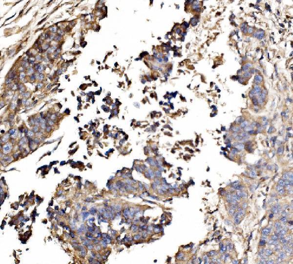
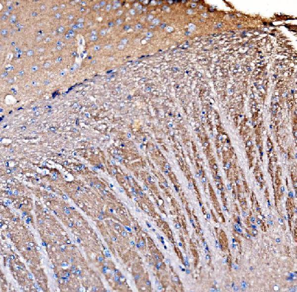
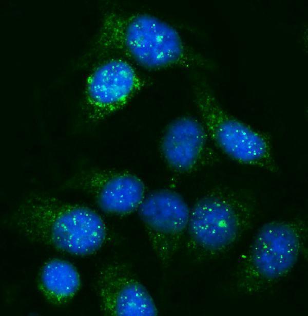
The catalog antibody has paraffin-section IHC images from human lung cancer and brain, mouse brain, and rat brain, plus IF/ICC data from HeLa cells (A09404-1 image captions).

Real IHC data IHC analysis of GNAL using anti-GNAL antibody (A09404-1). GNAL was detected in a paraffin-embedded section of human lung cancer tissue. Heat mediated antigen retrieval was performed in EDTA buffer (pH 8.0, epitope retrieval solution). The tissue section was blocked with 10% goat serum. The tissue section was then incubated with 2 μg/ml rabbit anti-GNAL Antibody (A09404-1) overnight at 4°C. Biotinylated goat anti-rabbit IgG was used as secondary antibody and incubated for 30 minutes at 37°C. The tissue section was developed using Strepavidin-Biotin-Complex (SABC) (Catalog # SA1022) with DAB as the chromogen.
Anti-GNAL Antibody ®
Cat # A09404-1

A09404-1 is the sole listed SKU; its IHC images show paraffin sections of human lung cancer and brain, mouse brain, and rat brain (A09404-1 IHC captions). Its IF image shows HeLa cells (A09404-1 IF caption), and its listed applications include IHC, ICC, and IF (catalog: A09404-1).

Which to pick: Choose A09404-1 for paraffin-section IHC using its documented heat retrieval in EDTA at pH 8.0 and primary antibody at 2 μg/ml (A09404-1 IHC captions); the fixative is unreported (A09404-1 IHC captions). For IF/ICC, the same SKU has a HeLa cell IF image at 5 μg/ml (A09404-1 IF caption). For work across species, its reactivity list includes human, mouse, and rat, with IHC images for all three (catalog: A09404-1 reactivity; A09404-1 IHC captions); clonality is unreported (catalog: A09404-1 clone field).

Each figure is that product's own IHC / IF validation image from its datasheet.

References

  1. UniProt Consortium. UniProt entry P38405 (GNAL_HUMAN, Guanine nucleotide-binding protein G(olf) subunit alpha).
  2. Human Protein Atlas. GNAL tissue IHC expression (reliability: Enhanced).
  3. Human Protein Atlas. GNAL subcellular location (ICC-IF): Localized to the plasma membrane and cytosol..
  4. Human Protein Atlas. GNAL antibody validation summary (1 antibodies).
  5. Multi-cohort comprehensive analysis unveiling the clinical value and therapeutic effect of GNAL in glioma. Oncology research 2024 — PMC11055992.
  6. Transcriptome Profiling of Anhidrotic Eccrine Sweat Glands Reveals that Olfactory Receptors on Eccrine Sweat Glands Regulate Perspiration in a Ligand-Dependent Manner. JID innovations : skin science from molecules to population health 2023 — PMC10392076.
  7. Effects of glucocorticoid on cardiac chronotropic responsiveness in cirrhotic rats: A possible role for dopamine receptors. Iranian journal of basic medical sciences 2026 — PMC13480706.
  8. Transcriptomic identification of HBx-associated hub genes in hepatocellular carcinoma. PeerJ 2021 — PMC8710059.
  9. PubMed PMID:8243272 — UniProt-cited evidence.
  10. PubMed PMID:14702039 — UniProt-cited evidence.
  11. PubMed PMID:16177791 — UniProt-cited evidence.