GNB1 / Guanine nucleotide-binding protein G(I)/G(S)/G(T) subunit beta-1 · IHC design guide

Design Immunohistochemistry for GNB1

Plan chromogenic GNB1 IHC on paraffin sections using the catalog antibody at 0.5–1 μg/mL (datasheet PB10067). Compare staining with high signal in cortical neuropil and kidney tubules, while accounting for the antibody's potential to detect proteins from more than one gene (HPA tissue IHC).

Evidence assembled Oct 2026 · For research use; verify linked source records and product datasheet before use
Immunohistochemistry protocol sheet for GNB1 (IHC for GNB1): expected localisation Cytoplasmic and membranous tissue staining (HPA tissue IHC), antibody PB10067, validated IHC image, and IHC protocol steps
Printable GNB1 IHC protocol sheet — expected localisation Cytoplasmic and membranous tissue staining (HPA tissue IHC), antibody PB10067, controls and protocol steps. Open the full GNB1 IHC guide →

GNB1 Immunohistochemistry Experimental Design Guide

Expected localisation, validated protocols, controls and antibodies — the at-a-glance facts below, then the full design guide.

Must know before staining
Expected localisation Cytoplasmic and membranous tissue staining (HPA tissue IHC)
Staining pattern High in cortical neuropil; variable cytoplasmic and membranous staining (HPA tissue IHC)
Antigen retrieval Citrate pH 6 HIER, heat-mediated (datasheet PB10067)
Positive control ⓘ Cerebral cortex+4 more · see all
Negative control ⓘ Bone marrow+4 more · see all
Important caveats
Reasons your staining may differ from the expected pattern.
Fixation Keep fixation consistent across sections (standard IHC practice; not target-specific)
Caveat Antibody may detect proteins from more than one gene (HPA tissue IHC)
Regulation No expression regulator annotated (UniProt)
Isoform / epitope 2 isoforms; epitope differences are unspecified (UniProt)
Section 1

Recommended GNB1 IHC & IF Protocols

The catalog antibody’s IHC-P protocol (datasheet: PB10067) is followed by three published GNB1 IHC protocols (PMC13565051; PMC10154882; PMC11515508).

Recommended immunohistochemistry (IHC-P) protocol parameters
SampleParaffin-embedded mouse kidney tissue; fixative not specified (datasheet PB10067)
FixationImage fixative and duration unreported (datasheet PB10067); verify before use.
Sectioning4–5 µm sections on charged slides (standard)
DeparaffinisationXylene, graded ethanol series to water (standard)
Antigen retrievalHeat retrieval: Citrate pH 6, 20 min (datasheet PB10067)
Peroxidase block3% H2O2, 10 min, room temperature (standard)
Blocking10% goat serum (datasheet PB10067)
Primary antibodyRabbit anti-GNB1, 0.5-1μg/ml (datasheet PB10067)
Primary incubationOvernight at 4 °C (datasheet PB10067)
DetectionStreptavidin-biotin complex (SABC), DAB chromogen (datasheet PB10067)
CounterstainHematoxylin, blue, dehydrate and mount (standard)
Expected resultGNB1-positive staining in neuropil of cerebral cortex (HPA tissue IHC: High). HPA tissue profile: Cytoplasmic and membranous expression at variable levels in several different tissue types, highly expressed in brain. No signal in the no-primary control.
💡Decision noteStart with citrate pH 6 heat retrieval (datasheet: PB10067); consider Tris/EDTA pH 9.0 if optimizing retrieval (PMC10154882: IHC methods).
Section 2

What Is the Expected GNB1 Staining Pattern?

GNB1 staining in paraffin sections is expected mainly in cytoplasmic and membranous compartments, with variable intensity across tissues (HPA: tissue IHC, Supported). Strong examples include cerebral cortex neuropil, kidney tubule cells and glandular cells in gallbladder and salivary gland (HPA: High). GNB1 has no transmembrane segment (UniProt P62873: topology). HPA reports medium agreement with RNA data and cautions that the antibodies may recognize proteins from more than one gene (HPA: reliability description).

What am I looking at on my slide?
Strong cytoplasmic or membranous chromogenic staining in cerebral cortex neuropil, kidney tubules, or gallbladder or salivary glandular cells (HPA: High).This matches the reported tissue and compartment pattern (HPA: tissue IHC). Score the named cell population and compartment, then compare intensity with a negative control on the same run (standard IHC practice). A matching pattern supports interpretation of the stain but does not resolve HPA's warning about recognition of proteins from more than one gene (HPA: reliability description).
Staining is exclusively nuclear, without the reported cytoplasmic or membranous pattern (HPA: tissue IHC).Treat an isolated nuclear pattern as discordant with the available GNB1 tissue evidence (HPA: tissue IHC). Check counterstain separation, detection background and the antibody's validation before assigning a biological explanation (standard IHC practice). HPA ICC-IF reports plasma membrane and additional Golgi localization; it does not establish a nuclear IHC pattern (HPA: subcellular ICC-IF).
Prominent signal appears in bone marrow hematopoietic cells, esophageal squamous cells or skeletal muscle myocytes (HPA: Not detected).These cell populations are useful low-signal comparators because HPA reports no detection in them (HPA: tissue IHC). Strong staining there may reflect nonspecific antibody binding or endogenous detection activity (standard IHC practice). Investigate it before scoring the slide as GNB1 positive; HPA's multiple-gene targeting caution limits specificity claims (HPA: reliability description).
Broad, even chromogen covers tissue and empty spaces, obscuring cell boundaries (standard IHC practice).This is a background problem rather than an interpretable compartment pattern (standard IHC practice). Review the no-primary control and detection steps, then reassess the named cells against HPA's cytoplasmic and membranous pattern (HPA: tissue IHC). Do not assign intensity scores where background prevents identification of the stained cells (standard IHC practice).
Little or no signal appears in cerebral cortex neuropil or kidney tubule cells (HPA: High).A failed expected-positive control makes a negative study section difficult to interpret (standard IHC practice). Verify tissue identity and run controls, then review retrieval, antibody dilution and detection using the catalog antibody's IHC-P instructions (standard IHC practice). The supplied HPA and UniProt records do not identify a GNB1-specific fixation effect.
💡Expected GNB1 appearanceA convincing positive is predominantly cytoplasmic or membranous staining in the named cells, including High signal in cerebral cortex neuropil or kidney tubules (HPA: tissue IHC); isolated nuclear signal or strong staining in HPA Not detected cell populations warrants investigation (HPA: tissue IHC; standard IHC practice).
How each factor affects the staining
Antibody specificity and evidence strengthThe tissue IHC assessment is Supported, with medium agreement between staining and RNA expression (HPA: reliability description). HPA cautions that the antibodies may target proteins from more than one gene, so a plausible tissue pattern alone cannot establish GNB1-specific binding (HPA: reliability description).
Isoforms and epitope coverageUniProt lists 2 GNB1 isoforms (UniProt P62873: isoforms). The supplied evidence does not locate the IHC antibody epitope or establish equal detection of both isoforms; interpret variation across cells without assigning it to a particular isoform.
Protein topology and processingGNB1 has no annotated transmembrane segment or signal peptide, and its annotated chain spans residues 2–340 (UniProt P62873: topology and processing). These facts do not by themselves determine how antigen retrieval affects staining in paraffin sections (standard IHC interpretation).
Antigen retrievalRetrieval choice can change visibility of epitopes in paraffin-section IHC (standard IHC practice). Follow the catalog antibody's IHC-P instructions and compare controls when optimizing; the supplied HPA and UniProt evidence gives no GNB1-specific retrieval response or fixation sensitivity.
IF/ICC: what pattern is reported?HPA reports a mainly plasma membrane location, with additional Golgi localization, in ICC-IF (HPA: subcellular ICC-IF, approved). Its multiple-gene targeting caution applies to that observation (HPA: subcellular ICC-IF). This cell-image result is context for localization, not a paraffin-section IHC scoring rule.
Why is my staining missing, weak or wrong?
SituationLikely causeNext action
Expected-positive cerebral cortex neuropil or kidney tubule cells are unstained (HPA: High).A failed control can indicate a problem with the IHC run or tissue identification (standard IHC practice); the supplied sources do not identify a GNB1-specific fixation cause.Confirm the positive-control tissue and staining run, then check retrieval, primary-antibody dilution and detection against the catalog antibody's IHC-P instructions (standard IHC practice). Interpret negative study tissue only after the expected-positive control works.
Bone marrow hematopoietic cells or esophageal squamous cells stain strongly (HPA: Not detected).Possible nonspecific binding or endogenous detection activity (standard IHC practice); HPA also cautions that the antibodies may recognize proteins from more than one gene (HPA: reliability description).Inspect a no-primary control and the distribution of signal; review blocking and detection steps as needed (standard IHC practice). Compare with a named HPA-positive cell population before calling the signal GNB1-associated (HPA: tissue IHC).
Chromogen obscures cells across the section (standard IHC practice).Excess background may arise during primary-antibody or detection steps (standard IHC practice); its appearance does not establish increased GNB1 expression.Compare the no-primary control, review reagent concentrations and wash steps, and repeat with controls if cell boundaries remain unreadable (standard IHC practice). Score compartment and intensity only where cells can be distinguished.
Only nuclei stain while cytoplasm and cell borders remain negative (HPA: tissue IHC).The distribution conflicts with the reported cytoplasmic and membranous tissue pattern (HPA: tissue IHC); counterstain or detection artifacts are possible (standard IHC practice).Check the no-primary control and whether chromogen can be distinguished from counterstain (standard IHC practice). Reassess localization in a named HPA-positive population before treating nuclear staining as target signal (HPA: tissue IHC).
A medium- or low-level cell population appears weaker than the positive control (HPA: tissue IHC).HPA reports variable expression, including Medium in cerebellar granular-layer cells and Low in caudate neuronal cells (HPA: tissue IHC). Weakness alone therefore does not show assay failure.Compare like cell populations across matched sections and retain a High HPA-positive control in the run (HPA: tissue IHC; standard IHC practice). Record compartment and relative intensity without converting weak staining into an unsupported negative call.
IF/ICC images suggest membrane or Golgi signal, but the paraffin IHC section shows a broader cytoplasmic pattern (HPA: subcellular ICC-IF; tissue IHC).The HPA observations come from different assay contexts: ICC-IF reports plasma membrane and Golgi, while tissue IHC describes cytoplasmic and membranous expression (HPA: subcellular ICC-IF; tissue IHC).Judge the paraffin section against the tissue IHC pattern and its named cell populations (HPA: tissue IHC). Keep the ICC-IF observation as localization context, with HPA's multiple-gene targeting caution attached (HPA: subcellular ICC-IF).

Sample controls for GNB1 IHC & IF

🧪Run kidney first: cells in tubules should stain (HPA: High in kidney tubule cells). Use esophagus squamous epithelial cells as the negative tissue (HPA: Not detected); on the kidney slide, cells outside stained tubules should show only background signal, but they are not an HPA-validated negative population.
Positive control tissue: Cerebral cortex (Neuropil, HPA High)
Negative control tissue: Bone marrow (HPA Not detected)
ICC-IF cell lines (HPA subcellular resource): HPA ICC-IF images show GNB1 in A-431, U-251MG, U2OS, MCF-7, SH-SY5Y, ASC52telo, RPTEC/TERT1, hTERT-RPE1 (serum starved), Sperm, with annotated localisation: Plasma membrane (approved) (HPA subcellular).
Technical controls: Include a no-primary (secondary-only) slide, a concentration-matched rabbit IgG isotype control, and GNB1-knockout tissue or peptide-blocked antibody where a validated blocking peptide is available (selected PB10067 IHC caption: rabbit primary antibody; standard IHC control practice). For kidney sections, quench endogenous peroxidase and check endogenous biotin before interpreting DAB signal from the biotinylated secondary and streptavidin-biotin detection system (selected PB10067 IHC caption: biotinylated secondary, SABC and DAB; standard IHC control practice).
⚠️Feasibility: A target-specific fixation window or fixation effect is unreported, and the fixative is unreported in the selected PB10067 paraffin-section caption (selected PB10067 IHC caption: fixative not stated). The demonstrated tissue-IHC procedure used heat retrieval in citrate buffer at pH 6 for 20 minutes, followed by 1 μg/ml primary antibody overnight at 4°C; this supports starting with retrieval but does not establish that retrieval is required (selected PB10067 IHC caption). HPA has ICC-IF images for GNB1, with a caution that its subcellular summary is based on antibodies targeting proteins from multiple genes; the supplied evidence does not show that IF or frozen sections are easier than paraffin IHC, and endogenous biotin can complicate the caption’s kidney DAB detection scheme (HPA: subcellular summary; selected PB10067 IHC caption: kidney, SABC and DAB; standard IHC control practice).

HPA tissue IHC evidence for GNB1

Comprehensive Human Protein Atlas IHC scoring per tissue (reliability: Supported — Medium consistency between antibody staining and RNA expression data. Caution, targets protein from more than one gene.). Rows are taken directly from the HPA tissue chart — click any row's HPA link to view the source.

Positive expression · recommended positive controls

TissueCell typeLevelEvidenceSource
Cerebral cortex Neuropil High Protein (IHC) HPA →
Gallbladder Glandular cells High Protein (IHC) HPA →
Kidney Cells in tubules High Protein (IHC) HPA →
Salivary gland Glandular cells High Protein (IHC) HPA →
Adrenal gland Glandular cells Medium Protein (IHC) HPA →

Undetected expression · recommended negative controls

TissueCell typeLevelEvidenceSource
Bone marrow Hematopoietic cells Not detected Protein (IHC) HPA →
Cervix Glandular cells Not detected Protein (IHC) HPA →
Esophagus Squamous epithelial cells Not detected Protein (IHC) HPA →
Lymph node Germinal center cells Not detected Protein (IHC) HPA →
Oral mucosa Squamous epithelial cells Not detected Protein (IHC) HPA →
Section 3

Advanced GNB1 IHC Tips

Troubleshoot GNB1 staining in paraffin sections using the PB10067 tissue image and expression references, while keeping antibody specificity and processing limits in view.

How should I adjust retrieval when GNB1 staining is weak in paraffin sections?
Start with heat-mediated citrate retrieval at pH 6 for 20 minutes (datasheet PB10067). The illustrated paraffin-section result used that treatment before overnight incubation with 1 μg/ml primary antibody at 4°C (datasheet PB10067). If staining remains weak, check that the section stayed covered during heating, then compare a modestly longer retrieval against the original condition on adjacent sections (standard IHC practice). Record heating and cooling conditions so any gain in signal can be distinguished from tissue damage (standard IHC practice). Judge improvement in kidney tubule cells alongside a no-primary control, because those cells have high reported staining but antibody specificity has a stated caveat (HPA: kidney tubules High; HPA: Supported, multiple-gene caution).
Could fixation explain weak or uneven GNB1 staining?
Target-specific sensitivity to fixative type or fixation duration is unknown from the supplied evidence (datasheet PB10067: fixative not stated). The product image establishes staining in a paraffin-embedded mouse kidney section, but its caption does not identify the fixative (datasheet PB10067). Compare sections with documented processing histories while holding retrieval at citrate pH 6 for 20 minutes and primary incubation at 1 μg/ml overnight at 4°C (datasheet PB10067; standard IHC practice). Examine morphology and staining across the full section before attributing patchiness to fixation (standard IHC practice). Include a consistently processed reference section in each run to reveal processing-related variation (standard IHC practice).
Where should convincing GNB1 staining appear in tissue?
Assess cytoplasmic and membranous staining in tissue; the reported tissue profile varies across cell types and is especially strong in brain (HPA: tissue IHC profile). Kidney tubule cells and cerebral-cortex neuropil are reported as High, whereas several other compartments are Not detected (HPA: kidney tubules High; HPA: cerebral-cortex neuropil High; HPA: tissue IHC negatives). GNB1 lacks a transmembrane segment and participates in G-protein complexes, so a crisp outline alone should not be treated as proof of an integral membrane protein (UniProt P62873: topology and subunit). The cell-imaging reference places signal mainly at the plasma membrane and additionally at the Golgi, with a multiple-gene antibody caution (HPA: subcellular). Compare staining with tissue architecture and a no-primary control before assigning a compartment (standard IHC practice).
Can this stain distinguish GNB1 isoforms or modified forms?
GNB1 has 2 recorded isoforms, but the supplied antibody evidence does not map its recognized epitope to either one (UniProt P62873: isoforms; datasheet PB10067: epitope not stated). Report chromogenic staining as GNB1-associated immunoreactivity unless antibody specificity for an isoform has been independently established (UniProt P62873: isoforms; standard IHC interpretation). The record lists N-acetylserine and phosphoserine at residue 2 and phosphohistidine at residue 266; no supplied evidence shows that PB10067 distinguishes these states (UniProt P62873: modified residues; datasheet PB10067: epitope not stated). Compare serial sections and controls under identical retrieval and detection conditions when assessing apparent differences (standard IHC practice). Avoid interpreting intensity changes as altered splicing or phosphorylation from this stain alone (UniProt P62873: isoforms and modified residues; standard IHC interpretation).
How can IF help check an ambiguous chromogenic GNB1 pattern?
Use IF as a separate localization check, pairing GNB1 with a marker that identifies the cell population under study (standard IF practice). For kidney sections, choose a marker that identifies tubule cells, since high staining is reported there (HPA: kidney tubules High; standard IF practice). Select fluorophores after inspecting tissue autofluorescence, and separate emission channels and single-stain controls to assess bleed-through (standard IF practice). Because GNB1 has no transmembrane segment and its antibody epitope is unspecified, compare gentle permeabilisation conditions before interpreting absent intracellular signal; do not assume which membrane side exposes the epitope (UniProt P62873: topology; datasheet PB10067: epitope not stated; standard IF practice). Keep IF observations distinct from the paraffin-section chromogenic result, whose fixative is unreported (datasheet PB10067).
What should I check when DAB appears throughout the section?
Inspect a no-primary section first to assess signal from the detection system, endogenous enzyme activity, or deposited chromogen (standard chromogenic IHC practice). The illustrated workflow used biotinylated goat anti-rabbit secondary antibody, a streptavidin-biotin complex, and DAB, so background may arise at several detection steps (datasheet PB10067). Include a peroxidase block and optimize blocking, washing, and DAB development while keeping the primary-antibody condition documented (standard chromogenic IHC practice; datasheet PB10067: 1 μg/ml overnight at 4°C). The image caption reports a 10% goat-serum block, which provides a starting condition for comparison (datasheet PB10067). Score structured cellular staining separately from diffuse precipitate or staining at section edges (standard IHC practice).
How should I quantify GNB1 staining across paraffin sections? ⚠ ANSWER MARKED FOR VERIFICATION
Define the tissue compartment and analysis region before scoring, because reported GNB1 staining varies by cell type (HPA: tissue IHC profile; standard IHC practice). For cell-rich regions, record percent positive cells and an H-score from 0–300 using intensity grades 0–3; for diffuse neuropil, consider positive area or optical-density measurements per measured area (standard IHC quantification; HPA: cerebral-cortex neuropil High). Normalize each result to the number of evaluable cells or analyzed tissue area, and use the same threshold across sections (standard IHC quantification). Keep retrieval, primary incubation, DAB development, imaging, and counterstain settings consistent across the comparison (standard IHC practice). Exclude folds, necrosis, and edge staining using predefined criteria (standard IHC quantification).
How do I separate plausible GNB1 staining from artefact?
Seek a reproducible cytoplasmic or membranous pattern in an appropriate cell compartment, then compare it with the no-primary control (HPA: tissue IHC profile; standard IHC practice). High reported staining in kidney tubule cells or cerebral-cortex neuropil supports those locations as reference patterns, while a uniformly dark section does not establish cellular specificity (HPA: kidney tubules High; HPA: cerebral-cortex neuropil High; standard IHC interpretation). Review unexpected nuclear-only signal, stained edges, necrotic areas, and diffuse DAB for processing or detection artefact (standard chromogenic IHC practice). A peroxidase-block control can help investigate endogenous enzyme signal in a DAB workflow (standard chromogenic IHC practice; datasheet PB10067: DAB detection). Treat agreement with the reported pattern as supporting evidence rather than definitive gene-level validation, because the tissue reference cautions that its antibodies target proteins from multiple genes (HPA: Supported, multiple-gene caution).
Boster reagents

Best GNB1 / Guanine nucleotide-binding protein G(I)/G(S)/G(T) subunit beta-1 IHC Antibodies

Real GNB1 IHC images cover paraffin sections of mouse kidney, rat kidney and human liver cancer; IF/ICC images cover human A549 and SiHa cells (PB10067 and M04650 image captions).

Real IHC data IHC analysis of GNB1 using anti-GNB1 antibody (PB10067). GNB1 was detected in paraffin-embedded section of mouse kidney tissue. Heat mediated antigen retrieval was performed in citrate buffer (pH6, epitope retrieval solution) for 20 mins. The tissue section was blocked with 10% goat serum. The tissue section was then incubated with 1μg/ml rabbit anti-GNB1 Antibody (PB10067) overnight at 4°C. Biotinylated goat anti-rabbit IgG was used as secondary antibody and incubated for 30 minutes at 37°C. The tissue section was developed using Strepavidin-Biotin-Complex (SABC)(Catalog # SA1022) with DAB as the chromogen.
Anti-GNB1 Antibody ®
Cat # PB10067
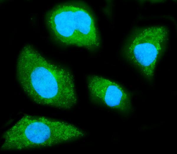
Real IF data IF analysis of GNB1 using anti-GNB1 antibody (M04650). GNB1 was detected in an immunocytochemical section of SiHa cells. Enzyme antigen retrieval was performed using IHC enzyme antigen retrieval reagent (AR0022) for 15 mins. The cells were blocked with 10% goat serum. And then incubated with 5 μg/mL mouse anti-GNB1 Antibody (M04650) overnight at 4°C. DyLight®488 Conjugated Goat Anti-Mouse IgG (BA1126) was used as secondary antibody at 1:100 dilution and incubated for 30 minutes at 37°C. The section was counterstained with DAPI. Visualize using a fluorescence microscope and filter sets appropriate for the label used.
Anti-GNB1 Antibody ® (monoclonal, 11F9)
Cat # M04650

PB10067 lists IHC and IF/ICC applications, with paraffin-section IHC images from mouse kidney, rat kidney and human liver cancer, plus IF/ICC in A549 cells (catalog applications; PB10067 image captions). M04650 lists IF/ICC and shows IF in SiHa cells; its catalog does not list IHC (catalog applications; M04650 IF image caption).

Which to pick: Choose PB10067 for paraffin-section tissue IHC: it is a rabbit antibody with listed human, mouse and rat IHC-P reactivity, and its IHC captions document citrate pH 6 retrieval for 20 minutes; the fixative is unreported (catalog applications/reactivity; PB10067 IHC image captions). Choose M04650 for human-cell IF/ICC when a mouse monoclonal is preferred: its SiHa IF image documents that use, while IHC is absent from its application list (catalog clone/applications; M04650 IF image caption). For cross-species tissue IHC, PB10067 has IHC images from human, mouse and rat samples; both SKUs list human, mouse and rat reactivity, but M04650’s shown IF result is in human cells (PB10067 IHC image captions; catalog reactivity; M04650 IF image caption).

Each figure is that product's own IHC / IF validation image from its datasheet.

References

  1. UniProt Consortium. UniProt entry P62873 (GBB1_HUMAN, Guanine nucleotide-binding protein G(I)/G(S)/G(T) subunit beta-1).
  2. Human Protein Atlas. GNB1 tissue IHC expression (reliability: Supported).
  3. Human Protein Atlas. GNB1 subcellular location (ICC-IF): Mainly localized to the plasma membrane. In addition localized to the Golgi apparatus. Caution: Based on antibodies targeting proteins from multiple genes..
  4. Human Protein Atlas. GNB1 antibody validation summary (2 antibodies).
  5. Multi-Organ Comparative Transcriptomic Study on Short-Term and Long-Term Hypoxia Adaptation in Cattle. Biology 2026 — PMC13565051.
  6. CircRNA circ_POLA2 Promotes Cervical Squamous Cell Carcinoma Progression via Regulating miR-326/GNB1. Frontiers in oncology 2020 — PMC7381119.
  7. GNB1 promotes hepatocellular carcinoma progression by targeting BAG2 to activate P38/MAPK signaling. Cancer science 2023 — PMC10154882.
  8. A multidimensional recommendation framework for identifying biological targets to aid the diagnosis and treatment of liver metastasis in patients with colorectal cancer. Molecular cancer 2024 — PMC11515508.
  9. PubMed PMID:3095147 — UniProt-cited evidence.
  10. PubMed PMID:16710414 — UniProt-cited evidence.
  11. PubMed PMID:15489334 — UniProt-cited evidence.