GNG7 / Guanine nucleotide-binding protein G(I)/G(S)/G(O) subunit gamma-7 · IHC design guide

Design Immunohistochemistry for GNG7

Plan GNG7 paraffin IHC using caudate neuronal cells as a positive reference (HPA tissue IHC). Start with the IHC-validated antibody at 2–5 μg/mL (datasheet A10690-1) and assess staining against a negative control.

Evidence assembled Oct 2026 · For research use; verify linked source records and product datasheet before use
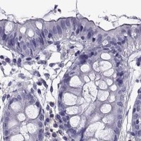
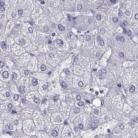
Immunohistochemistry protocol sheet for GNG7 (IHC for GNG7): expected localisation Cell membrane expected; tissue compartment unconfirmed (UniProt), antibody A10690-1, validated IHC image, and IHC protocol steps
Printable GNG7 IHC protocol sheet — expected localisation Cell membrane expected; tissue compartment unconfirmed (UniProt), antibody A10690-1, controls and protocol steps. Open the full GNG7 IHC guide →

GNG7 Immunohistochemistry Experimental Design Guide

Expected localisation, validated protocols, controls and antibodies — the at-a-glance facts below, then the full design guide.

Must know before staining
Expected localisation Cell membrane expected; tissue compartment unconfirmed (UniProt)
Staining pattern High staining in caudate neuronal cells; compartment unspecified (HPA tissue IHC)
Antigen retrieval EDTA pH 8.0 HIER, heat-mediated (datasheet A10690-1)
Positive control ⓘ Caudate
Negative control ⓘ Adipose tissue+4 more · see all
Important caveats
Reasons your staining may differ from the expected pattern.
Fixation Keep fixation consistent across sections (standard IHC practice; not target-specific)
Caveat HPA staining may include protein from more than one gene (HPA tissue IHC)
Regulation Reduced in pancreatic and esophageal cancer (UniProt)
Isoform / epitope No annotated isoforms; processed chain spans residues 2–65 (UniProt)
Section 1

Recommended GNG7 IHC & IF Protocols

Start with the catalog antibody’s IHC-P protocol, then compare the published paraffin-section protocols for head and neck squamous cell carcinoma and bladder cancer (datasheet A10690-1; PMC3334494; PMC13466384).

Recommended immunohistochemistry (IHC-P) protocol parameters
SampleParaffin-embedded human bladder cancer tissue; fixative not specified (datasheet A10690-1)
FixationImage fixative and duration unreported (datasheet A10690-1); verify before use.
Sectioning4–5 µm sections on charged slides (standard)
DeparaffinisationXylene, graded ethanol series to water (standard)
Antigen retrievalHeat retrieval: EDTA pH 8.0 (datasheet A10690-1); 20 min, 95–100 °C (standard)
Peroxidase block3% H2O2, 10 min, room temperature (standard)
Blocking10% goat serum (datasheet A10690-1)
Primary antibodyRabbit anti-GNG7, 2-5μg/ml (datasheet A10690-1)
Primary incubationOvernight at 4 °C (datasheet A10690-1)
DetectionStreptavidin-biotin complex (SABC), DAB chromogen (datasheet A10690-1)
CounterstainHematoxylin, blue, dehydrate and mount (standard)
Expected resultGNG7-positive staining in neuronal cells of caudate (HPA tissue IHC: High). HPA tissue profile: Expressed in the CNS, mainly in caudate nucleus. No signal in the no-primary control.
💡Decision noteTry heat-mediated EDTA, pH 8.0, first for the catalog antibody (datasheet A10690-1). Citrate, pH 6.0, is a published alternative using a different antibody (PMC13466384).
Section 2

What Is the Expected GNG7 Staining Pattern?

GNG7 is annotated at the cell membrane and has no transmembrane segment (UniProt O60262 topology). In paraffin tissue sections, expect the strongest IHC signal in caudate neuronal cells (HPA: High in caudate neuronal cells). HPA rates the tissue pattern Supported because staining is consistent with RNA expression, while cautioning that the antibody may target protein from more than one gene and awaits external verification (HPA: tissue IHC reliability).

What am I looking at on my slide?
Strong staining associated with caudate neuronal cells, especially at cell boundaries or processes (HPA: High in caudate neuronal cells; UniProt O60262: cell membrane).This fits the reported positive tissue and membrane annotation. Score the neuronal pattern and its distribution across the section; chromogenic IHC alone does not prove that every stained outline is exclusively GNG7 (HPA: Supported, with multiple-gene-target caution).
Predominantly nuclear staining, with little neuronal membrane-associated signal (UniProt O60262: cell membrane).A nuclear-dominant pattern conflicts with the annotated location and warrants review for nonspecific staining or a detection artefact. Recheck the antibody and detection controls before assigning it to GNG7 (UniProt O60262: cell membrane; standard IHC practice).
Prominent staining of adipocytes or cerebellar granular-layer cells (HPA: Not detected in those cell populations).These are discordant cell-type results, rather than proof that the whole tissue must be blank. Consider cross-reactivity or endogenous detection activity; HPA also cautions that the antibody may target protein from more than one gene (HPA: tissue IHC and reliability).
Widespread, fairly uniform chromogen over cells and surrounding tissue, without a recognizable neuronal pattern (HPA: High in caudate neuronal cells).Treat this as background until controls show otherwise. Review blocking, washes, primary-antibody concentration and the detection-only control; these are general IHC checks, not reported GNG7-specific sensitivities (standard IHC practice).
No convincing neuronal signal in a caudate section (HPA: High in caudate neuronal cells).A negative result in the reported positive tissue leaves the run inconclusive. Check tissue identity, staining controls, antigen retrieval and detection steps before concluding that GNG7 is absent from that specimen (HPA: tissue IHC; standard IHC practice).
💡Expected GNG7 appearanceA credible positive is high neuronal staining in caudate with a membrane-associated distribution; dominant nuclear staining or strong signal in HPA-listed undetected cell populations calls for validation (HPA: caudate High; HPA: listed Not detected populations; UniProt O60262: cell membrane).
How each factor affects the staining
Tissue and cell choice (HPA: tissue IHC)Caudate neuronal cells provide the clearest reported positive comparison; cerebral-cortex neuropil is Low. HPA lists adipocytes and cerebellar granular-layer cells as Not detected, which applies to those cells, not every cell in their tissues (HPA: tissue IHC).
Membrane association (UniProt O60262 topology)GNG7 is annotated at the cell membrane without a transmembrane segment. Interpret compartment at cellular resolution; the record does not define an IHC-specific epitope or require a continuous, sharp membrane rim (UniProt O60262 topology).
Antibody evidence (HPA: HPA057790 IHC Supported)HPA lists one rabbit polyclonal antibody with IHC Supported status. Its tissue-profile reliability is Supported, but HPA cautions that the target may include protein from more than one gene and that external verification is pending (HPA: antibody and tissue IHC).
Processing and epitope uncertainty (UniProt O60262)UniProt annotates a mature chain at residues 2–65 and a 66–68 propeptide, but the supplied record gives no antibody epitope. These annotations cannot establish whether processing changes this antibody's IHC signal (UniProt O60262 processing).
IF/ICC evidence (HPA: subcellular and antibody records)HPA supplies no ICC-IF images or main ICC-IF location, and lists no ICC validation for HPA057790. The reported REH RNA value, 48.7 nTPM, does not establish an IF staining pattern (HPA: subcellular; HPA: antibody record).
Why is my staining missing, weak or wrong?
SituationLikely causeNext action
Caudate neurons are unstained despite an otherwise completed IHC run (HPA: caudate High).The run may have lost sensitivity, or the sampled section may not contain the expected neuronal population; neither possibility establishes target absence (standard IHC practice).Confirm tissue identity and section quality, then review the positive control, retrieval conditions, primary incubation and detection controls in the IHC workflow (standard IHC practice).
Chromogen appears mainly in nuclei (UniProt O60262: cell membrane).The compartment conflicts with the annotated GNG7 location; nonspecific primary binding or detection background is possible (UniProt O60262; standard IHC practice).Compare with a detection-only control and inspect whether the caudate neuronal pattern remains after routine background checks; do not score nuclear-only signal as confirmed GNG7 (standard IHC practice).
Adipocytes stain strongly (HPA: adipocytes Not detected).The pattern conflicts with the reported adipocyte result. Cross-reactivity or endogenous detection activity may contribute, particularly given HPA's multiple-gene-target caution (HPA: tissue IHC reliability; standard IHC practice).Inspect cell identity and control sections; check endogenous enzyme blocking when enzyme-based chromogenic detection is used, and compare with the caudate positive pattern (standard IHC practice; HPA: caudate High).
Diffuse color obscures cell boundaries throughout the section (HPA: caudate neuronal pattern).Excess background can arise from staining or detection conditions; the supplied sources do not identify a GNG7-specific cause (standard IHC practice).Review blocking, washes, primary concentration and detection-only controls, then score only cellular signal that remains distinguishable from background (standard IHC practice).
Only a weak signal appears in cerebral-cortex neuropil (HPA: Low in cerebral-cortex neuropil).Low signal there agrees with the reported observation and is a weak basis for judging assay failure on its own (HPA: tissue IHC).Compare the same run with caudate neuronal cells, the reported High population, before changing IHC conditions or calling the assay negative (HPA: tissue IHC).
Can an IF/ICC image be used here to confirm the GNG7 compartment? (HPA: subcellular record).HPA provides no ICC-IF images or main ICC-IF location, and lists no ICC validation for the cited antibody (HPA: subcellular and antibody records).Treat IF/ICC localization as unverified by this evidence; use this page's caudate IHC pattern for the tissue interpretation and consult a separately validated IF/ICC guide for IF design (HPA: tissue IHC; HPA: subcellular record).

Sample controls for GNG7 IHC & IF

🧪Run caudate first and expect neuronal cells to stain (HPA: High in caudate neuronal cells). Use adipose tissue as the negative tissue, with adipocytes at background (HPA: Not detected in adipose adipocytes); on the caudate slide, compare neuronal staining with nearby non-neuronal cells, without assuming those cells are antigen-negative because their expression is unspecified (HPA: caudate neuronal cells only).
Positive control tissue: Caudate (Neuronal cells, HPA High)
Negative control tissue: Adipose tissue (HPA Not detected)
ICC-IF cell lines (HPA subcellular resource): HPA carries no ICC-IF cell line for GNG7; derive a cell-line control from the positive tissue's cell type (Neuronal cells) and confirm it by RNA or western blot first.
Technical controls: Include a no-primary, secondary-only control and a concentration-matched rabbit IgG isotype control (caption: rabbit primary antibody); use a validated GNG7 knockout specimen or immunizing-peptide competition as a biological specificity control (standard IHC practice). Quench endogenous peroxidase and check for endogenous biotin signal in caudate sections (caption: biotinylated secondary and SABC/DAB detection).
⚠️Feasibility: A target-specific fixation window and fixation effect are unreported, and the selected A10690-1 caption does not state a fixative (selected A10690-1 caption: fixative not stated). The reported paraffin-section method used heat retrieval in EDTA at pH 8.0, but retrieval dependence was not tested in the supplied evidence (selected A10690-1 caption: EDTA retrieval); no supplied frozen-section or IF result establishes either as easier (HPA: no ICC-IF images; selected A10690-1 caption: tissue IHC). In caudate, assess tissue pigment and detection background alongside the controls; no target-specific caudate artefact is reported (HPA: High in caudate neuronal cells; selected A10690-1 caption: bladder cancer tissue).

HPA tissue IHC evidence for GNG7

Comprehensive Human Protein Atlas IHC scoring per tissue (reliability: Supported — High consistency between antibody staining and RNA expression data. Caution, targets protein from more than one gene. Pending external verification.). Rows are taken directly from the HPA tissue chart — click any row's HPA link to view the source.

Positive expression · recommended positive controls

TissueCell typeLevelEvidenceSource
Caudate Neuronal cells High Protein (IHC) HPA →

Undetected expression · recommended negative controls

TissueCell typeLevelEvidenceSource
Adipose tissue Adipocytes Not detected Protein (IHC) HPA →
Adrenal gland Glandular cells Not detected Protein (IHC) HPA →
Appendix Glandular cells Not detected Protein (IHC) HPA →
Bone marrow Hematopoietic cells Not detected Protein (IHC) HPA →
Breast Adipocytes Not detected Protein (IHC) HPA →
Section 3

Advanced GNG7 IHC Tips

These questions address GNG7 staining in paraffin sections with chromogenic detection, with one question on interpreting a related IF experiment.

How should I troubleshoot weak GNG7 staining after antigen retrieval?
Start with heat-mediated retrieval in EDTA at pH 8.0 (datasheet A10690-1). The catalog antibody stained a paraffin section of human bladder cancer after this retrieval, followed by 2 µg/ml primary antibody overnight at 4°C (datasheet A10690-1). If staining is weak, check that sections were fully deparaffinised and that heating, cooling, and buffer preparation were consistent across slides (general IHC practice). Compare the suspect run with a concurrently processed section that previously stained well, while keeping detection and DAB development consistent (general IHC practice). A weak result alone does not establish that retrieval failed, because the caption does not report the fixative (datasheet A10690-1).
Could fixation explain weak or uneven GNG7 staining?
Target-specific fixation sensitivity is unknown: the selected paraffin-section caption does not state a fixative (datasheet A10690-1). Record the fixation method and duration for each specimen before comparing staining between cases, since fixation can change antigen accessibility in IHC (general IHC practice). If a batch stains unevenly, compare sections with similar processing history and inspect tissue preservation alongside the GNG7 signal (general IHC practice). Keep the documented EDTA pH 8.0 retrieval and 2 µg/ml primary concentration consistent during that comparison (datasheet A10690-1). Neither the reported tissue distribution nor GNG7’s membrane location establishes a fixation preference (HPA tissue IHC; UniProt O60262 subcellular location).
Where should convincing GNG7 staining appear in a paraffin section?
Assess GNG7 primarily for membrane-associated staining, because its annotated location is the cell membrane and it has no transmembrane segment (UniProt O60262 subcellular location and topology). In caudate, neuronal cells provide a useful expression reference, with high staining reported there (HPA: High in caudate neuronal cells). Low signal in cerebral cortex neuropil and absent staining in the listed cerebellar granular-layer cells show that brain regions should not be treated as interchangeable controls (HPA tissue IHC). Compare the apparent cellular compartment with morphology and the counterstain before scoring, and investigate diffuse nuclear staining or staining confined to damaged edges (general IHC practice; UniProt O60262 subcellular location).
How do GNG7 processing and epitope location affect staining interpretation?
GNG7 has no annotated isoforms, and its annotated mature chain spans residues 2–65, with residues 66–68 listed as a propeptide (UniProt O60262 processing and isoforms). Its listed modifications include N-acetylserine at residue 2 and a cysteine methyl ester at residue 65 (UniProt O60262 modified residues). The supplied caption does not identify the antibody epitope, so it cannot establish whether processing or either modification changes binding (datasheet A10690-1; UniProt O60262 processing). If staining differs across specimens, first compare fixation, retrieval, and controls before assigning the difference to epitope accessibility (general IHC practice). Record the antibody identifier when comparing results from different antibody preparations (general IHC practice).
What would an IF check add to a disputed chromogenic GNG7 result?
Use IF as a separate check on cell identity and localisation, rather than treating its signal as a direct equivalent of DAB intensity (general IHC/IF practice). Multiplex GNG7 with a validated neuronal marker when examining caudate, where neuronal cells show high GNG7 staining (HPA: High in caudate neuronal cells). Select fluorophores and image settings after inspecting tissue autofluorescence and single-label controls, particularly when faint membrane signal is being judged (general IF practice; UniProt O60262 subcellular location). Choose permeabilisation according to the antibody epitope’s side of the membrane; that side is not established by the supplied antibody caption or the membrane-location annotation (datasheet A10690-1; UniProt O60262 topology).
How can I reduce background without losing credible GNG7 signal?
Check a no-primary control alongside the stained section to distinguish detection-system background from antibody-dependent staining (general IHC practice). The reported paraffin-section method used 10% goat serum blocking, a biotinylated secondary antibody, a streptavidin–biotin complex, and DAB (datasheet A10690-1). Review peroxidase blocking and, where relevant, endogenous biotin background before increasing primary-antibody dilution or shortening DAB development (general IHC practice; datasheet A10690-1 detection method). Compare background in tissue compartments with expected expression, especially caudate neuronal cells, against listed low or undetected compartments (HPA tissue IHC). Keep section handling and counterstaining consistent so apparent background changes can be attributed to the step being tested (general IHC practice).
How should I score GNG7 staining across sections? ⚠ ANSWER MARKED FOR VERIFICATION
Define the cell population and tissue area before scoring, and exclude folds, necrosis, and damaged edges using the counterstain (general IHC practice). For a cellular readout, report the percentage of positive cells with an intensity grade of 0–3, or calculate an H-score of 0–300 from those grades (general IHC practice). If counting stained cells, express density per mm² of evaluable tissue and state whether the denominator includes all cells or only the selected population (general IHC practice). Normalise comparisons to comparable viable tissue area or the same defined cell population, and process reference sections consistently (general IHC practice). Report caudate neuronal-cell results separately from other compartments because expression varies by cell type and region (HPA tissue IHC).
When is a GNG7-positive DAB pattern likely to be artefactual?
A plausible positive result matches cell morphology and shows membrane-associated staining, consistent with GNG7’s annotated cell-membrane location (UniProt O60262 subcellular location; general IHC practice). High staining in caudate neuronal cells supports that context, whereas staining restricted to a cell type listed as undetected warrants closer review (HPA tissue IHC). Investigate signal limited to section edges or necrosis, and compare a no-primary control for endogenous enzyme or detection-system staining (general IHC practice). The selected bladder-cancer image shows that this antibody produced staining under its reported conditions, but the caption alone does not establish specificity in every specimen (datasheet A10690-1). Interpret isolated positives cautiously because the HPA tissue-IHC assessment notes staining of targets from more than one gene (HPA tissue IHC reliability description).
Boster reagents

Best GNG7 / Guanine nucleotide-binding protein G(I)/G(S)/G(O) subunit gamma-7 IHC Antibodies

A10690-1 has IHC images from human bladder cancer, liver cancer, and renal carcinoma paraffin sections (A10690-1 IHC image captions). Catalog reactivity includes human, mouse, and rat (A10690-1 catalog).

Real IHC data IHC analysis of GNG7 using anti-GNG7 antibody (A10690-1). GNG7 was detected in paraffin-embedded section of human bladder cancer tissue. Heat mediated antigen retrieval was performed in EDTA buffer (pH8.0, epitope retrieval solution). The tissue section was blocked with 10% goat serum. The tissue section was then incubated with 2μg/ml rabbit anti-GNG7 Antibody (A10690-1) overnight at 4°C. Biotinylated goat anti-rabbit IgG was used as secondary antibody and incubated for 30 minutes at 37°C. The tissue section was developed using Strepavidin-Biotin-Complex (SABC) (Catalog # SA1022) with DAB as the chromogen.
Anti-GNG7 Antibody ®
Cat # A10690-1

A10690-1 is listed for IHC and has images of human bladder cancer, liver cancer, and renal carcinoma paraffin sections (A10690-1 catalog; IHC image captions). Its listed reactivity is human, mouse, and rat, while the pictured IHC samples are human (A10690-1 catalog; IHC image captions).

Which to pick: Choose A10690-1 for human paraffin-section IHC; its captions report EDTA retrieval at pH 8.0 and 2 μg/ml primary antibody, but do not report the fixative (A10690-1 IHC image captions). No IF/ICC application or IF image is provided, so this payload does not establish an IF/ICC choice (A10690-1 catalog). A10690-1 lists mouse and rat reactivity, but cross-species tissue IHC needs verification because its IHC images show human samples only; the antibody is rabbit-hosted, with no clone specified (A10690-1 catalog; IHC image captions).

Each figure is that product's own IHC / IF validation image from its datasheet.

References

  1. UniProt Consortium. UniProt entry O60262 (GBG7_HUMAN, Guanine nucleotide-binding protein G(I)/G(S)/G(O) subunit gamma-7).
  2. Human Protein Atlas. GNG7 tissue IHC expression (reliability: Supported).
  3. Human Protein Atlas. GNG7 subcellular location (ICC-IF): Highest expression in REH: 48.7 nTPM.
  4. Human Protein Atlas. GNG7 antibody validation summary (1 antibodies).
  5. Establishment of a new prognostic risk model of GNG7 pathway-related molecules in clear cell renal cell carcinoma based on immunomodulators. BMC cancer 2023 — PMC10500784.
  6. Loss of protein expression and recurrent DNA hypermethylation of the GNG7 gene in squamous cell carcinoma of the head and neck. Journal of applied genetics 2012 — PMC3334494.
  7. RSPO3 induced by Helicobacter pylori extracts promotes gastric cancer stem cell properties through the GNG7/β-catenin signaling pathway. Cancer medicine 2024 — PMC10997846.
  8. H3K18 lactylation promotes glycolysis in bladder cancer via inhibiting GNG7 through regulating PI3K-AKT signaling pathway. World journal of surgical oncology 2026 — PMC13466384.
  9. PubMed PMID:9600093 — UniProt-cited evidence.
  10. PubMed PMID:14702039 — UniProt-cited evidence.
  11. PubMed PMID:15057824 — UniProt-cited evidence.