This website uses cookies to ensure you get the best experience on our website.
- Table of Contents
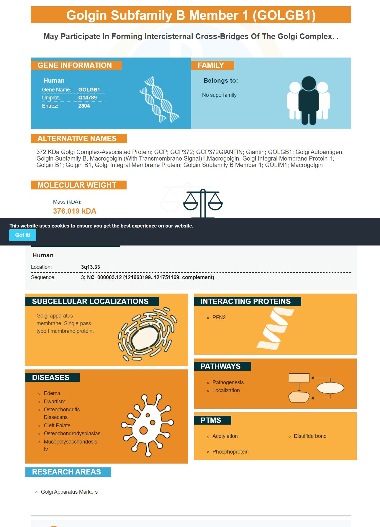
Facts about Golgin subfamily B member 1.
Human | |
---|---|
Gene Name: | GOLGB1 |
Uniprot: | Q14789 |
Entrez: | 2804 |
Belongs to: |
---|
No superfamily |
372 kDa Golgi complex-associated protein; GCP; GCP372; GCP372GIANTIN; Giantin; GOLGB1; golgi autoantigen, golgin subfamily b, macrogolgin (with transmembrane signal)1,macrogolgin; golgi integral membrane protein 1; golgin B1; golgin B1, golgi integral membrane protein; Golgin subfamily B member 1; GOLIM1; Macrogolgin
Mass (kDA):
376.019 kDA
Human | |
---|---|
Location: | 3q13.33 |
Sequence: | 3; NC_000003.12 (121663199..121751169, complement) |
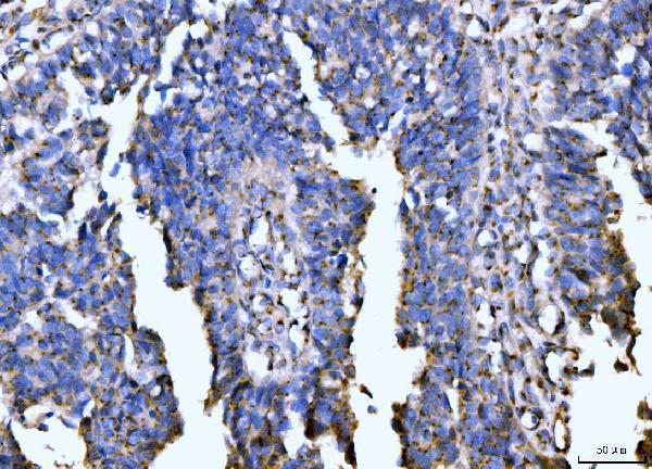
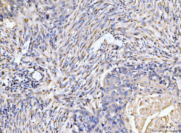
Golgi apparatus membrane; Single-pass type I membrane protein.
Here you will find information about the company as well as the GOLGB1 marker. Picogram sensitivity ELISA kits will be discussed as well as High-affinity primary antibody. The Picokine platform will also be discussed. Picokine enables heightened ELISA sensibilities. Picokine is approved for use in a variety samples. You can request validation images and the procedure. The Picoband platform utilizes insights from immunogen design and is backed by technical support from BeNeLux distributor Sanbio.
A Boster antibody for the GOLGB1 marker can be used for a wide range of applications, including immunohistochemistry, ELISA, and Western blotting. The antibody has been tested against a panel of over 250 tissues for its specificity, sensitivity, and affinity. The antibody was quantitatively validated by comparing it with known amounts of recombinant products. Sanbio, a BeNeLux distributor, provides technical support.
Boster Bio's Anti-Giantin/GOLGB1 antibody is part of the Picoband(tm) catalog. This antibody reacts with Human. It can be stored at -20°C for up to one year. It is formulated with Trehalose, which is good for long-term storage. Boster Bio's blocking peptide can be purchased separately, but the price will vary depending on the length of the immunogen.
The GOL-Gb1 marker is now the standard for immunofluorescence. It is cost-effective and allows you to test a wide array of proteins. This method has many advantages. It can identify as many 61 different types of antigens as one tissue section. It can also work with different imaging modalities.
GOL-Gb1 was created by immunizing BALB/c mice infected with the GB virus recombinant Glycoprotein E2. The antibody recognized sequence WSHPQFEK of amino acids. The antibody also recognized the VHH-E9 fusion protein, a Gb1 homogeneous and antigen with an identity constant of 9.1x10⁻¹⁰M.
Giantin is an integral component of the Golgi membrane and is associated with the cytoskeleton. It also may play a role in reorganizing Golgi complexes. It has been found to be an autoantigen in patients suffering from rheumatoid. The length of the human GOLGB1 Protein is 3259 amino Acids. The human GOLGB1 protein shares 92% of its amino acid sequence with that of its mouse and rat orthologs.
Previously, we identified several antibodies that were highly sensitive to SCAP. This antibody recognizes SCAP (a GOLGB1 protein). It also recognizes GFP–ATF6 (a GOLGB1 protein) and giantin in peroxisomes-deficient cells. However, the GFPATF6 protein does NOT colocalize in peroxisome-deficient tissues with giantin. Moreover, SCAP is specific to peroxisome-deficient cells. Defective ER-to–Golgi transport does not affect other proteins.
Picogram sensitivity ELISA for the GOLGB1 marker is a kit that allows researchers to detect low levels. The assay kit is suitable for research and can be used with native GOLGB1. Its half life is three to fourfold that of insulin making it a useful tool for measuring beta cell insulin production. Every batch of samples must be tested for reproducibility.
IHC-optimized monoclonals that use GOLGB1 markers are available to detect Giantin within cells. These antibodies are conjugated in goat anti-GOLGB1 antibodies and detect Giantin cells in human cervical epithelial carcinoma cell lines fixed in 2% paraformaldehyde. They were counterstained with DAPI for a more intense staining. GOLGB1/Giantin staining is localized to Golgi device vesicles within HeLa cells. NorthernLights (tm) 557 conjugated antibody anti-Sheep IgG is available for further testing.
Monoclonal antibodies recognize multiple epitopes on a single antigen. They are usually inexpensive and stable over a wide pH range and salt concentration. They are also less susceptible to batch-to-bath variability than polyclonal antibodies and are more stable against antigen variation. Monoclonal antibodies are produced from DNA sequences. They recognize specific epitopes of peptides that are unique for the antigen.
Polyclonal antibodies derived only from one B cell line are often more sensitive than monoclonal and have a higher sensitivity. Monoclonal antibodies can react with multiple epitopes and are less sensitive to cross-reactivity than polyclonal antibodies. Polyclonal antibodies recognize multiple epitopes so they can be used to detect multiple epitopes on a target protein.
Polyclonal antibodies used for IHC have fragment-specificity and can penetrate tissues more easily than monoclonal antibodies. Polyclonal antibodies are usually less concentrated than monoclonal. Temperature and incubation times affect the amount that the antibodies bind to each other. The non-specific signal will increase if the incubation period is longer.
Giantin antibody are available from multiple vendors. Giantin is also known as GOLGB1 in humans. It is a 3259-amino acid protein with a mass of 376.019 daltons. Suppliers will provide important details about the antibody. This antibody is available from several suppliers. For more information, take a look at the website of the supplier.
PMID: 8198703 by Seelig H.P., et al. Macrogolgin -- a new 376 kD Golgi complex outer membrane protein as target of antibodies in patients with rheumatic diseases and HIV infections.
PMID: 7802676 by Sohda M., et al. Molecular cloning and sequence analysis of a human 372-kDa protein localized in the Golgi complex.