This website uses cookies to ensure you get the best experience on our website.


- Table of Contents
Plan GOLGB1 chromogenic IHC scoring around cytoplasmic staining across cells (HPA tissue IHC). Use high staining in adrenal glandular cells and low staining in skeletal myocytes to guide tissue selection (HPA tissue IHC).

Expected localisation, validated protocols, controls and antibodies — the at-a-glance facts below, then the full design guide.
| Expected localisation | Cytoplasmic staining (HPA tissue IHC); Golgi membrane protein (UniProt) | |
| Staining pattern | Cytoplasmic staining in all cells (HPA tissue IHC) | |
| Antigen retrieval | EDTA pH 8.0 HIER, heat-mediated (datasheet A08226-1) | |
| Positive control | Adrenal gland+4 more · see all | |
| Negative control | None in HPA (detected in all 45 tissues); use no-primary + isotype controls |
| Fixation | Keep fixation consistent across sections (standard IHC practice; not target-specific) | |
| Caveat | Skeletal myocytes may stain weakly (HPA tissue IHC) | |
| Regulation | No expression regulator reported (UniProt) | |
| Isoform / epitope | 4 isoforms; epitope coverage unreported (UniProt) |
The catalog antibody’s IHC-P protocol uses EDTA retrieval (datasheet: EDTA pH 8.0); the published protocol below describes IHC staining of human nucleus pulposus tissue (PMC12887002: IHC methods).
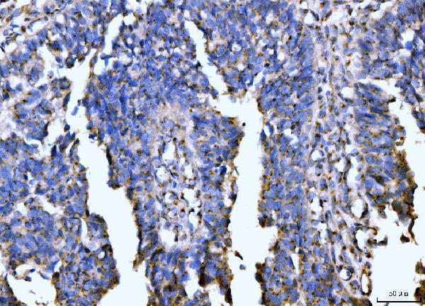
| Sample | Paraffin-embedded human colonic adenocarcinoma tissue; fixative not specified (datasheet A08226-1) |
| Fixation | Image fixative and duration unreported (datasheet A08226-1); verify before use. |
| Sectioning | 4–5 µm sections on charged slides (standard) |
| Deparaffinisation | Xylene, graded ethanol series to water (standard) |
| Antigen retrieval | Heat retrieval: EDTA pH 8.0 (datasheet A08226-1); 20 min, 95–100 °C (standard) |
| Peroxidase block | 3% H2O2, 10 min, room temperature (standard) |
| Blocking | 10% goat serum (datasheet A08226-1) |
| Primary antibody | Rabbit anti-GOLGB1, 1-2 μg/ml (datasheet A08226-1) |
| Primary incubation | Overnight at 4 °C (datasheet A08226-1) |
| Detection | Streptavidin-biotin complex (SABC), DAB chromogen (datasheet A08226-1) |
| Counterstain | Hematoxylin, blue, dehydrate and mount (standard) |
| Expected result | GOLGB1-positive staining in glandular cells of adrenal gland (HPA tissue IHC: High). HPA tissue profile: Cytoplasmic expression in all cells. No signal in the no-primary control. |
GOLGB1 is anchored in the Golgi membrane, with most of the protein facing the cytoplasm (UniProt Q14789 topology). In paraffin-section IHC, expect cytoplasmic staining in many cell types, often concentrated near the nucleus in the Golgi region (HPA: cytoplasmic expression in all cells; HPA: Golgi localization by ICC-IF). HPA rates its tissue staining reliability Enhanced, citing consistency between staining and RNA expression (HPA: tissue IHC reliability).
| A compact, asymmetric cytoplasmic signal near the nucleus appears in glandular or epithelial cells. | This fits Golgi-region staining (HPA: Golgi localization by ICC-IF; UniProt Q14789: Golgi membrane). HPA reports high staining in adrenal gland, appendix, breast, and cervix glandular cells and bronchial respiratory epithelial cells (HPA: tissue IHC). Chromogenic IHC may show a compact patch rather than individual Golgi structures (general IHC practice). |
| The strongest signal fills nuclei, outlines the plasma membrane, or forms a uniform cytoplasmic haze. | These distributions conflict with the expected Golgi-region pattern (HPA: Golgi localization by ICC-IF; UniProt Q14789: Golgi membrane). Treat them as possible artefact or nonspecific staining until control sections and antibody validation resolve the discrepancy (general IHC practice). |
| A cell population stains strongly while expected Golgi-region signal is absent from adjacent cells. | Check cell identity and detection controls before calling cross-reactivity or endogenous detection activity (general IHC practice). GOLGB1 is broadly expressed, so staining outside one highlighted cell type is not automatically wrong (HPA: cytoplasmic expression in all cells; low tissue specificity). The suspect signal is its unexpected distribution or persistence in controls (general IHC practice). |
| Brown signal spreads through tissue, stroma, or empty areas and obscures cell boundaries. | Diffuse staining cannot establish a Golgi-localized positive result (HPA: Golgi localization by ICC-IF). Examine background in the detection controls and review blocking, washes, and chromogen development as general IHC variables (general IHC practice). Do not score broad background as GOLGB1 expression. |
| No convincing signal appears in a section containing an HPA high-staining cell population. | First verify that the expected cells are present and preserved, then inspect assay controls (general IHC practice). HPA reports high staining in, for example, cerebral-cortex neurons, cerebellar granular-layer cells, and colon endothelial cells (HPA: tissue IHC). An absent result in one run does not establish biological absence. |
| Tissue and cell context | HPA reports cytoplasmic expression across cells and low tissue specificity, alongside high staining in selected populations and low staining in skeletal-muscle myocytes (HPA: tissue IHC). Compare the observed cell population with its stated HPA level; do not use skeletal muscle as an absolute negative control. |
| Golgi membrane topology | GOLGB1 has one transmembrane segment at residues 3236–3256; residues 1–3235 face the cytoplasm (UniProt Q14789 topology). This supports a cytoplasmic Golgi-region expectation, but the supplied record does not map the IHC antibody epitope (UniProt Q14789 topology; HPA: antibody summary). |
| Evidence behind the antibody pattern | HPA lists both HPA011008 and HPA011555 as Enhanced for IHC and ICC (HPA: antibody validation). HPA describes tissue-IHC reliability as consistency between staining and RNA expression (HPA: tissue IHC reliability). These summaries support pattern review; they do not prove that every brown deposit is specific. |
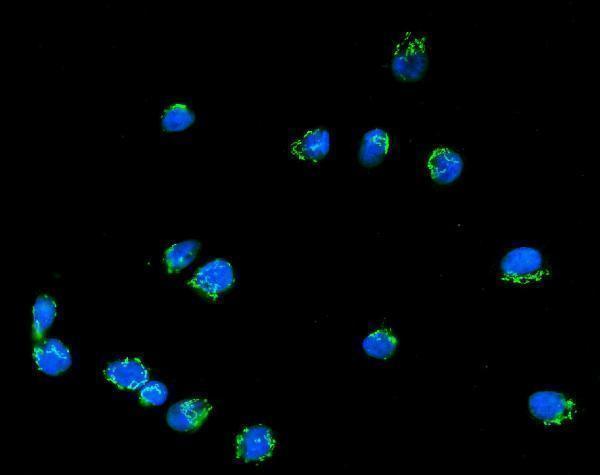
| IF/ICC Q: What localization should I expect? | A: Golgi-apparatus localization is reported as enhanced by HPA ICC-IF (HPA: subcellular). That observation helps interpret an IHC compartment pattern; IF/ICC methods and controls belong in its separate guide. No IF/ICC protocol choice follows from the supplied IHC evidence. |
| Situation | Likely cause | Next action |
|---|---|---|
| A known HPA high-staining population has no visible IHC signal. | The expected cells may be missing from the section, or the assay may have failed (HPA: tissue IHC; general IHC practice). The supplied sources do not establish GOLGB1-specific fixation sensitivity. | Confirm cell identity and section quality, then check a same-run positive control, primary-antibody step, and detection reagents (general IHC practice). Interpret a negative only after the assay performs as expected. |
| Signal is diffuse across tissue or extends into cell-free areas. | Background from blocking, washing, or detection can obscure a compact Golgi-region pattern (general IHC practice; HPA: Golgi localization by ICC-IF). | Compare with an appropriate detection control; review blocking, wash stringency, antibody concentration, and chromogen development according to the assay instructions (general IHC practice). Score only a resolved cellular pattern. |
| Staining is mainly nuclear or forms a continuous cell-surface rim. | That compartment does not match the Golgi membrane assignment (UniProt Q14789: subcellular location; HPA: Golgi localization by ICC-IF). | Check a known staining pattern and control sections, then reassess antibody specificity and detection background before assigning GOLGB1 positivity (general IHC practice). |
| A suspected cell population stains, but the expected cells lack Golgi-region signal. | Cross-reactivity or endogenous detection activity is possible (general IHC practice). Broad GOLGB1 expression means cell identity alone cannot establish an error (HPA: cytoplasmic expression in all cells). | Confirm cell identity, compare compartment patterns, and inspect a primary-antibody omission control for detection activity (general IHC practice). Compare with HPA tissue patterns where the same cell type is documented. |
| Skeletal-muscle myocytes stain faintly beside stronger cells. | HPA records myocytes as low, not negative, and reports low tissue specificity overall (HPA: tissue IHC). Faint cellular signal therefore needs pattern and control review. | Compare myocyte staining with background and the expected Golgi-region distribution; avoid using its low level as proof of assay failure or as an absolute negative control (HPA: tissue IHC; general IHC practice). |
| Golgi-region signal varies across sections or is hard to distinguish from background. | Interpretation may be limited by section quality or assay conditions (general IHC practice). Neither HPA tissue staining nor UniProt topology identifies a GOLGB1-specific fixation or retrieval effect. | Review section integrity, same-run controls, and the validated IHC workflow; document any retrieval or detection changes as general optimization rather than a proven GOLGB1-specific requirement (general IHC practice). |
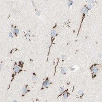


Comprehensive Human Protein Atlas IHC scoring per tissue (reliability: Enhanced — High consistency between antibody staining and RNA expression data.). Rows are taken directly from the HPA tissue chart — click any row's HPA link to view the source.
| Tissue | Cell type | Level | Evidence | Source |
|---|---|---|---|---|
| Adrenal gland | Glandular cells | High | Protein (IHC) | HPA → |
| Appendix | Glandular cells | High | Protein (IHC) | HPA → |
| Breast | Glandular cells | High | Protein (IHC) | HPA → |
| Bronchus | Respiratory epithelial cells | High | Protein (IHC) | HPA → |
| Cerebellum | Cells in granular layer | High | Protein (IHC) | HPA → |
| Tissue | Cell type | Level | Evidence | Source |
|---|---|---|---|---|
| None in HPA: GOLGB1 is detected in all 45 scored tissues. Use a no-primary (secondary-only) and an isotype control instead. | ||||
Use matched paraffin sections and compartment aware scoring to troubleshoot GOLGB1 chromogenic IHC; reserve fluorescence checks for the separate IF/ICC workflow.
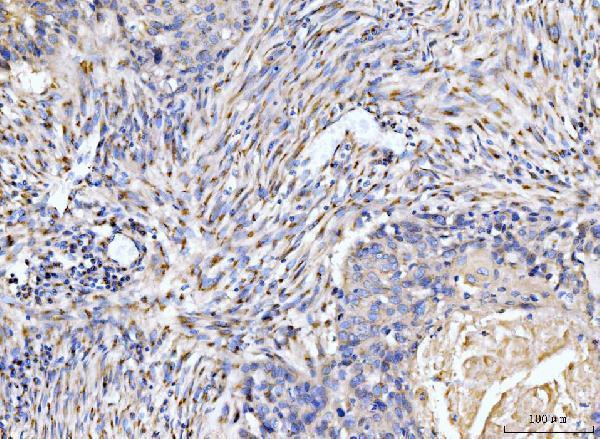
A08226-1 has real IHC data from human paraffin sections and IF data from RT4 cells (catalog IHC and IF image captions; catalog: Human reactivity).
A08226-1 is listed for human IHC, with paraffin-section images from colonic adenocarcinoma, esophageal squamous carcinoma, gall bladder adenosquamous carcinoma, and placenta (catalog applications and reactivity; A08226-1 IHC image captions). The same SKU is listed for IF/ICC and has an IF image from RT4 cells (catalog applications; A08226-1 IF image caption).
Which to pick: Choose A08226-1 for human tissue IHC on paraffin sections; its own captions document EDTA pH 8 retrieval and 2 μg/ml primary antibody (A08226-1 IHC image captions). For IF/ICC, A08226-1 lists both applications and shows IF in RT4 cells at 5 μg/ml (catalog applications; A08226-1 IF image caption). Its listed reactivity is Human, so the payload supports no cross-species recommendation; the IHC captions do not report a fixative (catalog: Human reactivity; A08226-1 IHC image captions).