GOT2 / Aspartate aminotransferase, mitochondrial · IHC design guide

Design Immunohistochemistry for GOT2

Plan paraffin-section GOT2 IHC around granular cytoplasmic staining (HPA tissue IHC) and mitochondrial matrix localisation (UniProt). Compare high-staining hepatocytes with adipocytes reported as undetected (HPA tissue IHC), and optimize the IHC-validated antibody within 2–5 μg/mL (datasheet A05998-1).

Evidence assembled Oct 2026 · For research use; verify linked source records and product datasheet before use
Immunohistochemistry protocol sheet for GOT2 (IHC for GOT2): expected localisation Granular cytoplasm (HPA tissue IHC); mitochondrial matrix (UniProt), antibody A05998-1, validated IHC image, and IHC protocol steps
Printable GOT2 IHC protocol sheet — expected localisation Granular cytoplasm (HPA tissue IHC); mitochondrial matrix (UniProt), antibody A05998-1, controls and protocol steps. Open the full GOT2 IHC guide →

GOT2 Immunohistochemistry Experimental Design Guide

Expected localisation, validated protocols, controls and antibodies — the at-a-glance facts below, then the full design guide.

Must know before staining
Expected localisation Granular cytoplasm (HPA tissue IHC); mitochondrial matrix (UniProt)
Staining pattern Granular cytoplasmic staining across cell types (HPA tissue IHC)
Antigen retrieval EDTA pH 8.0 HIER, heat-mediated (datasheet A05998-1)
Positive control ⓘ Cerebellum+4 more · see all
Negative control ⓘ Adipose tissue+3 more · see all
Important caveats
Reasons your staining may differ from the expected pattern.
Fixation Keep fixation conditions consistent across sections (standard IHC practice; not target-specific)
Caveat Alcohol exposure may shift GOT2 to the membrane (UniProt)
Regulation No expression regulator annotated (UniProt)
Isoform / epitope Two isoforms; epitope differences unspecified (UniProt)
Section 1

Recommended GOT2 IHC & IF Protocols

The catalog antibody’s IHC-P protocol is paired with published GOT2 staining methods for lymphoma arrays, spinal cord sections, and paraffin-embedded tissues (PMC5904148; PMC5673182; PMC9328765).

Recommended immunohistochemistry (IHC-P) protocol parameters
SampleParaffin-embedded human gastric cancer tissue; fixative not specified (datasheet A05998-1)
FixationImage fixative and duration unreported (datasheet A05998-1); verify before use.
Sectioning4–5 µm sections on charged slides (standard)
DeparaffinisationXylene, graded ethanol series to water (standard)
Antigen retrievalHeat retrieval: EDTA pH 8.0 (datasheet A05998-1); 20 min, 95–100 °C (standard)
Peroxidase block3% H2O2, 10 min, room temperature (standard)
Blocking10% goat serum (datasheet A05998-1)
Primary antibodyRabbit anti-GOT2, 2-5μg/ml (datasheet A05998-1)
Primary incubationOvernight at 4 °C (datasheet A05998-1)
DetectionStreptavidin-biotin complex (SABC), DAB chromogen (datasheet A05998-1)
CounterstainHematoxylin, blue, dehydrate and mount (standard)
Expected resultGOT2-positive staining in cells in granular layer of cerebellum (HPA tissue IHC: High). HPA tissue profile: General cytoplasmic expression with a granular pattern. No signal in the no-primary control.
💡Decision noteStart with heat-mediated EDTA pH 8.0 retrieval for the catalog antibody (datasheet A05998-1); use citrate pH 6.0 when reproducing the lymphoma protocol (PMC5904148).
Section 2

What Is the Expected GOT2 Staining Pattern?

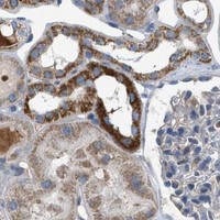
GOT2 is a mitochondrial matrix protein without a transmembrane segment; UniProt also reports alcohol-associated movement to the cell membrane (UniProt P00505). In paraffin-section IHC, expect granular cytoplasmic staining, especially in hepatocytes, cardiomyocytes, and selected neuronal cells (HPA tissue IHC). HPA rates the IHC evidence Enhanced, while reporting medium consistency between staining and RNA data (HPA tissue IHC).

What am I looking at on my slide?
Granular cytoplasmic stain in hepatocytes or cardiomyocytes.This matches HPA's general granular cytoplasmic profile and its High staining calls for these cells (HPA tissue IHC). The pattern is compatible with mitochondrial matrix localisation, although chromogenic IHC alone cannot prove that each granule is a mitochondrion (UniProt P00505; general IHC practice).
Predominantly nuclear staining, with little granular cytoplasmic signal.This differs from the reported tissue pattern and mitochondrial matrix localisation (HPA tissue IHC; UniProt P00505). Treat it as a possible staining artefact and check controls before interpreting it as GOT2 (general IHC practice).
Strong staining in adipocytes while hepatocytes stain weakly on a comparable run.HPA reports GOT2 as not detected in adipocytes and High in hepatocytes (HPA tissue IHC). Investigate unexpected staining for cross-reactivity or endogenous detection activity; one discordant slide cannot establish either cause (general IHC practice).
Uniform colour across cells and tissue spaces, with no clear granular pattern.This obscures the granular cytoplasmic profile reported by HPA (HPA tissue IHC). Diffuse colour may reflect nonspecific reagent binding, residual detection activity, or excess chromogen development; assess background controls (general IHC practice).
No stain in hepatocytes on a run that otherwise appears readable.Hepatocytes are an HPA High cell population, so a blank result is a reason to check the assay, section quality, and controls (HPA tissue IHC; general IHC practice). It does not by itself show that GOT2 is absent from the specimen.
💡Expected GOT2 appearanceA credible positive is High, granular cytoplasmic staining in hepatocytes or cardiomyocytes (HPA tissue IHC); predominant nuclear or uniform background colour is discordant with that pattern (HPA tissue IHC; general IHC practice).
How each factor affects the staining
Compartment and topologyMitochondrial matrix localisation and absence of a transmembrane segment support an intracellular granular readout (UniProt P00505; HPA tissue IHC). Because UniProt also reports alcohol-associated membrane translocation, membrane staining needs sample-context review rather than automatic rejection (UniProt P00505).
Choice of comparison cellsHPA calls hepatocytes, cardiomyocytes, neuronal cells in cerebral cortex and hippocampus, and kidney collecting ducts High (HPA tissue IHC). It calls adipocytes, marrow hematopoietic cells, and smooth muscle cells not detected; these are comparisons, not absolute biological negatives (HPA tissue IHC).
Strength of tissue evidenceHPA labels the tissue IHC assessment Enhanced but describes only medium consistency with RNA expression data (HPA tissue IHC). Interpret a discrepant sample with morphology and controls; the HPA category does not validate every specimen or staining condition (general IHC practice).
IF/ICC question: should a fluorescent image confirm mitochondrial localisation?UniProt predicts mitochondrial matrix localisation (UniProt P00505). HPA provides no main ICC-IF location or cell-line images here, so this payload cannot confirm an observed IF pattern or justify an IF/ICC protocol choice (HPA subcellular).
Retrieval and detection settingsNo GOT2-specific retrieval condition or dilution is supplied (UniProt P00505; HPA tissue IHC). Optimise these as general IHC variables with positive and background controls; do not infer fixation sensitivity or an epitope-masking mechanism for GOT2 (general IHC practice).
Why is my staining missing, weak or wrong?
SituationLikely causeNext action
Hepatocytes lack granular staining.A High HPA comparison population is unexpectedly blank (HPA tissue IHC); the cause is unresolved.Check a known-positive section, reagent performance, retrieval conditions, and chromogenic development within the assay workflow (general IHC practice). Record the outcome before scoring the sample as negative.
Adipocytes stain strongly.This conflicts with HPA's not-detected call for adipocytes (HPA tissue IHC); cross-reactivity or endogenous detection activity is possible (general IHC practice).Review morphology and a negative reagent control, then compare staining in an HPA High population on the same run where available (HPA tissue IHC; general IHC practice).
Most nuclei are intensely coloured.Predominant nuclear signal conflicts with the reported granular cytoplasmic pattern and mitochondrial matrix localisation (HPA tissue IHC; UniProt P00505).Check control sections and detection background; repeat with adjusted general IHC conditions if controls implicate the assay (general IHC practice). Do not assign nuclear GOT2 from this pattern alone.
Colour is diffuse across the section.Background can obscure the granular HPA pattern (HPA tissue IHC); nonspecific binding or detection background may contribute (general IHC practice).Compare a negative reagent control, assess blocking and washes, and review chromogen development (general IHC practice). Score cell-specific GOT2 only where the granular pattern remains distinguishable.
A thin rim of membrane colour appears with otherwise credible cytoplasmic staining.UniProt reports that alcohol exposure can promote GOT2 movement to the cell membrane (UniProt P00505); the image alone does not establish that this occurred.Check sample history and controls, and document membrane and granular cytoplasmic components separately (general IHC practice). Avoid treating membrane colour alone as confirmed GOT2 localisation.
An IF/ICC image is requested as confirmation.This payload has no HPA ICC-IF location or cell-line images for GOT2 (HPA subcellular).Treat an IF pattern as a separate validation question; use this section's IHC tissue comparisons for the paraffin assay and the dedicated IF/ICC guide for fluorescent assay design (HPA tissue IHC; HPA subcellular).

Sample controls for GOT2 IHC & IF

🧪Run heart muscle first: cardiomyocytes should stain (HPA: High in cardiomyocytes). Use smooth muscle as the negative tissue (HPA: Not detected in smooth muscle cells); non-cardiomyocytes on the heart slide should show only counterstain or background, but HPA does not identify a confirmed negative cell population within that slide.
Positive control tissue: Cerebellum (Cells in granular layer, HPA High)
Negative control tissue: Adipose tissue (HPA Not detected)
ICC-IF cell lines (HPA subcellular resource): HPA carries no ICC-IF cell line for GOT2; derive a cell-line control from the positive tissue's cell type (Cells in granular layer) and confirm it by RNA or western blot first.
Technical controls: Include a no-primary (secondary-only) control, rabbit IgG matched to the primary’s isotype and clonality once confirmed (caption: rabbit primary), and GOT2-knockout tissue or an immunizing-peptide block if available (standard IHC practice). In heart sections, check endogenous peroxidase and biotin background with the captioned SABC-DAB detection method (caption: SABC-DAB; standard IHC practice).
⚠️Feasibility: A target-specific fixation window or fixation effect is unreported, and the exact A05998-1 paraffin-section caption does not state a fixative (caption: fixative unreported). The caption uses heat retrieval in EDTA at pH 8.0, but it does not establish that retrieval is required (caption: EDTA pH 8.0). Whether frozen sections or IF/ICC are easier is unreported; heart autofluorescence can complicate IF assessment (standard IF practice), and no HPA ICC-IF images are supplied (HPA: no ICC-IF images).

HPA tissue IHC evidence for GOT2

Comprehensive Human Protein Atlas IHC scoring per tissue (reliability: Enhanced — Medium consistency between antibody staining and RNA expression data.). Rows are taken directly from the HPA tissue chart — click any row's HPA link to view the source.

Positive expression · recommended positive controls

TissueCell typeLevelEvidenceSource
Cerebellum Cells in granular layer High Protein (IHC) HPA →
Cerebral cortex Neuronal cells High Protein (IHC) HPA →
Epididymis Glandular cells High Protein (IHC) HPA →
Heart muscle Cardiomyocytes High Protein (IHC) HPA →
Hippocampus Neuronal cells High Protein (IHC) HPA →

Undetected expression · recommended negative controls

TissueCell typeLevelEvidenceSource
Adipose tissue Adipocytes Not detected Protein (IHC) HPA →
Bone marrow Hematopoietic cells Not detected Protein (IHC) HPA →
Smooth muscle Smooth muscle cells Not detected Protein (IHC) HPA →
Vagina Squamous epithelial cells Not detected Protein (IHC) HPA →
Section 3

Advanced GOT2 IHC Tips

Troubleshoot GOT2 staining in paraffin sections by checking retrieval, compartment, background and scoring against the catalog antibody’s tissue-IHC evidence and reference expression patterns.

How should I retrieve GOT2 in paraffin sections when staining is weak?
Start with heat-mediated retrieval in EDTA pH 8.0 (datasheet A05998-1). The selected antibody stained a paraffin section of human gastric cancer after this retrieval, followed by 2 μg/mL primary antibody overnight at 4°C (datasheet A05998-1). Keep the retrieval vessel, heating cycle and cooling conditions consistent across slides, because uneven processing can mimic variable GOT2 expression (standard IHC practice). If staining remains weak, test an alternative retrieval condition on matched sections as a fallback, recording tissue preservation and background alongside signal (standard IHC practice). Compare the result with granular cytoplasmic staining, the reported tissue pattern (HPA tissue IHC).
Could fixation explain weak or uneven GOT2 staining?
Target-specific fixation sensitivity is unknown: the selected paraffin-section caption does not report a fixative (datasheet A05998-1). Record the fixative, fixation duration and processing history for each specimen before attributing a weak result to GOT2 biology (standard IHC practice). Compare sections processed together, using the same EDTA pH 8.0 retrieval and staining run to reduce processing differences (datasheet A05998-1; standard IHC practice). Assess tissue morphology and staining at the section center as well as its edge, since processing artefacts can vary across a section (standard IHC practice). Do not infer fixation tolerance from expression maps or protein topology (standard IHC practice).
What staining pattern supports GOT2 localisation in tissue?
Look first for a granular cytoplasmic pattern, as reported in tissue IHC (HPA tissue IHC). GOT2 is annotated in the mitochondrial matrix and has no transmembrane segment, so diffuse nuclear staining needs independent validation before it is assigned to GOT2 (UniProt P00505 localisation; UniProt P00505 topology). Cell-membrane localisation is also annotated after alcohol exposure; document that context before interpreting a peripheral signal as biological translocation (UniProt P00505 localisation). Compare the granular signal with the morphology of the stained cells and with a negative control section (standard IHC practice). Score cytoplasmic and peripheral staining separately so a changed distribution does not appear to be a simple change in total expression (standard IHC practice).
Could epitope choice make GOT2 staining vary between specimens?
GOT2 has 2 annotated isoforms, and its annotated mature chain spans residues 30–430 (UniProt P00505 isoforms; UniProt P00505 processing). The record also lists modifications, including phosphothreonine at residue 48 and acetyllysine at residue 59 (UniProt P00505 modified residues). The selected caption does not identify this antibody’s epitope, so an isoform-specific or modification-sensitive staining claim cannot be made from that image (datasheet A05998-1). If specimens stain differently, compare the antibody’s documented immunogen or epitope with the isoform sequences when that information is available (standard antibody-validation practice). Use an independent epitope or orthogonal assay to investigate persistent discrepancies (standard IHC practice).
How should I investigate GOT2 localisation by multiplex IF?
Treat multiplex IF as a separate assay requiring its own controls; the supplied subcellular record lists no ICC/IF images (HPA subcellular). Pair GOT2 with a marker for the expected cell population, such as a hepatocyte marker in liver, where hepatocytes stain strongly by tissue IHC (HPA tissue IHC; standard IF practice). Choose a fluorophore in a channel with low measured tissue autofluorescence and include unstained and single-label controls (standard IF practice). Because GOT2 is annotated in the mitochondrial matrix without a transmembrane segment, test permeabilisation that permits antibody access to intracellular epitopes while preserving morphology (UniProt P00505 localisation; UniProt P00505 topology; standard IF practice). Validate any apparent membrane signal separately, particularly when alcohol exposure is relevant (UniProt P00505 localisation).
How can I separate GOT2 signal from chromogenic background?
Run a no-primary control through the complete secondary, detection and DAB sequence to locate background generated after primary incubation (standard IHC practice). The selected tissue image used 10% goat serum, a biotinylated goat anti-rabbit secondary and a streptavidin-biotin complex with DAB (datasheet A05998-1). If background persists, check endogenous peroxidase blocking and assess whether endogenous biotin contributes to signal under that detection scheme (standard IHC practice). Compare background at tissue edges, folds and damaged areas with intact regions before changing primary concentration (standard IHC practice). Preserve the reported 2 μg/mL starting condition while testing one background control at a time (datasheet A05998-1; standard IHC practice).
What is a defensible way to score GOT2 tissue staining? ⚠ ANSWER MARKED FOR VERIFICATION
Define the cell population and analysis region before scoring, then exclude folds, necrosis and section edges by a written rule (standard IHC practice). For granular cytoplasmic staining, report the percentage of positive cells and an intensity-based H-score, or report positive-cell density per mm² when cell counts vary substantially (HPA tissue IHC; standard IHC practice). Normalise positive counts to the number of evaluable cells, and compare intensity only among slides stained and imaged under matched conditions (standard IHC practice). Keep cytoplasmic and peripheral compartments separate because GOT2 has both matrix localisation and alcohol-associated membrane translocation annotated (UniProt P00505 localisation). Record the positivity threshold and control performance with every scored batch (standard IHC practice).
When should apparent GOT2 positivity be considered artefactual?
A convincing result has a granular cytoplasmic component in intact cells, consistent with the reported tissue pattern and mitochondrial-matrix annotation (HPA tissue IHC; UniProt P00505 localisation). Check unexpected positive cells against the tissue map: hepatocytes are reported high, whereas adipocytes are reported not detected (HPA tissue IHC). Isolated nuclear staining, edge accentuation or staining confined to necrotic areas warrants control review before biological interpretation (UniProt P00505 localisation; standard IHC practice). Signal in a no-primary control can indicate detection background, including endogenous peroxidase or biotin effects with the reported DAB and biotin-based workflow (datasheet A05998-1; standard IHC practice). Interpret peripheral staining in light of documented alcohol exposure and confirm it independently (UniProt P00505 localisation).
Boster reagents

Best GOT2 / Aspartate aminotransferase, mitochondrial IHC Antibodies

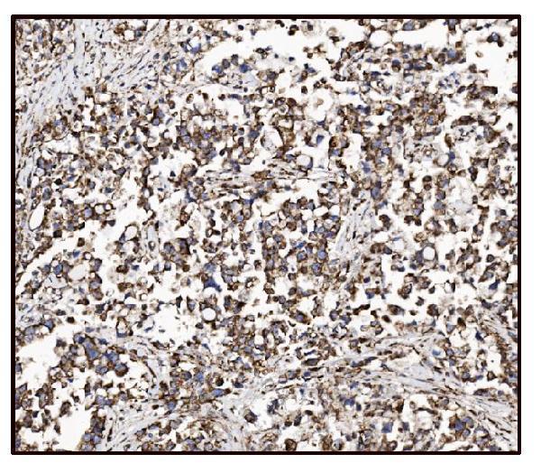
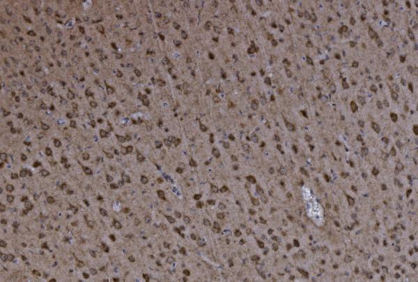
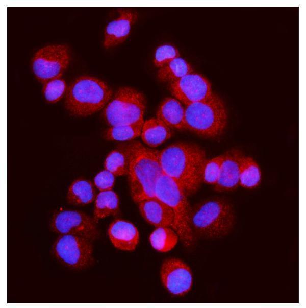
A05998-1 has paraffin-section IHC images from human gastric and lung cancer tissue and mouse and rat brain, plus an IF image from T-47D cells (catalog image captions).

Real IHC data IHC analysis of FABP-1/GOT2 using anti-FABP-1/GOT2 antibody (A05998-1). FABP-1/GOT2 was detected in paraffin-embedded section of human gastric cancer tissue. Heat mediated antigen retrieval was performed in EDTA buffer (pH8.0, epitope retrieval solution). The tissue section was blocked with 10% goat serum. The tissue section was then incubated with 2μg/ml rabbit anti-FABP-1/GOT2 Antibody (A05998-1) overnight at 4°C. Biotinylated goat anti-rabbit IgG was used as secondary antibody and incubated for 30 minutes at 37°C. The tissue section was developed using Strepavidin-Biotin-Complex (SABC) (Catalog # SA1022) with DAB as the chromogen.
Anti-FABP-1/GOT2 Antibody ®
Cat # A05998-1

A05998-1 is the SKU with a rendered IHC card; its captions show human gastric cancer tissue, human lung cancer tissue, mouse brain and rat brain (catalog IHC captions). Its IF caption shows T-47D cells, while M05998-1 lists IHC for human, mouse and rat but has no supplied IHC or IF image (catalog applications, reactivity and image captions).

Which to pick: Choose A05998-1 for paraffin-section IHC because its own captions document staining in human, mouse and rat tissue; the fixative is unreported (A05998-1 IHC captions). Choose A05998-1 for IF/ICC because its application list includes both and its IF caption shows T-47D cells; use the separate IF/ICC guide for that application (catalog applications and A05998-1 IF caption). For cross-species tissue IHC, A05998-1 has image evidence in all three listed species, whereas rabbit monoclonal M05998-1 lists human, mouse and rat IHC reactivity without a supplied tissue image (catalog reactivity, clone and IHC captions).

Each figure is that product's own IHC / IF validation image from its datasheet.

References

  1. UniProt Consortium. UniProt entry P00505 (AATM_HUMAN, Aspartate aminotransferase, mitochondrial).
  2. Human Protein Atlas. GOT2 tissue IHC expression (reliability: Enhanced).
  3. Human Protein Atlas. GOT2 subcellular location (ICC-IF): Highest expression in SiHa: 191.2 nTPM.
  4. Human Protein Atlas. GOT2 antibody validation summary (2 antibodies).
  5. Expression of GOT2 Is Epigenetically Regulated by DNA Methylation and Correlates with Immune Infiltrates in Clear-Cell Renal Cell Carcinoma. Current issues in molecular biology 2022 — PMC9222030.
  6. Cooperative STAT/NF-κB signaling regulates lymphoma metabolic reprogramming and aberrant GOT2 expression. Nature communications 2018 — PMC5904148.
  7. Febuxostat ameliorates secondary progressive experimental autoimmune encephalomyelitis by restoring mitochondrial energy production in a GOT2-dependent manner. PloS one 2017 — PMC5673182.
  8. Metabolic requirement for GOT2 in pancreatic cancer depends on environmental context. eLife 2022 — PMC9328765.
  9. PubMed PMID:3207426 — UniProt-cited evidence.
  10. PubMed PMID:14702039 — UniProt-cited evidence.
  11. PubMed PMID:15616553 — UniProt-cited evidence.