This website uses cookies to ensure you get the best experience on our website.


- Table of Contents
This GP1BA IHC-P guide covers selective cytoplasmic staining in platelets and megakaryocytes (HPA tissue IHC). It gives a catalog antibody starting dilution of 1:50 (datasheet A02073-4) and flags the glycocalicin fragment when choosing an epitope (UniProt).

Expected localisation, validated protocols, controls and antibodies — the at-a-glance facts below, then the full design guide.
| Expected localisation | Platelet cytoplasm observed; membrane expected (HPA tissue IHC; UniProt) | |
| Staining pattern | Selective cytoplasmic staining in platelets and megakaryocytes (HPA tissue IHC) | |
| Antigen retrieval | EDTA pH 8.0 HIER, heat-mediated (datasheet A02073-4) | |
| Positive control | Bone marrow+1 more · see all | |
| Negative control | Adipose tissue+4 more · see all |
| Fixation | Keep fixation consistent across sections (standard IHC practice; not target-specific) | |
| Caveat | Glycocalicin (17–506) may affect extracellular epitope staining (UniProt) | |
| Regulation | Expression regulation is not annotated (UniProt) | |
| Isoform / epitope | No isoforms; glycocalicin lacks the cytoplasmic tail (UniProt) |
The IHC-validated antibody’s paraffin-section protocol (datasheet: A02073-4) is accompanied by published GP1BA IHC protocols for cardiac and airway tissues (PMC12984341) and tumor tissue (PMC13397375).
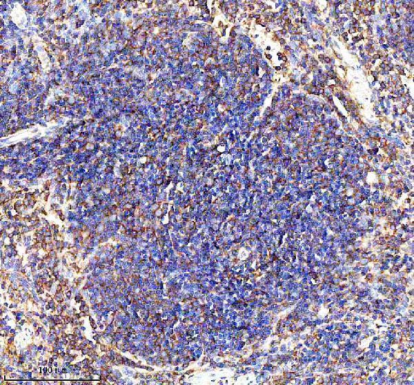
| Sample | Paraffin-embedded mouse spleen tissue; fixative not specified (datasheet A02073-4) |
| Fixation | Image fixative and duration unreported (datasheet A02073-4); verify before use. |
| Sectioning | 4–5 µm sections on charged slides (standard) |
| Deparaffinisation | Xylene, graded ethanol series to water (standard) |
| Antigen retrieval | Heat retrieval: EDTA pH 8.0 (datasheet A02073-4); 20 min, 95–100 °C (standard) |
| Peroxidase block | 3% H2O2, 10 min, room temperature (standard) |
| Blocking | 10% goat serum (datasheet A02073-4) |
| Primary antibody | Rabbit anti-GP1BA, 1:50 recommended; image 1:100 (datasheet A02073-4) |
| Primary incubation | Overnight at 4 °C (datasheet A02073-4) |
| Detection | HRP-conjugated secondary, DAB chromogen (datasheet A02073-4) |
| Counterstain | Hematoxylin, blue, dehydrate and mount (standard) |
| Expected result | GP1BA-positive staining in hematopoietic cells of bone marrow (HPA tissue IHC: Medium). HPA tissue profile: Selective cytoplasmic expression in platelets and megakaryocytes. No signal in the no-primary control. |
GP1BA is a platelet surface membrane protein with an extracellular region at residues 17–531, a transmembrane segment at 532–552, and a cytoplasmic tail at 553–652 (UniProt P07359 topology). In tissue IHC, expect selective staining of platelets and megakaryocytes; HPA describes the observed signal as cytoplasmic and rates its tissue profile Supported, citing consistency between staining and RNA expression (HPA tissue IHC).
| Selective staining of platelets and megakaryocytes, with signal appearing cytoplasmic in tissue sections (HPA tissue IHC). | This matches HPA’s reported pattern. The cytoplasmic appearance in IHC does not overturn GP1BA’s membrane topology; the two descriptions refer to observed tissue staining and protein location, respectively (HPA tissue IHC; UniProt P07359 topology). |
| Predominantly nuclear staining in cells scored as positive. | Nuclear localisation is inconsistent with the membrane location assigned to GP1BA (UniProt P07359). Review morphology, counterstain and detection controls before scoring this as target signal (general IHC practice). |
| Strong staining of adipocytes or respiratory epithelial cells. | These cell types were reported as Not detected in the sampled adipose tissue and bronchus, respectively (HPA tissue IHC). Consider nonspecific binding or endogenous detection activity; a control can help distinguish them (general IHC practice). |
| Diffuse colour across cells and surrounding tissue, obscuring platelet or megakaryocyte outlines. | This is difficult to reconcile with HPA’s selective cellular pattern (HPA tissue IHC). Inspect the no-primary control, blocking, washing and chromogen development before interpreting any cell as positive (general IHC practice). |
| No convincing signal in an otherwise interpretable bone marrow section. | HPA reports Medium staining in bone marrow hematopoietic cells and identifies platelets and megakaryocytes in its selective profile (HPA tissue IHC). Confirm that those elements are present, then check the assay controls and section processing (general IHC practice). |
| Tissue and cell selection (HPA tissue IHC) | Bone marrow hematopoietic cells and splenic red-pulp cells are listed at Medium staining, while the profile specifically identifies platelets and megakaryocytes. Interpret those tissue-level entries alongside cell morphology; they do not mean every hematopoietic or red-pulp cell expresses GP1BA (HPA tissue IHC). |
| Membrane topology and observed IHC pattern (UniProt P07359; HPA tissue IHC) | The extracellular region spans residues 17–531, followed by one transmembrane segment and a cytoplasmic tail (UniProt P07359 topology). HPA nevertheless calls its tissue staining cytoplasmic. Judge concordance against the reported slide pattern without claiming that this determines the antibody’s epitope (HPA tissue IHC). |
| Protein processing and epitope uncertainty (UniProt P07359) | UniProt annotates a full chain at residues 17–652 and glycocalicin at residues 17–506. An antibody’s exact epitope is not supplied here, so this record cannot establish whether it detects both products or whether processing explains a particular tissue pattern (UniProt P07359 processing). |
| Antibody evidence (HPA antibody validation) | HPA lists two IHC antibodies, HPA013316 and CAB002496, each with Supported status; neither has an ICC status in the supplied entries (HPA antibody validation). Supported tissue evidence helps anchor interpretation, but it is not a validation result for a different catalog antibody (HPA antibody validation). |
| IF/ICC question: is the tissue IHC pattern an IF/ICC reference? (HPA subcellular; HPA antibody validation) | HPA summarizes the subcellular location as Membrane but supplies no main location or ICC-IF images, and neither listed antibody has an ICC status (HPA subcellular; HPA antibody validation). Use the separate IF/ICC guide for assay design; this record alone cannot establish an expected image pattern. |
| Situation | Likely cause | Next action |
|---|---|---|
| Bone marrow lacks visible positive elements or shows no signal (HPA tissue IHC). | The sampled area may lack identifiable platelets or megakaryocytes, or the assay may have failed; HPA’s Medium bone marrow entry alone cannot separate these explanations (HPA tissue IHC). | Check cell morphology and section quality, then review the positive control, primary-antibody step, retrieval record and detection reagents (general IHC practice). |
| Only nuclei appear positive. | This differs from the reported cytoplasmic tissue pattern and membrane location (HPA tissue IHC; UniProt P07359). Counterstain or detection artefact is possible (general IHC practice). | Compare the no-primary control and counterstain, and inspect whether any platelet or megakaryocyte-associated signal remains (general IHC practice). |
| Adipocytes or bronchial epithelium stain prominently. | HPA reports Not detected in adipocytes from adipose tissue and respiratory epithelial cells from bronchus (HPA tissue IHC). Nonspecific or endogenous detection signal is possible (general IHC practice). | Run a no-primary control and review blocking and detection conditions; score only signal that also fits cell morphology and the expected tissue distribution (general IHC practice; HPA tissue IHC). |
| Brown signal is widespread and obscures cell boundaries. | Diffuse background prevents assessment of HPA’s selective platelet and megakaryocyte pattern (HPA tissue IHC). Excess detection signal or inadequate washing can contribute (general IHC practice). | Compare a no-primary section, check wash steps and chromogen development, and reassess only areas with interpretable cell boundaries (general IHC practice). |
| A positive cell looks cytoplasmic rather than sharply outlined at its membrane. | HPA expressly describes cytoplasmic expression in tissue IHC, while UniProt assigns GP1BA to the membrane (HPA tissue IHC; UniProt P07359). | Assess the cell type and selective distribution first. Do not reject a plausible tissue IHC result solely because its edge is indistinct (HPA tissue IHC). |
| IF/ICC shows an unexpected distribution. | The supplied HPA subcellular entry has no ICC-IF images, and both listed antibodies lack ICC status (HPA subcellular; HPA antibody validation). | Treat the IF/ICC pattern as unconfirmed by this tissue IHC record and consult the separate IF/ICC guide; compare localisation with membrane topology using assay controls (UniProt P07359; general IF practice). |
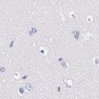
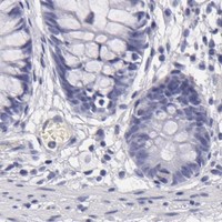

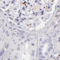
Comprehensive Human Protein Atlas IHC scoring per tissue (reliability: Supported — High consistency between antibody staining and RNA expression data.). Rows are taken directly from the HPA tissue chart — click any row's HPA link to view the source.
| Tissue | Cell type | Level | Evidence | Source |
|---|---|---|---|---|
| Bone marrow | Hematopoietic cells | Medium | Protein (IHC) | HPA → |
| Spleen | Cells in red pulp | Medium | Protein (IHC) | HPA → |
| Tissue | Cell type | Level | Evidence | Source |
|---|---|---|---|---|
| Adipose tissue | Adipocytes | Not detected | Protein (IHC) | HPA → |
| Adrenal gland | Glandular cells | Not detected | Protein (IHC) | HPA → |
| Appendix | Glandular cells | Not detected | Protein (IHC) | HPA → |
| Breast | Adipocytes | Not detected | Protein (IHC) | HPA → |
| Bronchus | Respiratory epithelial cells | Not detected | Protein (IHC) | HPA → |
Troubleshoot GP1BA staining in paraffin sections by checking retrieval, cell identity, membrane localisation and assay controls before comparing chromogenic signal across samples.
The catalog lists two IHC antibodies for GP1BA (catalog applications); only A02073-4 has an IHC image, from a paraffin-embedded mouse spleen section (A02073-4 image caption). Neither has IF/ICC data (catalog applications and images).
A02073-4 lists human and mouse reactivity and IHC use (catalog reactivity and applications); its image shows staining in a paraffin-embedded mouse spleen section (A02073-4 image caption). M02073-2 lists human reactivity and IHC use, with no IHC image supplied (catalog reactivity, applications and images).
Which to pick: Choose A02073-4 for tissue IHC when a demonstrated paraffin-section example matters; its mouse spleen caption reports antigen retrieval in EDTA at pH 8.0, but does not report the fixative (A02073-4 image caption). For a human-reactive monoclonal IHC option, M02073-2 is listed as clone 17G60, though no tissue image is supplied (M02073-2 catalog clone, applications, reactivity and images). For human-and-mouse reactivity, choose A02073-4 (catalog reactivity); neither SKU has listed IF/ICC validation, so no IF/ICC choice is supported here (catalog applications and images).