GPD1 / Glycerol-3-phosphate dehydrogenase [NAD(+)], cytoplasmic · IHC design guide

Design Immunohistochemistry for GPD1

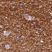
Plan chromogenic GPD1 IHC using the catalog antibody’s documented paraffin-section workflow (datasheet A03262-1). Assess cytoplasmic staining in hepatocytes, kidney proximal tubules and myocytes, using smooth muscle cells as a negative comparator (HPA tissue IHC).

Evidence assembled Oct 2026 · For research use; verify linked source records and product datasheet before use
Immunohistochemistry protocol sheet for GPD1 (IHC for GPD1): expected localisation Cytoplasmic tissue staining (HPA tissue IHC), antibody A03262-1, validated IHC image, and IHC protocol steps
Printable GPD1 IHC protocol sheet — expected localisation Cytoplasmic tissue staining (HPA tissue IHC), antibody A03262-1, controls and protocol steps. Open the full GPD1 IHC guide →

GPD1 Immunohistochemistry Experimental Design Guide

Expected localisation, validated protocols, controls and antibodies — the at-a-glance facts below, then the full design guide.

Must know before staining
Expected localisation Cytoplasmic tissue staining (HPA tissue IHC)
Staining pattern Cytoplasmic staining in myocytes and hepatocytes (HPA tissue IHC)
Antigen retrieval EDTA pH 8.0 HIER, heat-mediated (datasheet A03262-1)
Positive control ⓘ Adipose tissue+4 more · see all
Negative control ⓘ Smooth muscle
Important caveats
Reasons your staining may differ from the expected pattern.
Fixation Keep fixation consistent across sections. (standard IHC practice; not target-specific)
Caveat Presumed off-target staining was observed (HPA tissue IHC)
Regulation Tissue-enhanced RNA: adipose, breast, muscle (HPA tissue RNA)
Isoform / epitope 2 isoforms; epitope coverage is unresolved (UniProt)
Section 1

Recommended GPD1 IHC & IF Protocols

The catalog antibody’s IHC-P protocol is paired with published protocols for breast cancer sections (PMC5731876) and bladder cancer sections (PMC9284842).

Recommended immunohistochemistry (IHC-P) protocol parameters
SampleParaffin-embedded human bladder cancer tissue; fixative not specified (datasheet A03262-1)
FixationImage fixative and duration unreported (datasheet A03262-1); verify before use.
Sectioning4–5 µm sections on charged slides (standard)
DeparaffinisationXylene, graded ethanol series to water (standard)
Antigen retrievalHeat retrieval: EDTA pH 8.0 (datasheet A03262-1); 20 min, 95–100 °C (standard)
Peroxidase block3% H2O2, 10 min, room temperature (standard)
Blocking10% goat serum (datasheet A03262-1)
Primary antibodyRabbit anti-GPD1, 2-5 μg/ml (datasheet A03262-1)
Primary incubationOvernight at 4 °C (datasheet A03262-1)
DetectionHRP-conjugated secondary, DAB chromogen (datasheet A03262-1)
CounterstainHematoxylin, blue, dehydrate and mount (standard)
Expected resultGPD1-positive staining in adipocytes of adipose tissue (HPA tissue IHC: High). HPA tissue profile: Distinct cytoplasmic expression in skeletal muscle, breast, kidney, liver and gastrointestinal tract. No signal in the no-primary control.
💡Decision noteStart with heat-mediated EDTA retrieval at pH 8.0 (datasheet: A03262-1); the breast cancer protocol reports citrate at pH 6.0 (PMC5731876).
Section 2

What Is the Expected GPD1 Staining Pattern?

GPD1 is a cytoplasmic protein with no transmembrane segment (UniProt P21695: subcellular location and topology). In paraffin section IHC, expect cytoplasmic staining in hepatocytes, skeletal myocytes, adipocytes and kidney proximal tubule cells, among other HPA high staining populations (HPA: tissue IHC). HPA rates the tissue profile Enhanced while noting medium consistency with RNA data and presumed off target binding that was disregarded (HPA: tissue IHC reliability).

What am I looking at on my slide?
Distinct cytoplasmic stain in hepatocytes, skeletal myocytes, adipocytes or kidney proximal tubule cells.This matches GPD1's cytoplasmic location (UniProt P21695) and populations scored High by HPA tissue IHC. Compare intensity within the same run and assess cell identity from the section; a brown signal alone does not identify the stained cell (general IHC practice).
Predominantly nuclear or sharply membrane restricted signal in otherwise recognizable cells.That compartment conflicts with the UniProt cytoplasmic location and absence of a transmembrane segment (UniProt P21695). Check morphology, counterstain and detection controls before interpreting it as GPD1; the supplied sources do not establish a nuclear or membrane pool.
Strong signal in smooth muscle cells, or staining that follows a different cell population than expected.HPA reports GPD1 as Not detected in smooth muscle cells (HPA: tissue IHC). Treat staining there as a specificity concern, including possible cross reactivity or detection background; HPA itself notes presumed off target binding in its tissue assessment (HPA: reliability description).
Brown haze across cells, stroma or a no primary antibody control.A field wide signal is difficult to assign to GPD1 positive cells (general IHC practice). Consider nonspecific detection or endogenous chromogenic activity, then inspect the no primary control and the spatial pattern. HPA's cell specific tissue profile cannot validate uniform background (HPA: tissue IHC).
No cytoplasmic signal in a section containing intact hepatocytes or skeletal myocytes.Those cell populations are High in HPA tissue IHC, so an entirely blank result warrants a workflow check (HPA: tissue IHC). Review tissue preservation, antibody use and control staining as general IHC practice. A negative slide alone does not establish biological absence or a GPD1 specific fixation effect.
💡Expected GPD1 appearanceCall a result positive when distinct cytoplasmic signal is present in recognizable HPA High cells such as hepatocytes or skeletal myocytes (UniProt P21695: cytoplasm; HPA: tissue IHC); dominant nuclear, membrane or smooth muscle staining is suspect (UniProt P21695: topology; HPA: smooth muscle Not detected).
How each factor affects the staining
Cell and compartment referenceUniProt places GPD1 in the cytoplasm without a transmembrane segment (UniProt P21695). HPA reports High staining in adipocytes, hepatocytes, skeletal myocytes and kidney proximal tubule cells, while smooth muscle cells are Not detected (HPA: tissue IHC). Use these as pattern references, not a universal threshold for every section.
Strength of tissue evidenceHPA labels the tissue profile Enhanced, yet describes medium agreement between antibody staining and RNA expression and disregarded presumed off target binding (HPA: tissue IHC reliability). Its listed antibodies have IHC ratings Enhanced for HPA044620 and Supported for HPA058621 (HPA: antibody validation); these ratings do not validate an unrelated reagent.
Isoform and epitope scopeUniProt annotates two GPD1 isoforms, 1 and 2 (UniProt P21695). The supplied record does not locate this assay's epitope or show isoform specific tissue staining. Do not interpret a regional intensity difference as isoform selection without separate evidence.
IF/ICC: where should signal appear?Cytoplasm is the expected compartment from UniProt (UniProt P21695: subcellular location). HPA lists no main ICC/IF location or cell lines with ICC/IF images (HPA: subcellular record), so its IHC tissue pattern does not independently validate an IF image.
Why is my staining missing, weak or wrong?
SituationLikely causeNext action
A known HPA High tissue is blank or very weak.A failed staining run or an unsuitable antibody condition is possible (general IHC practice); HPA High is a reference observation, not a guarantee for every section (HPA: tissue IHC).Check the run's positive control, tissue morphology, antibody identity and IHC validation, then review retrieval and dilution against the antibody's documented IHC conditions (general IHC practice). Do not infer a GPD1 specific fixation sensitivity from this result.
Smooth muscle cells stain as strongly as nearby expected positive cells.HPA scores smooth muscle cells Not detected; a mismatch raises concern for off target or detection signal (HPA: tissue IHC and reliability description).Confirm cell identity on the counterstained section and compare a no primary control (general IHC practice). Reassess antibody specificity before scoring the smooth muscle signal as GPD1.
Signal appears mainly in nuclei or at cell borders.This conflicts with cytoplasmic GPD1 and its lack of a transmembrane segment (UniProt P21695). Background or misassigned cell boundaries may explain the appearance (general IHC practice).Inspect morphology and the no primary control, and compare with a known HPA High cell population (general IHC practice; HPA: tissue IHC). Record the discordance rather than calling the compartment positive.
Brown precipitate is widespread, including outside recognizable cells.Nonspecific detection or endogenous chromogenic activity can create diffuse signal (general IHC practice). That distribution does not match HPA's distinct cytoplasmic tissue profile (HPA: tissue IHC).Review no primary and detection controls, blocking, washes and development time (general IHC practice). Interpret only cell associated staining that can be assigned to a defined compartment.
Expected positive cells stain unevenly across the section.Uneven processing or detection is possible (general IHC practice); HPA reports tissue level cell patterns, not uniform intensity in every field (HPA: tissue IHC).Compare intact regions and run controls, then assess section quality and staining consistency (general IHC practice). Avoid attributing a gradient to GPD1 biology or target specific fixation behavior without evidence.
An IF/ICC image shows a compartment that differs from the IHC call.UniProt predicts cytoplasm, but HPA provides no ICC/IF images or main subcellular location for this record (UniProt P21695; HPA: subcellular record).Use the cytoplasmic expectation as a comparison and evaluate IF controls in its dedicated guide (general IF practice). Do not treat the HPA IHC rating as validation of that IF result (HPA: antibody validation).

Sample controls for GPD1 IHC & IF

🧪Run liver first: hepatocytes should show GPD1 staining (HPA: High in hepatocytes). Run smooth muscle as the negative tissue (HPA: Not detected in smooth muscle cells); on the liver slide, non-hepatocyte cells should serve as a background comparator and lack the hepatocyte-pattern cytoplasmic signal, without being presumed biologically GPD1-negative (HPA: High in hepatocytes; UniProt P21695: Cytoplasm).
Positive control tissue: Adipose tissue (Adipocytes, HPA High)
Negative control tissue: Smooth muscle (HPA Not detected)
ICC-IF cell lines (HPA subcellular resource): HPA carries no ICC-IF cell line for GPD1; derive a cell-line control from the positive tissue's cell type (Adipocytes) and confirm it by RNA or western blot first.
Technical controls: Include no-primary (secondary-only) and concentration-matched rabbit IgG isotype controls, plus GPD1-knockout tissue or peptide-blocked antibody if available (selected-SKU caption: rabbit primary antibody). For liver, quench endogenous peroxidase before HRP/DAB detection and check endogenous biotin if using a biotin-based system (selected-SKU caption: HRP/DAB detection; standard IHC practice).
⚠️Feasibility: A target-specific fixation window or fixation effect is unreported, and the selected-SKU paraffin-section caption does not state a fixative (selected-SKU caption: fixative not stated). The documented IHC workflow uses heat retrieval in EDTA at pH 8.0, so retrieval should be held consistent while controls are compared (selected-SKU caption: EDTA heat retrieval). Frozen sections and IF/ICC cannot be judged easier from the supplied application evidence; in liver, endogenous peroxidase can produce misleading chromogenic signal unless quenched (selected-SKU caption: HRP/DAB detection; standard IHC practice).

HPA tissue IHC evidence for GPD1

Comprehensive Human Protein Atlas IHC scoring per tissue (reliability: Enhanced — Medium consistency between antibody staining and RNA expression data. Presumed off target binding observed and disregarded.). Rows are taken directly from the HPA tissue chart — click any row's HPA link to view the source.

Positive expression · recommended positive controls

TissueCell typeLevelEvidenceSource
Adipose tissue Adipocytes High Protein (IHC) HPA →
Duodenum Endocrine cells High Protein (IHC) HPA →
Kidney Proximal tubules (cell body) High Protein (IHC) HPA →
Liver Hepatocytes High Protein (IHC) HPA →
Lung Alveolar cells type II High Protein (IHC) HPA →

Undetected expression · recommended negative controls

TissueCell typeLevelEvidenceSource
Smooth muscle Smooth muscle cells Not detected Protein (IHC) HPA →
Section 3

Advanced GPD1 IHC Tips

Use cytoplasmic staining, appropriate tissue controls, and the catalog antibody’s paraffin-section evidence to troubleshoot GPD1 chromogenic IHC.

How should I troubleshoot weak GPD1 staining after antigen retrieval?
Start with heat-mediated antigen retrieval in EDTA at pH 8.0 for paraffin sections (datasheet A03262-1). The selected tissue image used this retrieval before incubation with 2 μg/ml antibody overnight at 4°C (datasheet A03262-1). If staining is weak, first compare retrieval temperature and duration across sections processed from the same block, while keeping detection and antibody concentration constant (standard IHC practice). Include a high-expression control such as liver hepatocytes or kidney proximal tubules, and judge improvement by cytoplasmic signal rather than total brown staining (HPA: High in hepatocytes and proximal tubules; UniProt P21695: Cytoplasm).
Could fixation explain weak or uneven GPD1 staining in paraffin sections?
The selected paraffin-section caption does not report a fixative, so GPD1-specific fixation sensitivity is unknown for this antibody (datasheet A03262-1). Record the fixative, fixation duration, and tissue thickness for each specimen, then compare sections processed under matched conditions (standard IHC practice). Keep the documented EDTA pH 8.0 retrieval and 2 μg/ml overnight antibody incubation constant while assessing fixation as a variable (datasheet A03262-1). Use an intact positive-control area to distinguish broadly weak staining from patchy preservation, and avoid treating a negative field alone as evidence that GPD1 is absent (standard IHC practice; HPA: tissue expression varies).
What pattern should count as specific GPD1 staining?
Score GPD1 primarily as cytoplasmic staining because its annotated location is the cytoplasm and it has no transmembrane segment (UniProt P21695: subcellular location and topology). Hepatocytes, kidney proximal tubules, and skeletal-muscle myocytes provide expected positive cell populations for comparison (HPA: High in these cells). Inspect the cell body at sufficient magnification to separate cytoplasmic DAB from pigment, precipitate, or staining over an adjacent cell (standard IHC practice). If staining is confined to nuclei, cell borders, or extracellular material, review controls and detection background before assigning it to GPD1 (UniProt P21695: Cytoplasm; standard IHC practice).
Can the available evidence establish which GPD1 isoform this antibody detects?
GPD1 has 2 annotated isoforms, but the supplied tissue caption does not identify this antibody’s epitope or establish isoform selectivity (UniProt P21695: isoforms 1 and 2; datasheet A03262-1). Do not interpret a positive cytoplasmic stain as proof of either isoform without an epitope map and isoform-specific validation (UniProt P21695: Cytoplasm; standard IHC practice). The record lists phosphoserine 154, succinyllysine 289, and phosphotyrosine 326; it does not show whether any modification affects antibody binding (UniProt P21695: modified residues; datasheet A03262-1: epitope unreported). Compare staining with an independent validated assay if an isoform-level or modification-dependent conclusion is required (standard IHC practice).
How should I adapt an IHC finding for a GPD1 multiplex IF experiment?
Treat the catalog antibody’s paraffin-section result as IHC evidence; the supplied payload provides no GPD1 ICC/IF images or IF validation for it (datasheet A03262-1; HPA: no ICC/IF images listed). In a separate IF assay, pair GPD1 with a marker identifying an expected positive cell population, such as hepatocytes or proximal tubules, and include single-label controls (HPA: High in those cells; standard IF practice). Choose spectrally separated fluorophores after checking tissue autofluorescence, using a farther-red channel for a weak signal when the specimen permits (standard IF practice). Because GPD1 is cytoplasmic and lacks a transmembrane segment, validate permeabilisation that gives antibody access to the cytoplasm without disrupting cell outlines (UniProt P21695: location and topology; standard IF practice).
How can I separate GPD1 signal from chromogenic background?
The documented tissue workflow used 10% goat serum blocking, a peroxidase-conjugated secondary, and DAB development (datasheet A03262-1). For unexplained brown staining, compare a no-primary control and an appropriate positive tissue section, then check whether pigment or precipitate persists without primary antibody (standard IHC practice; HPA: High in hepatocytes). Include an endogenous-peroxidase block and inspect secondary-antibody background as general chromogenic IHC controls; neither step establishes GPD1 specificity by itself (standard IHC practice). Interpret a convincing result as cellular cytoplasmic staining in an expected population, while discounting diffuse deposits, section-edge intensification, and staining over damaged tissue (UniProt P21695: Cytoplasm; HPA: tissue profile; standard IHC practice).
How should I quantify GPD1 staining across tissue sections? ⚠ ANSWER MARKED FOR VERIFICATION
Define the anatomical region and cell population before scoring, because GPD1 expression differs among cell types within and across tissues (HPA: tissue IHC profile). For chromogenic IHC, record the percentage of positive cells and intensity by category, then calculate an H-score if that scale is specified in advance (standard IHC practice). Normalise to the number of evaluable cells in the same compartment, or report positive-cell density per mm² when cell counts are impractical (standard IHC practice). Apply one staining threshold across comparably processed sections, exclude folds and necrosis, and retain a positive-control section to track run-to-run variation (standard IHC practice; HPA: High in hepatocytes).
When is unexpected GPD1 staining more likely to be artefact?
GPD1 is annotated as cytoplasmic, and HPA reports high staining in hepatocytes, proximal tubules, and skeletal-muscle myocytes but no detection in smooth-muscle cells (UniProt P21695: Cytoplasm; HPA: tissue IHC). Predominantly nuclear or extracellular DAB, or strong staining limited to an unexpected cell population, therefore warrants review of specificity controls before biological interpretation (UniProt P21695: location; HPA: tissue profile; standard IHC practice). Check section edges, necrotic areas, pigment, and endogenous-peroxidase signal with a no-primary control (standard IHC practice). HPA rates its tissue evidence “Enhanced” but notes presumed off-target binding, so agreement with cell type and compartment strengthens an interpretation without proving antibody specificity (HPA: reliability description).
Boster reagents

Best GPD1 / Glycerol-3-phosphate dehydrogenase [NAD(+)], cytoplasmic IHC Antibodies

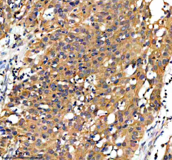
The catalog includes one anti-GPD1 antibody with real IHC data from a paraffin-embedded human bladder cancer section (A03262-1 image caption). Human, mouse, and rat reactivity is listed (catalog reactivity).

Real IHC data IHC analysis of GPD1 using anti-GPD1 antibody (A03262-1). GPD1 was detected in a paraffin-embedded section of human bladder cancer tissue. Heat mediated antigen retrieval was performed in EDTA buffer (pH 8.0, epitope retrieval solution). The tissue section was blocked with 10% goat serum. The tissue section was then incubated with 2 μg/ml rabbit anti-GPD1 Antibody (A03262-1) overnight at 4°C. Peroxidase Conjugated Goat Anti-rabbit IgG was used as secondary antibody and incubated for 30 minutes at 37°C. The tissue section was developed using HRP Conjugated Rabbit IgG Super Vision Assay Kit (Catalog # SV0002) with DAB as the chromogen.
Anti-GPD1 Antibody ®
Cat # A03262-1

A03262-1 has an IHC image from a paraffin-embedded human bladder cancer section (A03262-1 image caption). It is listed for IHC and as reactive with human, mouse, and rat samples (catalog applications; catalog reactivity).

Which to pick: Choose A03262-1 for chromogenic tissue IHC: its image caption documents EDTA pH 8.0 retrieval and DAB detection in a paraffin-embedded human section; the fixative is unreported (A03262-1 image caption). There is no supported IF/ICC pick because the catalog lists no IF/ICC application or IF image for A03262-1 (catalog applications; catalog IF images). For mouse or rat work, A03262-1 lists reactivity with both species, but its supplied tissue IHC image demonstrates human tissue only (catalog reactivity; A03262-1 image caption).

Each figure is that product's own IHC / IF validation image from its datasheet.

References

  1. UniProt Consortium. UniProt entry P21695 (GPDA_HUMAN, Glycerol-3-phosphate dehydrogenase [NAD(+)], cytoplasmic).
  2. Human Protein Atlas. GPD1 tissue IHC expression (reliability: Enhanced).
  3. Human Protein Atlas. GPD1 subcellular location (ICC-IF): Highest expression in Hep-G2: 15.1 nTPM.
  4. Human Protein Atlas. GPD1 antibody validation summary (2 antibodies).
  5. A HIF1α-GPD1 feedforward loop inhibits the progression of renal clear cell carcinoma via mitochondrial function and lipid metabolism. Journal of experimental & clinical cancer research : CR 2021 — PMC8185942.
  6. Identification of glycerol-3-phosphate dehydrogenase 1 as a tumour suppressor in human breast cancer. Oncotarget 2017 — PMC5731876.
  7. Allosteric activation of the metabolic enzyme GPD1 inhibits bladder cancer growth via the lysoPC-PAFR-TRPV2 axis. Journal of hematology & oncology 2022 — PMC9284842.
  8. Prognostic value of a novel glycolysis-related gene expression signature for gastrointestinal cancer in the Asian population. Cancer cell international 2021 — PMC7934443.
  9. PubMed PMID:7772607 — UniProt-cited evidence.
  10. PubMed PMID:14702039 — UniProt-cited evidence.
  11. PubMed PMID:16541075 — UniProt-cited evidence.