This website uses cookies to ensure you get the best experience on our website.


- Table of Contents
Plan paraffin-section GPR37L1 IHC around selective CNS cytoplasmic staining and cerebellar molecular-layer processes (HPA tissue IHC). Start the catalog antibody at 1:100–1:300 (datasheet: A11679-1), and consider adipocytes in adipose tissue as a negative reference (HPA tissue IHC).

Expected localisation, validated protocols, controls and antibodies — the at-a-glance facts below, then the full design guide.
| Expected localisation | CNS cytoplasmic staining (HPA tissue IHC); cell and cilium membrane (UniProt) | |
| Staining pattern | Selective CNS cytoplasm; cerebellar molecular-layer processes (HPA tissue IHC) | |
| Antigen retrieval | Citrate pH 6.0 HIER, 95–98 °C, 20 min (rule: cytoplasmic / membrane antigen) | |
| Positive control | Cerebellum+1 more · see all | |
| Negative control | Adipose tissue+4 more · see all |
| Fixation | Keep formalin fixation consistent across sections (standard IHC practice; not target-specific); Matched tissue-IHC evidence does not establish the fixation claim. Validate the specimen-specific method before use. (selected-SKU IHC image A11679-1) | |
| Caveat | Testis spermatids stain despite brain-enriched RNA (HPA tissue IHC) | |
| Regulation | Staining regulation is not established (UniProt) | |
| Isoform / epitope | 0 isoforms annotated; epitope side matters across the seven-transmembrane topology (UniProt) |
The catalog antibody’s IHC-P protocol is accompanied by 3 published GPR37L1 chromogenic IHC protocols (PMC7707515; PMC8357083; PMC7870391).
| Sample | Paraffin-embedded human brain tissue; fixative not specified (datasheet A11679-1) |
| Fixation | Image fixative and duration unreported (datasheet A11679-1); verify before use. |
| Sectioning | 4–5 µm sections on charged slides (standard) |
| Deparaffinisation | Xylene, graded ethanol series to water (standard) |
| Antigen retrieval | Heat-induced epitope retrieval in citrate buffer, pH 6.0, 20 min at 95–98 °C (standard rule: cytoplasmic / membrane antigen) |
| Peroxidase block | 3% H2O2, 10 min, room temperature (standard) |
| Blocking | 10% normal serum of the secondary host, 30 min, room temperature (standard) |
| Primary antibody | Rabbit anti-GPR37L1, 1:100-1:300 (datasheet A11679-1) |
| Primary incubation | Overnight at 4 °C (standard) |
| Detection | HRP-polymer secondary, DAB chromogen 5–10 min (standard) |
| Counterstain | Hematoxylin, blue, dehydrate and mount (standard) |
| Expected result | GPR37L1-positive staining in processes in molecular layer of cerebellum (HPA tissue IHC: Medium). HPA tissue profile: Selective cytoplasmic expression in CNS. No signal in the no-primary control. |
GPR37L1 is a seven-pass receptor assigned to the cell membrane and cilium membrane, including the basal membrane of Bergmann glial primary cilia (UniProt O60883: topology and subcellular location). In paraffin-section IHC, expect selective cytoplasmic staining in the CNS, especially cerebellar molecular-layer processes (HPA tissue IHC: selective cytoplasmic CNS pattern; cerebellum, medium). HPA rates its tissue IHC reliability Enhanced, with medium consistency between antibody staining and RNA data (HPA tissue IHC: reliability).
| Molecular-layer processes stain in cerebellum, with little staining outside the expected structures. | This matches the reported medium signal in cerebellar molecular-layer processes and the selective cytoplasmic CNS profile (HPA tissue IHC: cerebellum, medium; CNS profile). Interpret the slide by its tissue pattern: UniProt's membrane and cilium assignment does not require a resolvable membrane outline in chromogenic sections (UniProt O60883: subcellular location). |
| Strong nuclear staining dominates the cerebellar section. | Treat this as a compartment mismatch requiring validation against the reported cytoplasmic tissue pattern (HPA tissue IHC: selective cytoplasmic CNS profile). Check a matched negative control and the expected molecular-layer process staining before scoring it as GPR37L1; nuclear colour alone does not establish target localisation (general IHC practice). |
| Prominent staining appears in an unexpected cell population, such as adipocytes. | Adipocytes were not detected in the listed adipose-tissue and breast observations (HPA tissue IHC: adipose tissue and breast). An unexpected positive population may reflect antibody cross-reactivity or endogenous detection activity; compare the same cell population in controls and examine whether the expected cerebellar pattern is present (general IHC practice). |
| Colour spreads broadly across cells and tissue without a discernible process pattern. | Diffuse background makes the selective CNS pattern difficult to judge (HPA tissue IHC: CNS profile; cerebellar processes, medium). Inspect the no-primary control for detection background, then review blocking, wash stringency and primary-antibody concentration as general chromogenic IHC variables (general IHC practice). Do not score uniform haze as a positive compartment. |
| No staining appears in a cerebellar molecular layer used as a positive comparison. | This conflicts with the reported medium staining of molecular-layer processes (HPA tissue IHC: cerebellum, medium). First verify that the layer is present on the section and that detection controls worked; then review the antibody's validated IHC-P conditions. A blank section alone cannot distinguish weak detection from absent antigen (general IHC practice). |
| Tissue and cell selection | Use cerebellar molecular-layer processes as the principal positive reference; round or early spermatids in testis are another reported medium-staining population (HPA tissue IHC: cerebellum and testis, medium). The listed neuronal cells in cerebral cortex and glial cells in hippocampus and caudate have low staining, so faint signal there needs cautious scoring (HPA tissue IHC: low observations). |
| Topology and antibody epitope | GPR37L1 has seven transmembrane segments, extracellular and cytoplasmic regions, and an annotated signal peptide at residues 1–25 (UniProt O60883: topology and processing). The payload does not identify the antibody epitope. Use the antibody's own IHC-P instructions when choosing retrieval conditions; topology alone cannot specify retrieval or predict epitope accessibility (general IHC practice). |
| Strength of the tissue evidence | The tissue IHC profile is rated Enhanced, while antibody staining and RNA expression show medium consistency (HPA tissue IHC: reliability). The listed IHC Enhanced status belongs to HPA064454; it does not automatically validate another antibody (HPA antibodies: HPA064454). Judge an experiment against its own controls and the reported cell pattern (general IHC practice). |
| IF/ICC Q: Should nuclear fluorescence count as the expected IHC result? | A: No. HPA reports nucleoplasm as uncertain in ICC-IF, cell junctions as approved, and plasma membrane as supported; its paraffin-section tissue profile is selective cytoplasmic staining in the CNS (HPA subcellular ICC-IF; HPA tissue IHC). Evaluate IF/ICC on its separate guide page. This section provides no IF/ICC protocol option. |
| Situation | Likely cause | Next action |
|---|---|---|
| Cerebellar molecular-layer processes are blank. | The expected medium-staining structures may be missing from the section, or the staining run may have failed (HPA tissue IHC: cerebellum, medium; general IHC practice). | Confirm section anatomy and run controls, then check the catalog antibody's IHC-P instructions for retrieval, dilution and detection conditions (general IHC practice). |
| All tissue compartments show similar brown colour. | The reported staining is selective within the CNS, so uniform colour is inconsistent with that pattern (HPA tissue IHC: CNS profile). Background from the detection workflow is possible (general IHC practice). | Examine a no-primary control; review blocking, washes and chromogen development before interpreting the slide (general IHC practice). |
| Adipocytes or other listed negative populations stain strongly. | Adipocytes in adipose tissue and breast were not detected in the listed HPA observations; cross-reactivity or endogenous detection activity may explain unexpected colour (HPA tissue IHC: negative observations; general IHC practice). | Compare the same population with a no-primary control and with the cerebellar positive reference; avoid assigning target positivity from colour alone (general IHC practice). |
| Nuclear colour dominates while molecular-layer processes remain faint. | The nuclear-dominant pattern differs from the reported cytoplasmic CNS tissue pattern (HPA tissue IHC: CNS profile). Counterstain or detection background can also complicate compartment assignment (general IHC practice). | Inspect a no-primary control and reassess where chromogen sits relative to nuclei and processes; score only a reproducible expected tissue pattern (general IHC practice). |
| Cerebral cortex, hippocampus or caudate looks nearly negative. | HPA lists low staining in cortical neuronal cells and in hippocampal and caudate glial cells (HPA tissue IHC: low observations). Weak colour in these populations may be hard to separate from background (general IHC practice). | Compare them with the medium-staining cerebellar reference and matched controls before concluding that the run failed (HPA tissue IHC: cerebellum, medium; general IHC practice). |


Comprehensive Human Protein Atlas IHC scoring per tissue (reliability: Enhanced — Medium consistency between antibody staining and RNA expression data.). Rows are taken directly from the HPA tissue chart — click any row's HPA link to view the source.
| Tissue | Cell type | Level | Evidence | Source |
|---|---|---|---|---|
| Cerebellum | Processes in molecular layer | Medium | Protein (IHC) | HPA → |
| Testis | Round or early spermatids | Medium | Protein (IHC) | HPA → |
| Tissue | Cell type | Level | Evidence | Source |
|---|---|---|---|---|
| Adipose tissue | Adipocytes | Not detected | Protein (IHC) | HPA → |
| Adrenal gland | Glandular cells | Not detected | Protein (IHC) | HPA → |
| Appendix | Glandular cells | Not detected | Protein (IHC) | HPA → |
| Bone marrow | Hematopoietic cells | Not detected | Protein (IHC) | HPA → |
| Breast | Adipocytes | Not detected | Protein (IHC) | HPA → |
Troubleshoot GPR37L1 staining in paraffin sections by comparing signal location, tissue pattern and controls with the supplied receptor and IHC evidence.
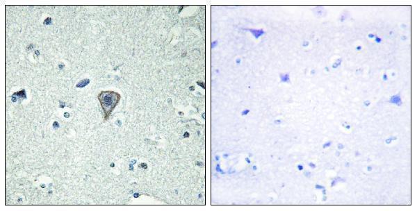
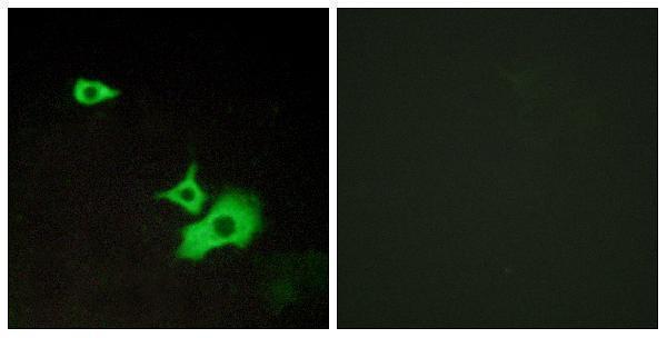
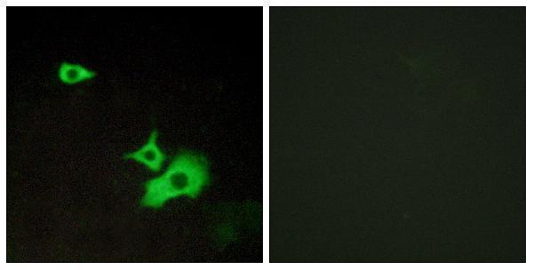
Two anti-GPR37L1 antibodies have pictured human tissue IHC or cell IF results (A11679-1 IHC image caption; A11679 IF image caption). Both list Human, Mouse and Rat reactivity (catalog: reactivity).
A11679-1 renders with paraffin-embedded human brain IHC and a peptide-blocked comparison (A11679-1 IHC image caption). A11679 renders with LOVO-cell IF and a peptide-blocked comparison (A11679 IF image caption); IHC and ICC are also listed applications (catalog: A11679 applications).
Which to pick: Choose A11679-1 for paraffin-section IHC because its own image shows human brain tissue; the fixative is unreported (A11679-1 IHC image caption). Choose A11679 for cell IF/ICC: its own image shows IF in LOVO cells, and ICC is a listed application (A11679 IF image caption; catalog: A11679 applications). For cross-species work, both rabbit polyclonals list Human, Mouse and Rat reactivity (catalog: host, dilution_raw, reactivity); the supplied images show human tissue or LOVO cells (A11679-1 IHC image caption; A11679 IF image caption).