This website uses cookies to ensure you get the best experience on our website.


- Table of Contents
Plan GRB14 staining in paraffin sections using the reported granular cytoplasmic and membranous tissue pattern (HPA tissue IHC). The guide covers controls, detection and scoring while accounting for the uncertain agreement between antibody staining and RNA expression (HPA tissue IHC).

Expected localisation, validated protocols, controls and antibodies — the at-a-glance facts below, then the full design guide.
| Expected localisation | Granular cytoplasm and membrane in tissues (HPA tissue IHC) | |
| Staining pattern | Granular cytoplasmic and membranous staining in glandular cells (HPA tissue IHC) | |
| Antigen retrieval | EDTA pH 8.0 HIER, heat-mediated (datasheet A04177-2) | |
| Positive control | Caudate+4 more · see all | |
| Negative control | Adipose tissue+4 more · see all |
| Fixation | Keep paraffin section fixation consistent (standard IHC practice; not target-specific) | |
| Caveat | Staining has low consistency with RNA expression (HPA tissue IHC) | |
| Regulation | Insulin shifts GRB14 to the plasma membrane (UniProt) | |
| Isoform / epitope | 2 isoforms; epitope coverage is unspecified (UniProt) |
Compare the catalog antibody’s EDTA pH 8.0 IHC protocol (datasheet: A04177-2) with published GRB14 staining of mouse embryos, gastric tissue and rodent retina (PMC11441139; PMC12627322; PMC2763493).
| Sample | Paraffin-embedded human renal cancer tissue; fixative not specified (datasheet A04177-2) |
| Fixation | Image fixative and duration unreported (datasheet A04177-2); verify before use. |
| Sectioning | 4–5 µm sections on charged slides (standard) |
| Deparaffinisation | Xylene, graded ethanol series to water (standard) |
| Antigen retrieval | Heat retrieval: EDTA pH 8.0 (datasheet A04177-2); 20 min, 95–100 °C (standard) |
| Peroxidase block | 3% H2O2, 10 min, room temperature (standard) |
| Blocking | 10% goat serum (datasheet A04177-2) |
| Primary antibody | Rabbit anti-GRB14, 2-5μg/ml (datasheet A04177-2) |
| Primary incubation | Overnight at 4 °C (datasheet A04177-2) |
| Detection | Streptavidin-biotin complex (SABC), DAB chromogen (datasheet A04177-2) |
| Counterstain | Hematoxylin, blue, dehydrate and mount (standard) |
| Expected result | GRB14-positive staining in neuronal cells of caudate (HPA tissue IHC: Medium). HPA tissue profile: Cytoplasmic expression with a granular pattern and membranous expression in several tissues. No signal in the no-primary control. |
GRB14 is a cytoplasmic protein also associated with endosome membranes; insulin stimulation can move it to the plasma membrane, and it has no transmembrane segment (UniProt Q14449). In tissue IHC, expect granular cytoplasmic and some membranous staining in selected cells, including epididymal glandular and neuronal cells (HPA: tissue IHC). Interpret cautiously: HPA rates the tissue pattern Uncertain because staining and RNA expression have low consistency (HPA: tissue IHC).
| Granular cytoplasmic staining, with some membrane staining, in epididymal glandular cells or neurons. | This fits the reported IHC pattern; epididymal glandular cells and neurons in the caudate or cerebral cortex have Medium staining (HPA: tissue IHC). Compare stained cells with tissue morphology and a counterstain. A plausible pattern supports identification, but the HPA tissue result remains Uncertain (HPA: tissue IHC). |
| Predominantly nuclear staining in a paraffin section, with little cytoplasmic signal. | That differs from the reported tissue IHC pattern and UniProt cytoplasmic location (HPA: tissue IHC; UniProt Q14449). Check morphology, detection controls and staining conditions before calling it GRB14. Nucleoplasm is an approved ICC-IF location, but that cell-based observation does not establish a nuclear tissue IHC pattern (HPA: subcellular ICC-IF). |
| Strong staining in adipocytes or lymph-node germinal-center cells. | Those cell populations were Not detected in the supplied tissue IHC data (HPA: tissue IHC). Unexpected signal raises a specificity or endogenous detection concern; review the cell identity and controls. Because HPA rates tissue IHC Uncertain, the discrepancy warrants investigation rather than an automatic false-positive label (HPA: tissue IHC). |
| Uniform color across cells, extracellular spaces or the whole section. | A field-wide haze is less persuasive than the reported cellular, granular cytoplasmic and membranous pattern (HPA: tissue IHC). General IHC practice: compare a no-primary control and examine blocking, washes and chromogen development. These checks identify possible detection background; they do not establish a GRB14-specific cause. |
| No staining in epididymal glandular cells despite an intact section. | HPA reports Medium staining in these cells, so absence merits a technical and tissue-identity check (HPA: tissue IHC). Confirm the expected cells are present, inspect the run control and review the IHC-validated antibody instructions. A negative section alone is not proof of absent GRB14, given the Uncertain tissue reliability (HPA: tissue IHC). |
| Localization and stimulation (UniProt Q14449) | GRB14 is cytoplasmic and associated with endosome membranes; insulin stimulation can shift it to the plasma membrane (UniProt Q14449). A membrane component can therefore be biologically plausible, but stimulation status is not provided for the HPA tissue sections. Do not use membrane staining alone to infer insulin activation. |
| Tissue and cell selection (HPA: tissue IHC) | HPA reports Medium staining in epididymal glandular cells and several neuronal populations, while adipocytes and lymph-node germinal-center cells were Not detected (HPA: tissue IHC). Select and score the named cell populations, rather than treating a whole organ as uniformly positive or negative. |
| Evidence strength and antibody agreement (HPA: tissue IHC; HPA: antibodies) | The overall tissue IHC reliability is Uncertain because antibody staining and RNA expression show low consistency (HPA: tissue IHC). Both supplied antibodies with IHC assessments, HPA035053 and CAB022294, are individually rated Uncertain (HPA: antibodies). Treat intensity and unexpected compartments as provisional until controls and an independent approach support them. |
| Does ICC-IF predict the paraffin IHC pattern? (HPA: subcellular ICC-IF) | ICC-IF reports approved nucleoplasm and cytosol locations and images from A-549, HEK293 and U2OS (HPA: subcellular ICC-IF). Tissue IHC instead reports granular cytoplasmic and membranous expression (HPA: tissue IHC). Read the two applications in their own context; the ICC-IF result is not a paraffin-section protocol or a reason to require nuclear IHC staining. |
| Protein forms and epitope knowledge (UniProt Q14449) | UniProt lists two GRB14 isoforms, no transmembrane segment and no signal peptide (UniProt Q14449). The supplied evidence does not map the IHC antibody epitope or its recognition of each isoform. Accordingly, do not assign a staining difference to isoform selectivity, shedding or a specific retrieval condition. |
| Situation | Likely cause | Next action |
|---|---|---|
| Expected epididymal cells show no signal (HPA: Medium in glandular cells). | The target cells may be absent from the examined area, or the IHC run may have failed; the result alone does not distinguish these explanations. | Confirm glandular-cell morphology and inspect a run control. Review the catalog antibody's IHC-P instructions for retrieval, dilution and detection; change one general workflow variable at a time. |
| Most of the section has diffuse color. | Nonspecific primary or detection binding, endogenous detection activity, or excessive chromogen development are general IHC possibilities. | Compare a no-primary control, check the detection system's endogenous-activity controls, and review blocking, washes and development. Seek a cellular pattern matching HPA's granular cytoplasmic or membranous description (HPA: tissue IHC). |
| Nuclear staining dominates the tissue section. | This differs from the supplied tissue IHC profile, although nucleoplasm is an approved ICC-IF location (HPA: tissue IHC; HPA: subcellular ICC-IF). | Check the counterstain, cell boundaries and no-primary control. Reassess with an independent specificity control before assigning nuclear GRB14 in tissue; keep the ICC-IF observation separate. |
| Adipocytes or germinal-center cells stain strongly. | HPA reports these cell populations as Not detected, so cell identification, cross-reactivity or detection background may explain the discrepancy (HPA: tissue IHC). | Verify the cell type and compare controls and another region of the section. Report the mismatch with HPA's Uncertain tissue reliability instead of converting a single stain into a new expression claim (HPA: tissue IHC). |
| Only a sharp membrane rim stains. | Plasma-membrane translocation is possible after insulin stimulation, but the stimulus status of the section is unknown (UniProt Q14449). | Check whether granular cytoplasmic signal or a credible cell-specific pattern accompanies the rim (HPA: tissue IHC). Review edge and background artifacts, then describe the observed compartment without claiming insulin activation. |
| Ovarian follicle cells are negative despite a high-expression tissue description. | UniProt describes high expression in ovary, whereas HPA reports follicle cells as Not detected by tissue IHC; the statements concern different levels of observation (UniProt Q14449; HPA: tissue IHC). | Record the sampled cell population and the IHC result separately from organ-level expression. Check controls before interpreting the discrepancy, and avoid using ovarian follicle cells as a guaranteed positive control. |


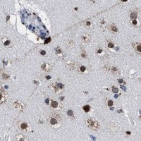
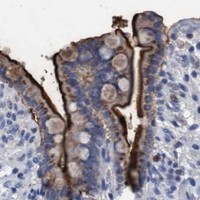
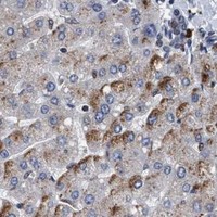
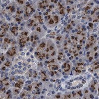


Comprehensive Human Protein Atlas IHC scoring per tissue (reliability: Uncertain — Low consistency between antibody staining and RNA expression data.). Rows are taken directly from the HPA tissue chart — click any row's HPA link to view the source.
| Tissue | Cell type | Level | Evidence | Source |
|---|---|---|---|---|
| Caudate | Neuronal cells | Medium | Protein (IHC) | HPA → |
| Cerebellum | Cells in granular layer | Medium | Protein (IHC) | HPA → |
| Cerebral cortex | Neuronal cells | Medium | Protein (IHC) | HPA → |
| Cervix | Squamous epithelial cells | Medium | Protein (IHC) | HPA → |
| Duodenum | Glandular cells | Medium | Protein (IHC) | HPA → |
| Tissue | Cell type | Level | Evidence | Source |
|---|---|---|---|---|
| Adipose tissue | Adipocytes | Not detected | Protein (IHC) | HPA → |
| Appendix | Glandular cells | Not detected | Protein (IHC) | HPA → |
| Bone marrow | Hematopoietic cells | Not detected | Protein (IHC) | HPA → |
| Bronchus | Respiratory epithelial cells | Not detected | Protein (IHC) | HPA → |
| Lymph node | Germinal center cells | Not detected | Protein (IHC) | HPA → |
Troubleshoot GRB14 staining in paraffin sections by checking retrieval, compartment pattern, controls and scoring against the supplied IHC evidence.
A04177-2 has IHC data from human renal cancer paraffin sections and IF data from A549 cells (catalog image captions); its listed reactivity is human (catalog reactivity).
A04177-2 is listed for IHC and IF/ICC (catalog applications). Its IHC image shows human renal cancer paraffin sections, and its IF image shows A549 cells (catalog image captions).
Which to pick: For tissue IHC, choose A04177-2: its image shows paraffin sections with EDTA retrieval at pH 8.0; the fixative is unreported (catalog IHC image caption). For IF/ICC, the same SKU has an A549 cell IF image and an IF/ICC application listing (catalog IF image caption; catalog applications). No cross-species option is supported by this catalog: A04177-2 lists human reactivity only, and clonality is unreported (catalog reactivity; catalog clone field).