GRB2 / Growth factor receptor-bound protein 2 · IHC design guide

Design Immunohistochemistry for GRB2

Plan chromogenic GRB2 IHC in paraffin sections using the nuclear and cytoplasmic tissue pattern as a reference (HPA tissue IHC). This guide covers fixation consistency, antibody titration and interpretation of strong germinal center staining (HPA tissue IHC).

Evidence assembled Oct 2026 · For research use; verify linked source records and product datasheet before use
Immunohistochemistry protocol sheet for GRB2 (IHC for GRB2): expected localisation Nuclear and cytoplasmic staining in most tissues (HPA tissue IHC), antibody A00351, validated IHC image, and IHC protocol steps
Printable GRB2 IHC protocol sheet — expected localisation Nuclear and cytoplasmic staining in most tissues (HPA tissue IHC), antibody A00351, controls and protocol steps. Open the full GRB2 IHC guide →

GRB2 Immunohistochemistry Experimental Design Guide

Expected localisation, validated protocols, controls and antibodies — the at-a-glance facts below, then the full design guide.

Must know before staining
Expected localisation Nuclear and cytoplasmic staining in most tissues (HPA tissue IHC)
Staining pattern Nuclear/cytoplasmic signal; strong in germinal centers (HPA tissue IHC)
Antigen retrieval Tris-EDTA pH 9.0 HIER, 95–98 °C, 20 min (rule: nuclear antigen)
Positive control ⓘ Appendix+4 more · see all
Negative control ⓘ Heart muscle
Important caveats
Reasons your staining may differ from the expected pattern.
Fixation Keep formalin fixation consistent across sections (standard IHC practice; not target-specific)
Caveat Staining and RNA show medium consistency (HPA tissue IHC)
Regulation No expression regulator annotated (UniProt)
Isoform / epitope 2 isoforms; epitope differences unspecified (UniProt)
Section 1

Recommended GRB2 IHC & IF Protocols

The catalog antibody’s IHC-P protocol is paired with published GRB2 staining methods for endometrium, liver and a pancreatic cancer tissue microarray (PMC11880417; PMC3877332; PMC7901901).

Recommended immunohistochemistry (IHC-P) protocol parameters
SampleFFPE human lung carcinoma tissue (datasheet A00351)
FixationImage formalin-fixed; duration unreported (datasheet A00351); verify before use.
Sectioning4–5 µm sections on charged slides (standard)
DeparaffinisationXylene, graded ethanol series to water (standard)
Antigen retrievalHeat-induced epitope retrieval in Tris-EDTA buffer, pH 9.0, 20 min at 95–98 °C (standard rule: nuclear antigen)
Peroxidase block3% H2O2, 10 min, room temperature (standard)
Blocking10% normal serum of the secondary host, 30 min, room temperature (standard)
Primary antibodyRabbit anti-GRB2, 1:10-1:50 (datasheet A00351)
Primary incubationOvernight at 4 °C (standard)
DetectionHRP-polymer secondary, DAB chromogen 5–10 min (standard)
CounterstainHematoxylin, blue, dehydrate and mount (standard)
Expected resultGRB2-positive staining in lymphoid tissue of appendix (HPA tissue IHC: High). HPA tissue profile: Nuclear and cytoplasmic expression in most tissues, including lymphoid tissue and CNS. No signal in the no-primary control.
💡Decision noteStart with Tris-EDTA pH 9.0 retrieval at 95–98 °C for 20 min (page retrieval); published citrate pH 6.0 methods offer a comparison (PMC3877332; PMC7901901).
Section 2

What Is the Expected GRB2 Staining Pattern?

In paraffin-section IHC, expect nuclear and cytoplasmic GRB2 staining across many tissues, including lymphoid tissue and CNS (HPA: tissue IHC profile). Germinal center cells, Purkinje cells and decidual cells are reported high (HPA: tissue IHC). GRB2 has no transmembrane segment (UniProt P62993). HPA rates the tissue IHC pattern Approved, with medium consistency between staining and RNA expression (HPA: tissue IHC reliability).

What am I looking at on my slide?
Nuclear and cytoplasmic staining in germinal center cells of lymph node or tonsil.This matches the reported compartments and high staining in those cells (HPA: tissue IHC). Judge intensity against cells on the same section; HPA levels are descriptive, not an absolute scoring threshold.
Staining appears exclusively as a sharp cell-surface rim or in extracellular material.Recheck specificity and section morphology. GRB2 lacks a transmembrane segment (UniProt P62993), while HPA reports nuclear and cytoplasmic tissue staining (HPA: tissue IHC). Receptor association alone does not predict a continuous membrane rim (UniProt P62993: subunit).
Strong signal predominates in cells outside the expected pattern, such as cardiomyocytes.Consider cross-reactivity or endogenous chromogen activity (standard IHC practice). HPA reports cardiomyocytes as not detected (HPA: heart muscle IHC), but that observation is an assay-specific comparator, not proof that every cardiomyocyte must be negative.
A broad haze covers cells and extracellular space without readable cell boundaries.Treat this as background until controls resolve it (standard IHC practice). It cannot establish the nuclear and cytoplasmic pattern reported for GRB2 (HPA: tissue IHC); inspect a no-primary control and adjust detection or washing as needed.
No interpretable signal appears in a lymph node germinal center or cerebellar Purkinje cells.A run failure or insufficient assay sensitivity is possible (standard IHC practice), because both sites are reported high (HPA: tissue IHC). Check tissue preservation, antibody conditions and detection controls before calling the sample GRB2-negative.
💡Expected GRB2 appearanceA convincing positive shows nuclear and cytoplasmic signal in expected cells, with high staining possible in germinal center cells or Purkinje cells (HPA: tissue IHC); diffuse haze or an isolated extracellular signal is suspect (standard IHC practice).
How each factor affects the staining
Tissue and cell selectionUse reported high sites, such as tonsil germinal center cells or cerebellar Purkinje cells, as pattern comparators (HPA: tissue IHC). Medium staining in bone marrow hematopoietic cells is also reported (HPA: tissue IHC); do not demand identical intensity across tissues.
Strength of IHC validationHPA calls tissue IHC Approved and describes medium staining–RNA consistency (HPA: tissue IHC reliability). Its listed IHC antibody, CAB002589, is Approved (HPA: antibody validation); this supports interpretation with controls, not a claim of Enhanced IHC validation.
Topology and receptor associationGRB2 has no transmembrane segment and associates with activated, phosphorylated receptors (UniProt P62993: topology, subunit). A local receptor-associated signal may be plausible, but a continuous membrane-only pattern does not match HPA's tissue IHC summary (HPA: tissue IHC).
Isoforms and processingUniProt lists 2 isoforms, no signal peptide or propeptide, and one annotated chain spanning residues 1–217 (UniProt P62993). The payload gives no antibody epitope, so it cannot establish whether the IHC antibody detects both isoforms.
IF/ICC cross-check: should the pattern look identical?HPA IF/ICC reports mainly cytosol, with an additional centrosome location (HPA: subcellular). That evidence uses a separate ICC-Supported antibody, HPA003991 (HPA: antibody validation); compare compartment trends without treating IF/ICC as an IHC protocol or an identical readout.
Why is my staining missing, weak or wrong?
SituationLikely causeNext action
No signal in a reported high-staining control tissue.The assay or detection may have failed; a negative result conflicts with the HPA comparator (HPA: tissue IHC).Check the control section and detection reagents, then review the antibody dilution and the chosen retrieval conditions (standard IHC practice). No GRB2-specific retrieval requirement is supplied.
Diffuse brown haze obscures tissue structure.Nonspecific binding, excess detection signal or inadequate washing can raise background (standard IHC practice).Compare a no-primary control; review blocking, washes and detection exposure (standard IHC practice). Score GRB2 only where cellular compartments remain distinguishable.
Signal is nuclear only throughout the section.This omits the cytoplasmic component of the reported tissue pattern (HPA: tissue IHC); counterstain or detection conditions may affect visual interpretation (standard IHC practice).Inspect higher magnification and compare a reported high-staining tissue with the same staining run (HPA: tissue IHC; standard IHC practice).
Cardiomyocytes stain strongly while expected positive cells do not.This reverses the reported HPA comparison: cardiomyocytes are not detected, while several other cell groups stain high (HPA: tissue IHC). Cross-reactivity or endogenous detection activity is possible (standard IHC practice).Run no-primary and appropriate detection controls, then reassess antibody specificity before assigning a biological explanation (standard IHC practice).
A crisp membrane rim is the only positive feature.A membrane-only pattern conflicts with HPA's nuclear and cytoplasmic IHC summary (HPA: tissue IHC), although GRB2 can associate with phosphorylated receptors (UniProt P62993: subunit).Check cell boundaries, compare reported positive tissue and examine no-primary staining before scoring the rim as GRB2 (HPA: tissue IHC; standard IHC practice).
IF/ICC shows a centrosomal spot that is hard to recognize in chromogenic IHC.The centrosome is an additional IF/ICC location, while tissue IHC is summarized as nuclear and cytoplasmic (HPA: subcellular; HPA: tissue IHC).Interpret the IHC section against its tissue IHC pattern and controls; use the separate IF/ICC evidence only as a compartment cross-check (HPA: tissue IHC, subcellular).

Sample controls for GRB2 IHC & IF

🧪Run tonsil first: germinal center cells should stain for GRB2 (HPA: High in tonsil germinal center cells). Use heart muscle cardiomyocytes as the negative tissue (HPA: Not detected); on the tonsil slide, cells without specific staining should show only background, although HPA does not identify a defined internal negative cell population.
Positive control tissue: Appendix (Lymphoid tissue, HPA High)
Negative control tissue: Heart muscle (HPA Not detected)
ICC-IF cell lines (HPA subcellular resource): HPA ICC-IF images show GRB2 in HEL, THP-1, U2OS, with annotated localisation: Cytosol (supported) (HPA subcellular).
Technical controls: Run a no-primary, secondary-only control and a control matched to the primary antibody’s host and isotype, or host immunoglobulin for a polyclonal antibody; use GRB2-knockout material or immunizing-peptide competition where available (standard IHC practice). Quench endogenous peroxidase for tonsil DAB staining, and block endogenous biotin if using avidin–biotin detection (standard IHC practice).
⚠️Feasibility: The A00351 tissue-IHC caption explicitly describes formalin-fixed, paraffin-embedded lung carcinoma, but reports no target-specific fixation window or fixation effect (catalog antibody caption: A00351). Retrieval dependency is unreported, and the supplied evidence does not establish whether frozen sections or IF would be easier (supplied target/application evidence). In tonsil, endogenous peroxidase can create misleading DAB background; assess the no-primary section alongside the stained section (standard IHC practice).

HPA tissue IHC evidence for GRB2

Comprehensive Human Protein Atlas IHC scoring per tissue (reliability: Approved — Medium consistency between antibody staining and RNA expression data.). Rows are taken directly from the HPA tissue chart — click any row's HPA link to view the source.

Positive expression · recommended positive controls

TissueCell typeLevelEvidenceSource
Appendix Lymphoid tissue High Protein (IHC) HPA →
Cerebellum Purkinje cells High Protein (IHC) HPA →
Lymph node Germinal center cells High Protein (IHC) HPA →
Placenta Decidual cells High Protein (IHC) HPA →
Tonsil Germinal center cells High Protein (IHC) HPA →

Undetected expression · recommended negative controls

TissueCell typeLevelEvidenceSource
Heart muscle Cardiomyocytes Not detected Protein (IHC) HPA →
Section 3

Advanced GRB2 IHC Tips

Troubleshoot GRB2 staining in paraffin sections by checking retrieval, compartment patterns, controls, and scoring before interpreting biological differences.

How should I retrieve GRB2 in paraffin sections when nuclear staining is weak?
Start with Tris-EDTA at pH 9.0 for heat-induced epitope retrieval at 95–98 °C for 20 min (page retrieval specification). If nuclear staining remains weak, compare a milder retrieval condition on adjacent sections while keeping antibody detection and imaging constant (standard IHC practice). GRB2 has documented nuclear and cytoplasmic localisation, so assess both compartments against tissue morphology rather than treating a nuclear signal alone as proof of specificity (UniProt P62993 localisation; HPA tissue IHC). Include a known positive section and a no-primary control in each comparison; excessive retrieval can damage morphology and make apparent gains in staining difficult to interpret (standard IHC practice).
How can I troubleshoot uneven GRB2 staining after tissue fixation?
GRB2-specific sensitivity to fixation duration is unknown from the supplied evidence; assess uneven staining without assigning it to a target-specific fixation effect (supplied fixation evidence). Record fixative, time before fixation, fixation duration, section thickness, and processing batch, then compare sections processed together (standard IHC practice). Examine whether weak areas coincide with poor nuclear detail, tissue folds, or uneven reagent coverage, using the counterstain and a no-primary control to separate morphology problems from detection background (standard IHC practice). Reprocess or recut affected material where feasible, and retain a consistently processed positive control before interpreting differences between specimens as changes in GRB2 expression (standard IHC practice).
Should GRB2 staining be nuclear, cytoplasmic, or concentrated near intracellular structures?
Assess nuclear and cytoplasmic staining separately: both occur in tissue IHC, while UniProt also lists endosome and Golgi localisation (HPA tissue IHC; UniProt P62993 localisation). GRB2 has no transmembrane segment, so a continuous membrane-only outline warrants scrutiny rather than automatic assignment to GRB2 (UniProt P62993 topology). Review the counterstain and compare intact cells with tissue edges and folds, where deposited chromogen can obscure compartment boundaries (standard IHC practice). HPA cell imaging reports mainly cytosolic signal with an additional centrosomal location, but use that observation as context rather than a required pattern for every paraffin section (HPA subcellular).
Could epitope location explain different GRB2 staining patterns between antibodies?
GRB2 has 2 annotated isoforms and contains SH3 domains at residues 1–58 and 156–215, with an SH2 domain at 60–152 (UniProt P62993 isoforms and domains). Its listed modifications include acetylation and phosphorylation at Tyr209, but the supplied evidence does not map the catalog antibody’s epitope or establish modification-dependent staining (UniProt P62993 modified residues; supplied antibody evidence). Compare each antibody’s stated immunogen, isoform coverage, and IHC validation before treating differences as biology (standard IHC practice). If those details are unavailable, test antibodies on adjacent sections with matched retrieval and controls, and describe discordant staining as unresolved antibody-dependent evidence (standard IHC practice).
How should I adapt GRB2 localisation checks to multiplex IF?
Use a marker for the expected cell population in a separate channel, then assess whether GRB2 signal occurs in those cells and in plausible intracellular compartments (standard IF practice; UniProt P62993 localisation). HPA reports high tissue IHC staining in lymph-node germinal-center cells, which can guide cell identification where that tissue is studied (HPA tissue IHC). Choose fluorophores and acquisition channels after checking the specimen’s autofluorescence, and include single-stain controls to identify spectral bleed-through (standard IF practice). Because GRB2 lacks a transmembrane segment and has cytosolic and nuclear localisation, optimise permeabilisation for access to intracellular epitopes, then verify that cell boundaries and nuclear morphology remain intact (UniProt P62993 topology and localisation; standard IF practice).
What should I check when GRB2 DAB staining appears diffuse or widespread?
Compare stained sections with a no-primary control and inspect whether brown signal tracks tissue edges, folds, necrotic areas, or pigment rather than intact cell compartments (standard IHC practice). Apply a peroxidase block before chromogenic detection and check the control for endogenous enzyme activity; these are general DAB workflow steps, not GRB2-specific validation (standard IHC practice). Optimise blocking, antibody concentration, incubation, and washes on matched sections while keeping retrieval and detection conditions recorded (standard IHC practice). GRB2 can show nuclear and cytoplasmic expression across many tissues, so widespread staining alone cannot establish background; judge it alongside cellular morphology and control staining (HPA tissue IHC; standard IHC practice).
How should I quantify GRB2 staining across paraffin sections? ⚠ ANSWER MARKED FOR VERIFICATION
Define the cell population and score nuclear and cytoplasmic staining separately if both are present, because tissue IHC reports expression in both compartments (HPA tissue IHC; standard IHC practice). Use the percentage of positive cells or an H-score based on staining intensity and positive-cell fraction; for spatial questions, report positive-cell density per mm² of viable tissue (standard IHC practice). Normalise comparisons to the same compartment, cell type, viable tissue area, and staining batch, and apply fixed thresholds using control sections (standard IHC practice). Exclude folds, necrosis, and section edges from scored regions, and report how many cells or regions contributed to each specimen’s estimate (standard IHC practice).
How do I distinguish credible GRB2 staining from artefact?
Look for reproducible staining in intact cells with nuclear or cytoplasmic distribution, consistent with reported tissue patterns, and compare it with a no-primary control (HPA tissue IHC; standard IHC practice). A membrane-only outline is questionable for GRB2 because it has no transmembrane segment, although its interactions with activated receptors can influence where it accumulates (UniProt P62993 topology and subunit). Treat staining confined to edges, folds, or necrotic tissue as suspect, and investigate diffuse DAB signal with a peroxidase-blocked control (standard IHC practice). Interpret cell identity explicitly: HPA reports high staining in germinal-center cells but no detected staining in cardiomyocytes, with medium consistency between antibody staining and RNA data (HPA tissue IHC).
Boster reagents

Best GRB2 / Growth factor receptor-bound protein 2 IHC Antibodies

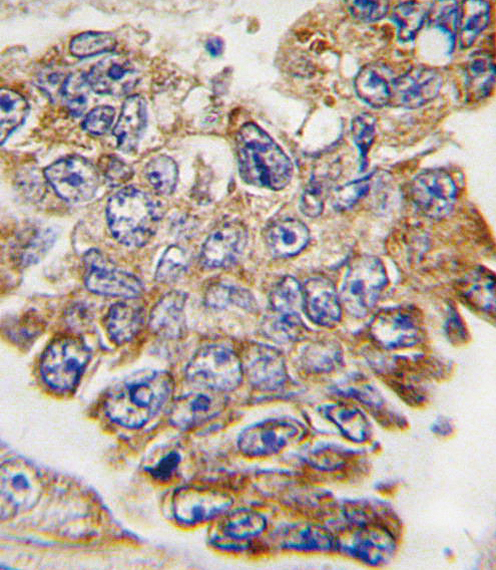
A00351 has a DAB IHC image from formalin-fixed, paraffin-embedded human lung carcinoma (catalog image caption). M00351 and M00351-2 list IHC and IF/ICC for human, mouse, and rat (catalog applications/reactivity).

Real IHC data Formalin-fixed and paraffin-embedded human lung carcinoma tissue reacted with GRB2 Antibody (Y209), which was peroxidase-conjugated to the secondary antibody, followed by DAB staining. This data demonstrates the use of this antibody for immunohistochemistry; clinical relevance has not been evaluated.
Anti-GRB2 Antibody (Y209)
Cat # A00351

A00351 lists human IHC-P and has an IHC image from formalin-fixed, paraffin-embedded human lung carcinoma (catalog applications/reactivity; catalog image caption). M00351 and M00351-2 list IHC and IF/ICC for human, mouse, and rat, but have no IHC or IF images in the payload (catalog applications/reactivity; catalog image arrays).

Which to pick: Choose A00351 for human paraffin-section DAB IHC: its own image documents that preparation and detection, and its IHC-P dilution is 1:10–1:50 (catalog image caption; catalog dilution). For IF/ICC, consider M00351 or M00351-2; both are rabbit monoclonals that list IF/ICC and human, mouse, and rat reactivity, without a supplied IF image (catalog host/clone/applications/reactivity; catalog image arrays). For mouse or rat tissue IHC, those same two SKUs list IHC and cross-species reactivity; their IHC listings do not specify fixation or paraffin processing, and neither has an IHC image in the payload (catalog applications/reactivity; catalog image arrays).

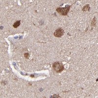
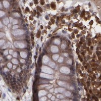
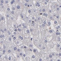
Each figure is that product's own IHC / IF validation image from its datasheet.

References

  1. UniProt Consortium. UniProt entry P62993 (GRB2_HUMAN, Growth factor receptor-bound protein 2).
  2. Human Protein Atlas. GRB2 tissue IHC expression (reliability: Approved).
  3. Human Protein Atlas. GRB2 subcellular location (ICC-IF): Mainly localized to the cytosol. In addition localized to the centrosome..
  4. Human Protein Atlas. GRB2 antibody validation summary (2 antibodies).
  5. GRB2 regulation of essential signaling pathways in the endometrium is critical for implantation and decidualization. Nature communications 2025 — PMC11880417.
  6. Identification of GRB2 and GAB1 coexpression as an unfavorable prognostic factor for hepatocellular carcinoma by a combination of expression profile and network analysis. PloS one 2013 — PMC3877332.
  7. ECT2 Increases the stability of EGFR and Tumorigenicity by Inhibiting Grb2 Ubiquitination in Pancreatic Cancer. Frontiers in oncology 2020 — PMC7901901.
  8. GRB2 Promotes Sorafenib Resistance in Hepatocellular Carcinoma Cells Under Hypoxia by Activating the PI3K/AKT Signaling Pathway. Journal of hepatocellular carcinoma 2026 — PMC13157854.
  9. PubMed PMID:1322798 — UniProt-cited evidence.
  10. PubMed PMID:1384039 — UniProt-cited evidence.
  11. PubMed PMID:8178156 — UniProt-cited evidence.