This website uses cookies to ensure you get the best experience on our website.


- Table of Contents
Plan chromogenic GRHL2 IHC on paraffin sections using the IHC-validated antibody at 2–5 μg/ml (datasheet A04120-2). Assess granular nuclear and cytoplasmic staining with appropriate controls, bearing in mind that the tissue staining profile has uncertain reliability (HPA tissue IHC).

Expected localisation, validated protocols, controls and antibodies — the at-a-glance facts below, then the full design guide.
| Expected localisation | Granular nuclear and cytoplasmic staining (HPA tissue IHC) | |
| Staining pattern | Granular nuclear and cytoplasmic signal in several tissues (HPA tissue IHC) | |
| Antigen retrieval | EDTA pH 8.0 HIER, heat-mediated (datasheet A04120-2) | |
| Positive control | Appendix+4 more · see all | |
| Negative control | Adipose tissue+4 more · see all |
| Fixation | Keep fixation conditions consistent across paraffin sections. (standard IHC practice; not target-specific) | |
| Caveat | Tissue staining has low consistency with RNA expression (HPA tissue IHC) | |
| Regulation | RNA expression is tissue enhanced in skin (HPA tissue RNA) | |
| Isoform / epitope | Two isoforms; confirm epitope coverage (UniProt) |
The catalog antibody protocol uses EDTA pH 8.0 heat retrieval (datasheet A04120-2). The published options below cover mouse intestinal, human pancreatic, and human bladder tissue (PMC13414807; PMC6341798; PMC7127877).
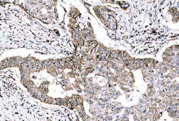
| Sample | Paraffin-embedded human adenocarcinoma of the right colon tissue; fixative not specified (datasheet A04120-2) |
| Fixation | Image fixative and duration unreported (datasheet A04120-2); verify before use. |
| Sectioning | 4–5 µm sections on charged slides (standard) |
| Deparaffinisation | Xylene, graded ethanol series to water (standard) |
| Antigen retrieval | Heat retrieval: EDTA pH 8.0 (datasheet A04120-2); 20 min, 95–100 °C (standard) |
| Peroxidase block | 3% H2O2, 10 min, room temperature (standard) |
| Blocking | 10% goat serum (datasheet A04120-2) |
| Primary antibody | Rabbit anti-GRHL2, 2-5 μg/ml (datasheet A04120-2) |
| Primary incubation | Overnight at 4 °C (datasheet A04120-2) |
| Detection | HRP-conjugated secondary, DAB chromogen (datasheet A04120-2) |
| Counterstain | Hematoxylin, blue, dehydrate and mount (standard) |
| Expected result | GRHL2-positive staining in glandular cells of appendix (HPA tissue IHC: Medium). HPA tissue profile: Granular cytoplasmic and nuclear expression in several tissues. No signal in the no-primary control. |
GRHL2 is primarily nuclear, with detection also reported at cell–cell contacts (UniProt Q6ISB3: subcellular location). In paraffin IHC, expect staining in selected epithelial cells, including bronchial respiratory epithelium and intestinal glands (HPA: medium staining in bronchus, colon and duodenum). HPA also describes granular cytoplasmic and nuclear staining across several tissues, but rates its tissue IHC profile uncertain because staining and RNA expression show low consistency (HPA: tissue IHC).
| Nuclear staining in bronchial respiratory epithelial cells or colon glands, with adjacent cells providing contrast. | This fits GRHL2’s nuclear location and HPA’s medium staining in those cell types (UniProt Q6ISB3: subcellular location; HPA: bronchus and colon). Record the stained cell type and compartment separately. A plausible pattern supports interpretation, but HPA’s tissue IHC reliability is uncertain (HPA: tissue IHC). |
| Predominantly cytoplasmic staining with little or no nuclear staining in an expected epithelial population. | Treat this as a localisation discrepancy: nucleoplasm is the supported ICC-IF location, although HPA tissue IHC describes granular cytoplasmic as well as nuclear staining (HPA: subcellular; HPA: tissue IHC). Review morphology and controls before calling the cytoplasmic signal specific; the tissue profile alone cannot settle it. |
| Strong staining in alveolar cells, adipocytes or liver cholangiocytes. | Those cell populations are listed as not detected in HPA tissue IHC (HPA: lung alveolar cells; adipose adipocytes; liver cholangiocytes). Consider nonspecific antibody binding or endogenous chromogen activity as general IHC possibilities. Check the relevant negative control and confirm which cells carry the signal; absence in HPA is not proof that every specimen must be negative. |
| Diffuse colour across epithelium, stroma and blank areas, without a discernible cellular pattern. | A field-wide deposit is difficult to reconcile with the cell-resolved HPA observations (HPA: tissue IHC). In general IHC practice, background can arise from insufficient blocking, excess antibody or chromogen development, or inadequate washing. Evaluate a no-primary control before interpreting weak compartmental staining. |
| No staining in bronchial respiratory epithelium or colon glands. | HPA reports medium staining in these populations, so their absence warrants a run-level check (HPA: bronchus and colon). Confirm that the expected cells are present, then inspect positive controls, retrieval, detection and antibody dilution as general IHC practice. Because HPA rates tissue IHC uncertain, one negative specimen does not establish biological absence (HPA: tissue IHC). |
| Compartment and cell identity | UniProt places GRHL2 in the nucleus and reports detection at cell–cell contacts; HPA tissue IHC describes granular nuclear and cytoplasmic staining (UniProt Q6ISB3: subcellular location; HPA: tissue IHC). Score nuclear, cytoplasmic and junctional patterns separately, and identify the stained cell population. UniProt lists no transmembrane segment, so a membrane pattern should not be assumed to represent a membrane-spanning form (UniProt Q6ISB3: topology). |
| Tissue-specific evidence and its limit | HPA records medium staining in bronchial respiratory epithelial cells and several glandular populations, but low staining in kidney tubule cells and not detected in lung alveolar cells (HPA: tissue IHC). Specify the cell population when choosing controls; a whole-organ label can hide this distinction. UniProt reports high kidney expression, so kidney should not be treated as an unambiguous strong IHC control (UniProt Q6ISB3: tissue specificity; HPA: kidney). |
| Antibody validation | The HPA tissue IHC profile is rated uncertain for low agreement between staining and RNA data; HPA004820 is also IHC uncertain (HPA: tissue IHC; HPA: antibody validation). This lowers confidence in a tissue-only positive call. HPA062839 has supported ICC-IF validation, which addresses a different application and does not establish IHC-P performance (HPA: antibody validation). |
| Isoforms and epitope information | UniProt lists 2 GRHL2 isoforms and a 1–625 protein chain, with no signal peptide, propeptide or annotated glycosylation sites in this record (UniProt Q6ISB3: isoforms, processing and glycosylation). These facts do not identify either antibody’s epitope or predict retrieval sensitivity. If staining differs between samples, consult the antibody’s documented immunogen and IHC-P instructions before proposing an isoform explanation. |
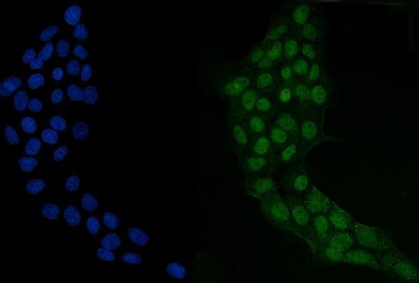
| IF/ICC relevance? | Yes, as a localisation cross-check: HPA supports nucleoplasmic localisation in ICC-IF and lists images from HaCaT, RT-4 and U2OS cells (HPA: subcellular). That evidence helps assess whether an IHC pattern is plausible, but the supported ICC-IF antibody status and cell images do not validate a paraffin IHC result (HPA: antibody validation; HPA: subcellular). |
| Situation | Likely cause | Next action |
|---|---|---|
| The expected epithelium is present, but the slide has no specific signal. | The run may have insufficient detectable signal; HPA reports medium staining in bronchial respiratory epithelium and colon glands, subject to uncertain tissue IHC reliability (HPA: bronchus, colon and tissue IHC). | General IHC practice: check the positive control and detection reagents, then verify retrieval and dilution against the IHC-validated antibody’s instructions. Reassess only after the control performs as expected. |
| Colour appears mainly in cytoplasm and nuclei remain pale. | This differs from supported nucleoplasmic ICC-IF localisation, although granular cytoplasmic tissue staining is reported (HPA: subcellular; HPA: tissue IHC). | Score compartments separately. General IHC practice: compare a no-primary control, review counterstain and cell boundaries, and repeat with an independently validated IHC antibody if the localisation remains decisive. |
| Alveolar cells or adipocytes appear strongly positive. | HPA lists those populations as not detected; nonspecific binding or endogenous detection activity are general IHC possibilities (HPA: lung alveolar cells; adipose adipocytes). | Confirm cell identity and compare no-primary and appropriate detection controls. In general IHC practice, address endogenous enzyme activity when using enzyme-based chromogenic detection. |
| Weak colour covers both cells and blank slide regions. | The lack of a cell-resolved pattern limits interpretation against HPA’s tissue observations (HPA: tissue IHC). Excess reagent, incomplete blocking or inadequate washing are general IHC causes. | Use a no-primary control to locate background. General IHC practice: review blocking, washes, antibody dilution and chromogen development before assigning any weak cellular signal. |
| Kidney tubules stain weakly despite an expectation of high kidney expression. | UniProt reports high kidney expression, while HPA tissue IHC records low staining in tubule cells; the measurements do not imply the same IHC intensity (UniProt Q6ISB3: tissue specificity; HPA: kidney). | Record the observed tubule pattern and compare a separate epithelial positive control. Avoid treating kidney RNA or broad expression descriptions as a fixed staining-intensity threshold. |
| A positive field contains mixed stained and unstained cell populations. | HPA reports GRHL2 staining by cell type within tissues, and its tissue IHC reliability is uncertain (HPA: tissue IHC). Apparent heterogeneity can also reflect cell identification or local technical variation in general IHC practice. | Identify the stained cells on the counterstain, score their compartment and intensity separately, and compare another area plus the run controls before calling biological heterogeneity. |
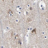


Comprehensive Human Protein Atlas IHC scoring per tissue (reliability: Uncertain — Low consistency between antibody staining and RNA expression data.). Rows are taken directly from the HPA tissue chart — click any row's HPA link to view the source.
| Tissue | Cell type | Level | Evidence | Source |
|---|---|---|---|---|
| Appendix | Glandular cells | Medium | Protein (IHC) | HPA → |
| Bronchus | Respiratory epithelial cells | Medium | Protein (IHC) | HPA → |
| Caudate | Neuronal cells | Medium | Protein (IHC) | HPA → |
| Cerebellum | Cells in granular layer | Medium | Protein (IHC) | HPA → |
| Cerebral cortex | Neuronal cells | Medium | Protein (IHC) | HPA → |
| Tissue | Cell type | Level | Evidence | Source |
|---|---|---|---|---|
| Adipose tissue | Adipocytes | Not detected | Protein (IHC) | HPA → |
| Adrenal gland | Glandular cells | Not detected | Protein (IHC) | HPA → |
| Fallopian tube | Glandular cells | Not detected | Protein (IHC) | HPA → |
| Liver | Cholangiocytes | Not detected | Protein (IHC) | HPA → |
| Lung | Alveolar cells | Not detected | Protein (IHC) | HPA → |
Troubleshoot GRHL2 staining in paraffin sections by checking retrieval, nuclear localisation, cell type and controls before interpreting chromogenic signal (UniProt Q6ISB3; HPA tissue IHC).
A04120-2 has human paraffin-section IHC images and A431-cell IF data (catalog: A04120-2 applications and image captions).
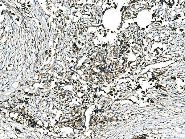
A04120-2 has IHC images from human paraffin-embedded right-colon adenocarcinoma, breast cancer, placenta, and stomach cancer sections (catalog: A04120-2 IHC captions). The same SKU has an IF image from A431 cells (catalog: A04120-2 IF caption).
Which to pick: Choose A04120-2 for human paraffin-section IHC; its right-colon adenocarcinoma caption documents 2 μg/ml primary antibody after heat retrieval in EDTA at pH 8.0, but does not report the fixative (catalog: A04120-2 IHC caption). For IF/ICC, A04120-2 is also listed for both applications, with A431-cell IF shown at 5 μg/ml (catalog: A04120-2 applications and IF caption). No cross-species choice is supported because the catalog lists human reactivity only; the antibody’s clonality is unreported (catalog: A04120-2 reactivity and clone fields).