GRK2 / G protein-coupled receptor kinase 2 · IHC design guide

Design Immunohistochemistry for GRK2

Plan chromogenic GRK2 IHC on paraffin sections using the IHC-validated antibody A01473-1 at 0.5–1 μg/mL (datasheet: A01473-1). Use immune-rich tissue as a positive reference and score cytoplasmic staining by cell type (HPA tissue IHC).

Evidence assembled Oct 2026 · For research use; verify linked source records and product datasheet before use
Immunohistochemistry protocol sheet for GRK2 (IHC for GRK2): expected localisation Cytoplasmic staining in tissue sections (HPA tissue IHC), antibody A01473-1, validated IHC image, and IHC protocol steps
Printable GRK2 IHC protocol sheet — expected localisation Cytoplasmic staining in tissue sections (HPA tissue IHC), antibody A01473-1, controls and protocol steps. Open the full GRK2 IHC guide →

GRK2 Immunohistochemistry Experimental Design Guide

Expected localisation, validated protocols, controls and antibodies — the at-a-glance facts below, then the full design guide.

Must know before staining
Expected localisation Cytoplasmic staining in tissue sections (HPA tissue IHC)
Staining pattern Cytoplasmic staining, most abundant in immune cells (HPA tissue IHC)
Antigen retrieval Citrate pH 6 HIER, heat-mediated (datasheet A01473-1)
Positive control ⓘ Appendix+4 more · see all
Negative control ⓘ Adipose tissue+4 more · see all
Important caveats
Reasons your staining may differ from the expected pattern.
Fixation Keep fixation consistent across sections (standard IHC practice; not target-specific); Matched tissue-IHC evidence does not establish the fixation claim. Validate the specimen-specific method before use. (selected-SKU IHC image A01473-1)
Caveat Immune-cell abundance can skew tissue-wide scoring (HPA tissue IHC)
Regulation Signal varies with immune-cell content (HPA tissue IHC)
Isoform / epitope 0 annotated isoforms; one 1–689 chain (UniProt)
Section 1

Recommended GRK2 IHC & IF Protocols

The catalog antibody’s IHC-P protocol (datasheet A01473-1) is accompanied by three published GRK2 IHC protocols (PMC10182096; PMC7931601; PMC6337949).

Recommended immunohistochemistry (IHC-P) protocol parameters
SampleParaffin-embedded human Lung cancer tissues; fixative not specified (datasheet A01473-1)
FixationImage fixative and duration unreported (datasheet A01473-1); verify before use.
Sectioning4–5 µm sections on charged slides (standard)
DeparaffinisationXylene, graded ethanol series to water (standard)
Antigen retrievalHeat retrieval: Citrate pH 6, 20 min (datasheet A01473-1)
Peroxidase block3% H2O2, 10 min, room temperature (standard)
Blocking10% goat serum (datasheet A01473-1)
Primary antibodyRabbit anti-GRK2, 0.5-1μg/ml (datasheet A01473-1)
Primary incubationOvernight at 4 °C (datasheet A01473-1)
DetectionStreptavidin-biotin complex (SABC), DAB chromogen (datasheet A01473-1)
CounterstainHematoxylin, blue, dehydrate and mount (standard)
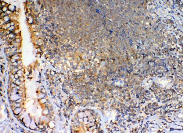
Expected resultGRK2-positive staining in lymphoid tissue of appendix (HPA tissue IHC: High). HPA tissue profile: Cytoplasmic expression in all tissues, most abundant in immune cells. No signal in the no-primary control.
💡Decision noteStart with heat-mediated citrate pH 6 retrieval (datasheet A01473-1); the mouse pancreas protocol also uses citrate pH 6 retrieval (PMC7931601).
Section 2

What Is the Expected GRK2 Staining Pattern?

GRK2 should show predominantly cytoplasmic staining across tissues, with the strongest signal in immune cells (HPA tissue IHC). Peripheral blood leukocyte expression supports this pattern (UniProt P25098). GRK2 has no transmembrane segment, although UniProt also lists cell membrane, cilium membrane and synaptic locations (UniProt P25098 topology and subcellular location). HPA rates its tissue IHC profile Enhanced, with medium consistency between staining and RNA data (HPA tissue IHC).

What am I looking at on my slide?
Strong cytoplasmic staining in lymphoid or hematopoietic cells.This fits High staining in appendix lymphoid tissue, bone marrow hematopoietic cells and lymph node germinal center cells (HPA tissue IHC). Compare the same cell population across sections before judging intensity (standard IHC practice).
Predominantly nuclear staining, with little cytoplasmic signal.This conflicts with the cytoplasmic tissue IHC profile (HPA tissue IHC). Check the negative control and repeat staining before assigning a nuclear GRK2 pattern (standard IHC practice).
Strong staining in adipocytes or parathyroid glandular cells.Both cell populations are listed as Not detected (HPA tissue IHC). Consider nonspecific antibody binding or endogenous detection activity; assess each with an appropriate control (standard IHC practice).
Diffuse color across cells and surrounding tissue, without clear boundaries.An uninterpretable background pattern does not establish GRK2 localization (standard IHC practice). Review blocking, washes and the negative control before scoring cytoplasmic staining (standard IHC practice).
No signal in a lymph node germinal center or bone marrow hematopoietic cells.These are High staining cell populations (HPA tissue IHC). Check tissue preservation, retrieval and detection controls before interpreting a study sample as negative (standard IHC practice).
💡Expected GRK2 appearanceA convincing positive is predominantly cytoplasmic staining, strongest in the specified immune cell populations (HPA tissue IHC); dominant nuclear color or strong staining in HPA Not detected cells warrants control checks (HPA tissue IHC; standard IHC practice).
How each factor affects the staining
Cell population and tissueHigh staining is recorded in appendix lymphoid tissue, bone marrow hematopoietic cells, lymph node and tonsil germinal center cells, and splenic red pulp cells (HPA tissue IHC). Score the named cells, rather than treating every cell in those tissues as equally positive (standard IHC practice).
Reference low and negative populationsCardiomyocytes and skeletal myocytes are Low, while adipocytes and parathyroid glandular cells are Not detected (HPA tissue IHC). These are cell-specific reference levels, not whole-tissue negative calls (HPA tissue IHC).
Compartment and molecular formThe tissue IHC profile is cytoplasmic (HPA tissue IHC). GRK2 has no transmembrane segment, signal peptide or propeptide, and the listed chain spans residues 1–689 (UniProt P25098); the record supplies no separate processed form to score.
Strength of antibody evidenceHPA assigns IHC Enhanced to CAB005021 and IHC Approved to CAB037248 (HPA antibody records). Those grades apply to the listed antibodies; they do not validate an unlisted catalog antibody (HPA antibody records).
IF/ICC Q&A: what pattern is expected?HPA reports mainly vesicular GRK2, with additional Golgi and cytosolic localization in ICC-IF (HPA subcellular). This is an IF localization reference; tissue IHC should be judged against the cytoplasmic tissue profile (HPA tissue IHC).
Why is my staining missing, weak or wrong?
SituationLikely causeNext action
High reference cells are blank.The result conflicts with High staining in bone marrow hematopoietic or lymph node germinal center cells (HPA tissue IHC).Verify the tissue contains those cells, then check retrieval and detection with appropriate controls (standard IHC practice).
Signal is mainly nuclear.The compartment conflicts with the cytoplasmic tissue profile (HPA tissue IHC).Review the negative control and repeat with an independently validated IHC antibody if available (standard IHC practice).
Adipocytes stain strongly.Adipocytes are listed as Not detected (HPA tissue IHC); nonspecific binding or detection background is possible (standard IHC practice).Compare adjacent morphology and negative controls before calling the signal GRK2 (standard IHC practice).
Color develops in a control lacking primary antibody.Endogenous detection activity or reagent background can generate chromogenic signal (standard IHC practice).Check the detection control and address endogenous activity according to the detection system (standard IHC practice).
Diffuse background obscures cytoplasm.Residual detection reagent or insufficient blocking can reduce contrast (standard IHC practice).Review blocking, washes and detection exposure; rescore only where cells remain distinguishable (standard IHC practice).
A low-intensity population is called negative.Cardiomyocytes and skeletal myocytes are Low, rather than Not detected (HPA tissue IHC).Use the same scoring threshold across sections and compare with a High immune-cell reference (HPA tissue IHC; standard IHC practice).

Sample controls for GRK2 IHC & IF

🧪Run spleen first: cells in its red pulp should stain for GRK2 (HPA: High in cells in red pulp). Use adipose tissue as the negative tissue, focusing on adipocytes (HPA: Not detected in adipocytes). On the spleen slide, internal negative cells should show counterstain without specific chromogen; the supplied HPA rows do not identify a validated internal negative cell type.
Positive control tissue: Appendix (Lymphoid tissue, HPA High)
Negative control tissue: Adipose tissue (HPA Not detected)
ICC-IF cell lines (HPA subcellular resource): HPA ICC-IF images show GRK2 in CACO-2, REH, U2OS, with annotated localisation: Vesicles (approved) (HPA subcellular).
Technical controls: Include a no-primary, secondary-only control; a rabbit IgG isotype control matched to the primary’s class, clonality and concentration where known; and GRK2 knockout tissue or peptide competition if available (caption: rabbit primary; standard IHC controls). Block endogenous peroxidase and check for blood-associated background in splenic red pulp before interpreting DAB staining (standard IHC practice).
⚠️Feasibility: A target-specific fixation window or fixation effect is unreported, and the selected A01473-1 paraffin-section caption does not state a fixative (selected-SKU caption). That caption used heat retrieval in citrate buffer at pH 6 for 20 minutes, but it does not establish that retrieval is required (selected-SKU caption). The supplied evidence does not establish frozen sections or IF as easier; splenic red pulp warrants attention to endogenous peroxidase background (standard IHC practice).

HPA tissue IHC evidence for GRK2

Comprehensive Human Protein Atlas IHC scoring per tissue (reliability: Enhanced — Medium consistency between antibody staining and RNA expression data.). Rows are taken directly from the HPA tissue chart — click any row's HPA link to view the source.

Positive expression · recommended positive controls

TissueCell typeLevelEvidenceSource
Appendix Lymphoid tissue High Protein (IHC) HPA →
Bone marrow Hematopoietic cells High Protein (IHC) HPA →
Lymph node Germinal center cells High Protein (IHC) HPA →
Spleen Cells in red pulp High Protein (IHC) HPA →
Tonsil Germinal center cells High Protein (IHC) HPA →

Undetected expression · recommended negative controls

TissueCell typeLevelEvidenceSource
Adipose tissue Adipocytes Not detected Protein (IHC) HPA →
Caudate Glial cells Not detected Protein (IHC) HPA →
Hippocampus Glial cells Not detected Protein (IHC) HPA →
Parathyroid gland Glandular cells Not detected Protein (IHC) HPA →
Salivary gland Glandular cells Not detected Protein (IHC) HPA →
Section 3

Advanced GRK2 IHC Tips

Use the catalog antibody’s paraffin section result and GRK2 expression patterns to guide chromogenic IHC controls, localisation checks, and scoring.

What retrieval should I start with for GRK2 in paraffin sections?
Start with heat-mediated citrate buffer at pH 6 for 20 minutes (datasheet A01473-1). The selected paraffin-section image used this retrieval before an overnight incubation at 4°C with 1 μg/mL antibody, so reproduce those conditions when assessing an initial result (datasheet A01473-1). If staining is weak, compare retrieval duration on adjacent sections while keeping antibody concentration and chromogenic detection constant; include a known positive immune-cell region to judge recovery (HPA: cytoplasmic expression most abundant in immune cells; standard IHC practice). Excessively strong retrieval can damage section morphology, so assess tissue integrity and background alongside signal (standard IHC practice).
How should I troubleshoot fixation-related loss of GRK2 staining?
Target-specific fixation sensitivity is unknown: the selected image identifies a paraffin section but does not report its fixative (datasheet A01473-1: fixative not stated). Record the fixative and fixation duration for each specimen, then compare sections processed under matched conditions before changing retrieval or antibody concentration (standard IHC practice). Use the documented citrate pH 6, 20-minute retrieval as the starting point, and include a positive-control region in each run (datasheet A01473-1; HPA: high staining in lymphoid tissue of appendix). If a specimen remains weak, evaluate its morphology and processing history before assigning the result to low GRK2 expression (standard IHC practice).
Where should convincing GRK2 staining appear in tissue sections?
Expect predominantly cytoplasmic staining across tissues, with stronger expression in immune cells (HPA: tissue IHC profile). GRK2 is also annotated in the cytoplasm, at the cell membrane, and at synaptic and ciliary sites; it has no transmembrane segment (UniProt P25098: subcellular location and topology). Vesicles are the main approved location in cell imaging, with Golgi and cytosolic signal also reported, so a granular cytoplasmic pattern can be plausible (HPA: subcellular). Compare suspected membrane-associated staining with cytoplasmic signal and tissue architecture, and avoid calling an isolated DAB rim specific without appropriate controls (standard IHC practice; UniProt P25098: subcellular location).
Could epitope choice explain discordant GRK2 staining patterns?
The supplied record lists 0 isoforms and one 1–689 protein chain, so an isoform-specific staining explanation lacks support here (UniProt P25098: isoforms and processing). GRK2 contains RGS, kinase, AGC-kinase C-terminal, and PH regions, plus a reported phosphoserine at residue 670 (UniProt P25098: domains and modified residues). The catalog antibody’s recognized epitope is not supplied, so these features cannot establish whether fixation or retrieval masks its binding site (datasheet A01473-1: epitope not specified; standard IHC practice). For discordant regions, compare an independently validated antibody and appropriate loss-of-target material, if available, under matched staining conditions (standard IHC practice).
How can IF help assess an ambiguous GRK2 IHC pattern?
Use IF as a separate localisation check when chromogenic IHC leaves the stained cell population uncertain (standard IHC/IF practice). Multiplex GRK2 with an immune-cell marker such as CD45 when assessing immune-rich regions, since tissue IHC reports the greatest GRK2 abundance in immune cells (HPA: tissue IHC profile; standard IF practice). Choose a far-red fluorophore and inspect an unstained section in the same channels to assess tissue autofluorescence before interpreting overlap (standard IF practice). GRK2 lacks a transmembrane segment, but the antibody epitope is unspecified; optimise permeabilisation for a cytosolic epitope only if that epitope assignment is established (UniProt P25098: topology; datasheet A01473-1: epitope not specified; standard IF practice).
How do I distinguish GRK2 signal from chromogenic background?
The selected result used 10% goat serum blocking, a biotinylated secondary antibody, an avidin-biotin detection complex, and DAB (datasheet A01473-1). Include a no-primary control and check endogenous peroxidase blocking when diffuse DAB appears; these are general chromogenic IHC controls, not GRK2-specific findings (standard IHC practice). Because the demonstrated workflow uses biotin-based detection, assess endogenous biotin contribution if control sections retain staining, especially before increasing antibody concentration (datasheet A01473-1; standard IHC practice). Compare background with the expected cytoplasmic, immune-cell-enriched distribution and preserve identical development conditions across comparison sections (HPA: tissue IHC profile; standard IHC practice).
How should I score GRK2 staining across specimens? ⚠ ANSWER MARKED FOR VERIFICATION
Define the cell population and viable tissue region before scoring, because immune cells can contribute disproportionately to GRK2 staining (HPA: cytoplasmic expression most abundant in immune cells; standard IHC practice). For cellular intensity, report an H-score from 0–300, calculated as 1 × % weak + 2 × % moderate + 3 × % strong, with thresholds fixed across specimens (standard IHC practice). Alternatively report percent positive cells within each cell type or positive-cell density per mm² of viable tissue (standard IHC practice). Normalise counts to the corresponding total viable cell count or analysed viable area, and exclude necrosis and section edges consistently (standard IHC practice).
When should an apparent GRK2-positive region be considered artefactual?
A credible result should fit the reported cytoplasmic, immune-cell-enriched tissue pattern, while allowing for annotated membrane-associated GRK2 (HPA: tissue IHC profile; UniProt P25098: subcellular location). Strong staining confined to section edges or necrotic areas warrants suspicion because these regions can accumulate nonspecific chromogen (standard IHC practice). Nuclear-only staining or signal in a cell population unexpected from the tissue context needs independent validation before assignment to GRK2 (HPA: tissue IHC profile; standard IHC practice). Check a no-primary section for endogenous enzyme or detection-system signal, then compare candidate positives with tissue morphology and an appropriate positive-control region processed in the same run (standard IHC practice).
Boster reagents

Best GRK2 / G protein-coupled receptor kinase 2 IHC Antibodies

GRK2 antibody data cover human lung cancer and mouse and rat spleen paraffin sections (A01473-1 IHC captions), plus U20S immunocytochemical samples (A01473-1 and A32388-1 IF captions).

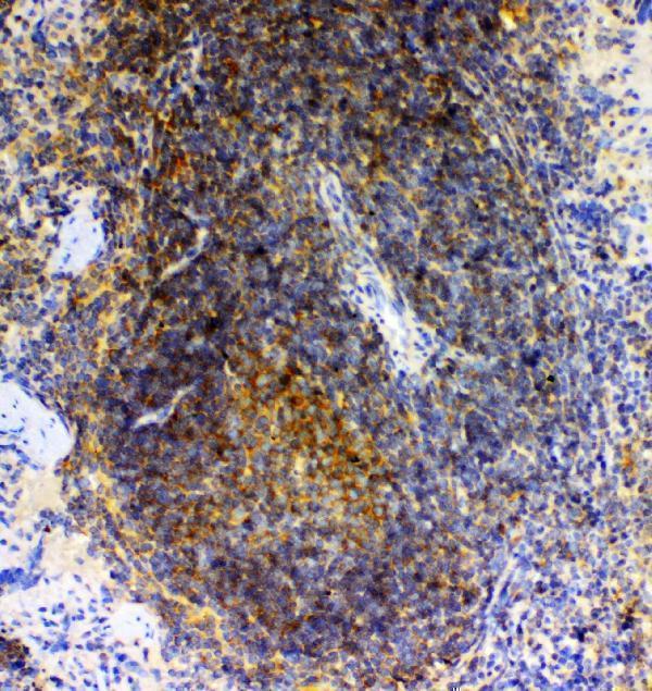
Real IHC data IHC analysis of GRK2 using anti-GRK2 antibody (A01473-1). GRK2 was detected in paraffin-embedded section of human Lung cancer tissues. Heat mediated antigen retrieval was performed in citrate buffer (pH6, epitope retrieval solution) for 20 mins. The tissue section was blocked with 10% goat serum. The tissue section was then incubated with 1μg/ml rabbit anti-GRK2 Antibody (A01473-1) overnight at 4°C. Biotinylated goat anti-rabbit IgG was used as secondary antibody and incubated for 30 minutes at 37°C. The tissue section was developed using Strepavidin-Biotin-Complex (SABC)(Catalog # SA1022) with DAB as the chromogen.
Anti-GRK2/GRK2 Antibody ®
Cat # A01473-1
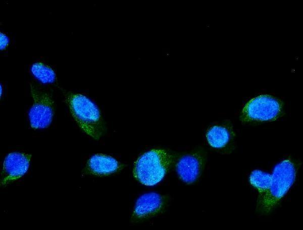
Real IF data IF analysis of GRK2 using anti-GRK2 antibody (A32388-1). GRK2 was detected in immunocytochemical section of U20S cells. Enzyme antigen retrieval was performed using IHC enzyme antigen retrieval reagent (AR0022) for 15 mins. The cells were blocked with 10% goat serum. And then incubated with 5μg/mL rabbit anti-GRK2 Antibody (A32388-1) overnight at 4°C. DyLight®488 Conjugated Goat Anti-Rabbit IgG (BA1127) was used as secondary antibody at 1:100 dilution and incubated for 30 minutes at 37°C. The section was counterstained with DAPI. Visualize using a fluorescence microscope and filter sets appropriate for the label used.
Anti-GRK2 Antibody ®
Cat # A32388-1

A01473-1 has IHC images from human lung cancer and mouse and rat spleen paraffin sections, plus an IF image from U20S cells (A01473-1 image captions). A32388-1 has an IF image from U20S cells and lists IF/ICC, but no IHC application or image (A32388-1 catalog and IF caption).

Which to pick: Choose A01473-1 for tissue IHC: its captions document paraffin sections with citrate retrieval, while the fixative is unreported (A01473-1 IHC captions). For IF/ICC, both SKUs list those applications and show U20S IF images; A32388-1 also lists monkey reactivity, though its IF image is from U20S cells (catalog applications, reactivity and IF captions). For IHC across human, mouse and rat samples, A01473-1 has matching tissue images; clonality is unreported for both SKUs (catalog IHC captions and clone fields).

Each figure is that product's own IHC / IF validation image from its datasheet.

References

  1. UniProt Consortium. UniProt entry P25098 (GRK2_HUMAN, G protein-coupled receptor kinase 2).
  2. Human Protein Atlas. GRK2 tissue IHC expression (reliability: Enhanced).
  3. Human Protein Atlas. GRK2 subcellular location (ICC-IF): Mainly localized to vesicles. In addition localized to the Golgi apparatus and cytosol..
  4. Human Protein Atlas. GRK2 antibody validation summary (3 antibodies).
  5. Overexpression of GRK2 in vascular smooth muscle leads to inappropriate hypertension and acute heart failure as in clinical scenario 1. Scientific reports 2023 — PMC10182096.
  6. Alteration of β-Adrenoceptor Signaling in Left Ventricle of Acute Phase Takotsubo Syndrome: a Human Study. Scientific reports 2018 — PMC6109068.
  7. GRK2 regulates GLP-1R-mediated early phase insulin secretion in vivo. BMC biology 2021 — PMC7931601.
  8. G protein-coupled receptor kinase 2 inhibition improves erectile function through amelioration of endothelial dysfunction and oxidative stress in a rat model of type 2 diabetes. Asian journal of andrology 2019 — PMC6337949.
  9. PubMed PMID:2037065 — UniProt-cited evidence.
  10. PubMed PMID:1339451 — UniProt-cited evidence.
  11. PubMed PMID:8195124 — UniProt-cited evidence.