This website uses cookies to ensure you get the best experience on our website.


- Table of Contents
Plan GRM2 chromogenic IHC in paraffin sections using the catalog antibody’s IHC-P conditions (datasheet A06123-1). Compare cerebral cortex neuropil staining with a non-detected tissue and interpret the cytoplasmic tissue pattern alongside GRM2’s synaptic membrane location (HPA tissue IHC; UniProt).

Expected localisation, validated protocols, controls and antibodies — the at-a-glance facts below, then the full design guide.
| Expected localisation | CNS cytoplasmic staining; synaptic membrane expected (HPA tissue IHC; UniProt) | |
| Staining pattern | Cytoplasmic CNS staining, including cerebral cortex neuropil (HPA tissue IHC) | |
| Antigen retrieval | EDTA pH 8.0 HIER, heat-mediated (datasheet A06123-1) | |
| Positive control | Cerebral cortex | |
| Negative control | Adipose tissue+4 more · see all |
| Fixation | Keep fixation consistent across sections (standard IHC practice; not target-specific) | |
| Caveat | Antibody staining has low concordance with RNA expression (HPA tissue IHC) | |
| Regulation | No specific expression regulator reported (UniProt) | |
| Isoform / epitope | 0 annotated isoforms; epitope side matters across 7 membrane spans (UniProt) |
The catalog antibody’s IHC-P protocol (datasheet A06123-1) is followed by one published paraffin-section GRM2 protocol (PMC9536148).
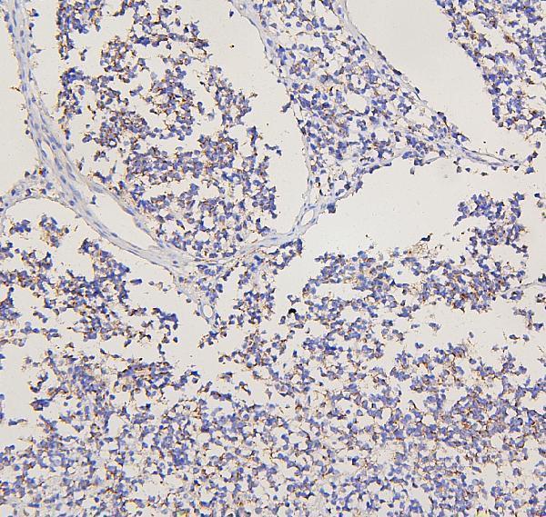
| Sample | Paraffin-embedded human glioma tissue; fixative not specified (datasheet A06123-1) |
| Fixation | Image fixative and duration unreported (datasheet A06123-1); verify before use. |
| Sectioning | 4–5 µm sections on charged slides (standard) |
| Deparaffinisation | Xylene, graded ethanol series to water (standard) |
| Antigen retrieval | Heat retrieval: EDTA pH 8.0 (datasheet A06123-1); 20 min, 95–100 °C (standard) |
| Peroxidase block | 3% H2O2, 10 min, room temperature (standard) |
| Blocking | 10% goat serum (datasheet A06123-1) |
| Primary antibody | Rabbit anti-GRM2, 0.5-1μg/ml recommended; image 2 μg/ml (datasheet A06123-1) |
| Primary incubation | Overnight at 4 °C (datasheet A06123-1) |
| Detection | HRP-conjugated secondary, DAB chromogen (datasheet A06123-1) |
| Counterstain | Hematoxylin, blue, dehydrate and mount (standard) |
| Expected result | GRM2-positive staining in neuropil of cerebral cortex (HPA tissue IHC: Medium). HPA tissue profile: Cytoplasmic expression in CNS. No signal in the no-primary control. |
GRM2 is a seven-pass membrane receptor at synapses and dendrites (UniProt Q14416 topology/localisation). In IHC, expect staining in cerebral cortex neuropil, with weaker staining reported in hippocampal neurons and cerebellar granular-layer cells (HPA tissue IHC: Medium/Low). HPA also describes cytoplasmic expression in the CNS; its tissue IHC reliability is Approved, with low consistency between antibody staining and RNA expression (HPA tissue IHC).
| Cerebral cortex neuropil has a moderate chromogenic signal. | This matches the strongest supplied tissue observation (HPA tissue IHC: Medium in cerebral cortex neuropil). Read a neuropil-rich pattern alongside GRM2's synaptic and dendritic localisation; IHC may not resolve receptor-bearing membranes individually (UniProt Q14416 localisation; general IHC practice). |
| Hippocampal neuronal cells or cerebellar granular-layer cells show weak staining. | Low staining is reported in these cells (HPA tissue IHC: Low). Compare intensity within the same staining run; a weak result here is less decisive than a cerebral cortex neuropil result (HPA tissue IHC: Medium; general IHC practice). |
| Signal is confined to nuclei, with no corresponding CNS neuropil pattern. | An exclusively nuclear pattern is unsupported by the supplied localisation evidence (UniProt Q14416 localisation; HPA tissue IHC). Treat it as suspect and inspect controls before assigning it to GRM2 (general IHC practice). HPA's broad ‘cytoplasmic expression in CNS’ description alone does not make all non-membrane-looking signal erroneous (HPA tissue IHC). |
| Prominent staining appears in adrenal glandular cells or adipocytes. | Those sampled cell types were not detected by HPA tissue IHC (HPA tissue IHC: adrenal glandular cells and adipocytes, Not detected). Investigate cross-reactivity or endogenous detection activity, especially if the expected cortex neuropil pattern is absent (general IHC practice; HPA tissue IHC: cerebral cortex neuropil, Medium). |
| The whole section has diffuse colour, or cortex neuropil has no signal. | Diffuse colour obscures the expected cell pattern and suggests background; absent cortex signal needs a run-level check because cortex neuropil was Medium in HPA (general IHC practice; HPA tissue IHC). Neither finding alone establishes that the tissue lacks GRM2, particularly given HPA's low staining–RNA consistency (HPA tissue IHC). |
| Tissue and cell selection | Cerebral cortex neuropil is the clearest supplied positive comparison; hippocampal neurons and cerebellar granular-layer cells have Low staining (HPA tissue IHC). Sampled adipocytes and adrenal glandular cells were Not detected, providing pattern comparisons rather than proof that every cell of those tissues is negative (HPA tissue IHC). |
| Membrane topology and antibody epitope | GRM2 has 7 transmembrane segments, a large extracellular region at residues 19–567, and a cytoplasmic C terminus at 820–872 (UniProt Q14416 topology). Epitope location can affect how an antibody reads a section, but the supplied record gives no catalog-antibody epitope; do not assume which face it recognises (UniProt Q14416 topology; supplied evidence). |
| Maturation and glycosylation | The annotated signal peptide spans residues 1–18; the receptor chain begins at residue 19, with 5 glycosylation sites annotated (UniProt Q14416 processing/glycosylation). These features describe the protein; the supplied sources do not establish a retrieval setting, epitope loss, or staining change caused by them. |
| Antibody validation and interpretation limit | Two listed antibodies have different HPA IHC statuses: HPA027868 is Supported and HPA065166 is Approved (HPA antibodies). HPA tissue IHC is Approved but reports low consistency with RNA expression; assess the observed cell pattern and controls before making a strong specificity claim (HPA tissue IHC; general IHC practice). |
| Antigen retrieval choice | Retrieval is a general variable in paraffin-section IHC and should follow the chosen IHC-validated antibody's instructions, with a known-positive section alongside it (general IHC practice). UniProt and HPA provide no GRM2-specific retrieval condition or fixation-sensitivity result in the supplied evidence. |
| Situation | Likely cause | Next action |
|---|---|---|
| No signal in cerebral cortex neuropil. | The expected Medium HPA pattern is missing; staining-run failure, unsuitable detection, or a specimen issue are possibilities (HPA tissue IHC; general IHC practice). | Check the IHC-validated antibody's stated paraffin-section conditions, a concurrent known-positive section, and detection controls; then reassess the neuropil pattern (general IHC practice). |
| Only faint hippocampal or cerebellar staining is visible. | Low staining is reported for hippocampal neuronal cells and cerebellar granular-layer cells (HPA tissue IHC: Low). | Include cerebral cortex neuropil as the stronger supplied comparison, then judge whether the weak signal follows the reported cells (HPA tissue IHC: cortex Medium; general IHC practice). |
| Strong nuclear staining dominates the section. | Exclusive nuclear localisation lacks support in UniProt's membrane/synapse/dendrite annotation and HPA's CNS IHC description (UniProt Q14416 localisation; HPA tissue IHC). | Review a no-primary detection control and the tissue pattern; avoid scoring nuclear colour as GRM2 without independent support (general IHC practice). |
| Colour appears in negative-comparison cell types. | Adrenal glandular cells and adipocytes were Not detected in the supplied HPA samples; cross-reactivity or endogenous chromogenic activity may contribute (HPA tissue IHC; general IHC practice). | Compare a no-primary control and the positive cortex section; check that the detection system's endogenous-activity blocking step was performed (general IHC practice). |
| Diffuse background hides neuropil detail. | Nonspecific antibody binding, incomplete blocking, or excess chromogenic development can reduce contrast (general IHC practice). | Inspect no-primary and tissue controls; review blocking, antibody dilution, washes, and development against the chosen antibody's IHC instructions (general IHC practice). |
| Can ICC-IF establish the expected IHC-P pattern? | HPA lists a Membrane subcellular summary but no ICC-IF image-bearing cell lines here; neither listed HPA antibody has an ICC status in the supplied payload (HPA subcellular; HPA antibodies). | Use this page's cortex neuropil IHC result as the paraffin-section reference (HPA tissue IHC: Medium); consult the separate IF/ICC guide for that application. Do not treat the HPA subcellular summary as an ICC-IF validation result (HPA subcellular). |
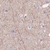
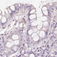
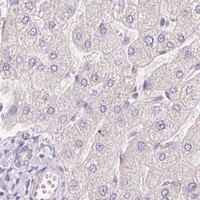
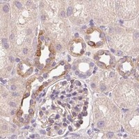
Comprehensive Human Protein Atlas IHC scoring per tissue (reliability: Approved — Low consistency between antibody staining and RNA expression data.). Rows are taken directly from the HPA tissue chart — click any row's HPA link to view the source.
| Tissue | Cell type | Level | Evidence | Source |
|---|---|---|---|---|
| Cerebral cortex | Neuropil | Medium | Protein (IHC) | HPA → |
| Tissue | Cell type | Level | Evidence | Source |
|---|---|---|---|---|
| Adipose tissue | Adipocytes | Not detected | Protein (IHC) | HPA → |
| Adrenal gland | Glandular cells | Not detected | Protein (IHC) | HPA → |
| Appendix | Glandular cells | Not detected | Protein (IHC) | HPA → |
| Bone marrow | Hematopoietic cells | Not detected | Protein (IHC) | HPA → |
| Breast | Adipocytes | Not detected | Protein (IHC) | HPA → |
Troubleshoot GRM2 staining in paraffin sections by checking retrieval, localisation and controls before interpreting chromogenic signal.
Anti-GRM2 options list human, mouse and rat reactivity (catalog reactivity: A06123-1, M06123); the supplied IHC image shows a human glioma paraffin section (A06123-1 image caption).
A06123-1 is listed for IHC (catalog applications: A06123-1), with an IHC image from human glioma paraffin tissue (A06123-1 image caption). M06123 is listed for IHC and ICC/IF (catalog applications: M06123) and human, mouse and rat reactivity (catalog reactivity: M06123); no IHC or IF image is supplied (catalog image alts: M06123).
Which to pick: For tissue IHC, choose A06123-1 because its image documents staining in a human glioma paraffin section; the fixative is unreported (A06123-1 image caption). For IF/ICC, choose the rabbit monoclonal M06123 based on its listed applications, while recognizing that no IF image is supplied (catalog host/clone, applications and image alts: M06123). Both list human, mouse and rat reactivity for cross-species work (catalog reactivity: A06123-1, M06123), but the supplied tissue image documents human staining only (A06123-1 image caption).