GRPEL2 / GrpE protein homolog 2, mitochondrial · IHC design guide

Design Immunohistochemistry for GRPEL2

Plan chromogenic paraffin IHC with the catalog antibody at a starting dilution of 1:50–1:100 (datasheet A15757). Compare cytoplasmic staining in colon glandular cells with undetected staining in adipocytes (HPA tissue IHC), and use the mitochondrial matrix annotation as molecular context (UniProt).

Evidence assembled Oct 2026 · For research use; verify linked source records and product datasheet before use
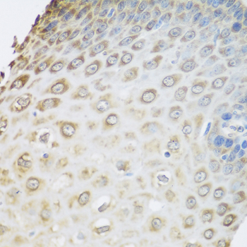
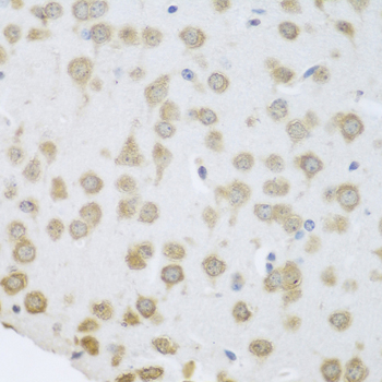
Immunohistochemistry protocol sheet for GRPEL2 (IHC for GRPEL2): expected localisation Cytoplasmic tissue staining; mitochondrial matrix annotation (HPA tissue IHC) (UniProt), antibody A15757, validated IHC image, and IHC protocol steps
Printable GRPEL2 IHC protocol sheet — expected localisation Cytoplasmic tissue staining; mitochondrial matrix annotation (HPA tissue IHC) (UniProt), antibody A15757, controls and protocol steps. Open the full GRPEL2 IHC guide →

GRPEL2 Immunohistochemistry Experimental Design Guide

Expected localisation, validated protocols, controls and antibodies — the at-a-glance facts below, then the full design guide.

Must know before staining
Expected localisation Cytoplasmic tissue staining; mitochondrial matrix annotation (HPA tissue IHC) (UniProt)
Staining pattern General cytoplasmic staining in colon glandular cells (HPA tissue IHC)
Antigen retrieval Citrate pH 6.0 HIER, heat-mediated (datasheet A15757)
Positive control ⓘ Adrenal gland+4 more · see all
Negative control ⓘ Adipose tissue
Important caveats
Reasons your staining may differ from the expected pattern.
Fixation Keep fixation consistent across samples (standard IHC practice; not target-specific)
Caveat Staining has low consistency with RNA expression (HPA tissue IHC)
Regulation No expression regulator established (UniProt)
Isoform / epitope 2 isoforms; mature chain spans aa 33–225; check epitope coverage (UniProt)
Section 1

Recommended GRPEL2 IHC & IF Protocols

The catalog antibody IHC-P protocol (datasheet A15757) is followed by three published GRPEL2 IHC procedures (PMC8657957; PMC12020167; PMC9850107).

Recommended immunohistochemistry (IHC-P) protocol parameters
SampleParaffin-embedded rat kidney tissue; fixative not specified (datasheet A15757)
FixationImage fixative and duration unreported (datasheet A15757); verify before use.
Sectioning4–5 µm sections on charged slides (standard)
DeparaffinisationXylene, graded ethanol series to water (standard)
Antigen retrievalHeat retrieval: Citrate pH 6.0 (datasheet A15757); 20 min, 95–100 °C (standard)
Peroxidase block3% H2O2, 10 min, room temperature (standard)
Blocking10% normal serum of the secondary host, 30 min, room temperature (standard)
Primary antibodyRabbit anti-GRPEL2, 1:50-1:100 (datasheet A15757)
Primary incubationOvernight at 4 °C (standard)
DetectionHRP-polymer secondary, DAB chromogen 5–10 min (standard)
CounterstainHematoxylin, blue, dehydrate and mount (standard)
Expected resultGRPEL2-positive staining in glandular cells of adrenal gland (HPA tissue IHC: High). HPA tissue profile: General cytoplasmic expression. No signal in the no-primary control.
💡Decision noteStart with citrate pH 6.0 heat retrieval (datasheet A15757); the published procedures provide tissue-specific starting conditions (PMC8657957; PMC9850107).
Section 2

What Is the Expected GRPEL2 Staining Pattern?

GRPEL2 resides in the mitochondrial matrix and has no transmembrane segment (UniProt Q8TAA5). In paraffin section IHC, expect cytoplasmic staining in cell populations reported by HPA, including adrenal glandular cells, bronchial respiratory epithelial cells and cortical neurons (HPA: general cytoplasmic expression; High in these cells). Interpret intensity cautiously: HPA rates its tissue IHC data Approved but reports low consistency between antibody staining and RNA expression (HPA: reliability).

What am I looking at on my slide?
Cytoplasmic staining in adrenal glandular cells, bronchial respiratory epithelial cells or cortical neurons (HPA: High in each).This matches reported tissue IHC and the expected compartment (HPA: general cytoplasmic expression; UniProt Q8TAA5: mitochondrial matrix). A chromogenic section may show cytoplasmic detail without resolving individual mitochondria (general IHC practice).
Predominantly nuclear or cell-surface staining, with little cytoplasmic signal.This conflicts with the reported cytoplasmic pattern and matrix location (HPA: tissue IHC profile; UniProt Q8TAA5: mitochondrial matrix). Treat it as a possible staining artefact; review controls and antibody specificity before assigning GRPEL2.
Strong staining of adipocytes, although HPA reports them as Not detected (HPA: adipose tissue IHC).Check for cross-reactivity or endogenous detection activity (general IHC practice). This is a discrepancy, not proof of either cause: HPA reports low staining–RNA consistency, and UniProt describes GRPEL2 tissue specificity as ubiquitous.
Diffuse color across cells and surrounding tissue, with no clear cellular pattern.This cannot establish the reported cytoplasmic distribution (HPA: tissue IHC profile). Consider nonspecific background or endogenous detection activity; inspect a negative reagent control and the localization of signal in a known-positive tissue (general IHC practice).
No signal in a sampled HPA High cell population.A negative result conflicts with the reported pattern for that cell population (HPA: tissue IHC). Check the catalog antibody’s IHC-P instructions and detection controls before interpreting absence; HPA’s Approved rating does not guarantee every specimen will stain (HPA: reliability).
💡Expected GRPEL2 appearanceCall a result positive when cytoplasmic staining is clear in a reported High population, such as colon glandular cells, while recognizing that dominant nuclear staining or strong adipocyte staining warrants investigation (HPA: High in colon glandular cells; general cytoplasmic expression; adipocytes Not detected; UniProt Q8TAA5: mitochondrial matrix).
How each factor affects the staining
Compartment and topologyGRPEL2 is a mitochondrial matrix protein with no transmembrane segment (UniProt Q8TAA5). Assess its cytoplasmic distribution in IHC; do not score membrane outlining as the expected pattern (HPA: general cytoplasmic expression).
Tissue and cell selectionHPA reports High staining in placental trophoblasts and kidney glomerular cells, Low staining in salivary glandular cells and smooth muscle cells, and Not detected in adipocytes (HPA: tissue IHC). Select comparison areas by cell population.
Processing and isoformsUniProt lists two isoforms and an annotated chain spanning residues 33–225 (UniProt Q8TAA5). Epitope recognition across isoforms or the processed chain cannot be inferred without the antibody’s epitope information.
Evidence strengthBoth listed antibodies, HPA018173 and HPA023211, have Approved IHC status (HPA: antibody validation). HPA also reports low consistency with RNA expression; Approved does not establish that every observed signal is specific (HPA: tissue IHC reliability).
IF/ICC Q: Where should signal appear?HPA reports enhanced mitochondrial localization in ICC-IF; UniProt places GRPEL2 in the mitochondrial matrix (HPA: subcellular; UniProt Q8TAA5). That localization supports interpretation, while IF/ICC procedure belongs in its separate guide.
Why is my staining missing, weak or wrong?
SituationLikely causeNext action
The selected positive area is blank.The sampled cells may differ from the HPA High population, or the staining run may have failed (HPA: cell-specific tissue IHC; general IHC practice).Confirm the cell type and compare a reported High population on the same run; check the catalog antibody’s IHC-P instructions and detection controls (HPA: tissue IHC; general IHC practice).
Signal is mainly nuclear or outlines cell membranes.The distribution disagrees with GRPEL2’s matrix location and HPA’s cytoplasmic profile (UniProt Q8TAA5; HPA: tissue IHC).Review the staining controls and antibody specificity; score the discordant compartment separately rather than counting it as expected GRPEL2 staining (general IHC practice).
Adipocytes stain strongly.HPA reports adipocytes as Not detected; cross-reactivity or endogenous detection activity are possibilities, not established causes (HPA: adipose tissue IHC; general IHC practice).Compare adipocytes with nearby reported positive cell populations and a negative reagent control; interpret the discrepancy in light of HPA’s low staining–RNA consistency (HPA: reliability; general IHC practice).
Color is diffuse across tissue and obscures cell boundaries.Nonspecific background or endogenous detection activity can obscure localization (general IHC practice).Inspect the negative reagent control and detection blocking steps, then reassess whether cellular cytoplasmic staining remains discernible (general IHC practice; HPA: cytoplasmic profile).
A Low population appears nearly unstained.HPA reports Low staining in several populations, including prostate glandular cells and ovarian follicle cells (HPA: tissue IHC).Compare a reported High population before treating faint signal in the Low population as a failed run; record which cells were scored (HPA: tissue IHC; general IHC practice).
IHC looks cytoplasmic, but IF/ICC images look more punctate.HPA describes general cytoplasmic tissue IHC and enhanced mitochondrial ICC-IF localization; the methods display different spatial detail (HPA: tissue IHC and subcellular; general microscopy practice).Judge each image against its method’s reported pattern; use the separate IF/ICC guide for that application (HPA: tissue IHC and subcellular).

Sample controls for GRPEL2 IHC & IF

🧪Run colon first and require staining in glandular cells (HPA: High in colon glandular cells); run adipose tissue as the negative and assess its adipocytes (HPA: Not detected in adipocytes). On the colon slide, treat unstained non-glandular cells as provisional internal negatives only, since GRPEL2 is described as ubiquitous (UniProt Q8TAA5: tissue specificity); nuclei should lack the mitochondrial-pattern signal (UniProt Q8TAA5: matrix; HPA subcellular: mitochondria).
Positive control tissue: Adrenal gland (Glandular cells, HPA High)
Negative control tissue: Adipose tissue (HPA Not detected)
ICC-IF cell lines (HPA subcellular resource): HPA ICC-IF images show GRPEL2 in A-431, U-251MG, U2OS, KOLF2.1J, with annotated localisation: Mitochondria (enhanced) (HPA subcellular).
Technical controls: Include no-primary and secondary-only controls, a concentration-matched rabbit polyclonal isotype control (selected product caption: Rabbit pAb), and a GRPEL2 knockout specimen as a biological negative. For chromogenic IHC in colon, quench endogenous peroxidase and check for residual background, particularly in inflammatory cells (standard IHC practice).
⚠️Feasibility: A target-specific fixation window or fixation effect is unreported, and the selected A15757 rat-kidney paraffin-section caption does not state the fixative. That caption reports high-pressure retrieval in 10 mM citrate, pH 6.0, followed by antibody at 1:200 (selected product caption); it does not establish a retrieval requirement for colon. Frozen sections have no supported ease advantage in the supplied evidence; IF/ICC offers a way to assess the expected mitochondrial localization (HPA subcellular: mitochondria), while endogenous peroxidase in inflammatory cells can complicate colon chromogenic scoring (standard IHC practice).

HPA tissue IHC evidence for GRPEL2

Comprehensive Human Protein Atlas IHC scoring per tissue (reliability: Approved — Low consistency between antibody staining and RNA expression data.). Rows are taken directly from the HPA tissue chart — click any row's HPA link to view the source.

Positive expression · recommended positive controls

TissueCell typeLevelEvidenceSource
Adrenal gland Glandular cells High Protein (IHC) HPA →
Bronchus Respiratory epithelial cells High Protein (IHC) HPA →
Cerebral cortex Neuronal cells High Protein (IHC) HPA →
Cervix Glandular cells High Protein (IHC) HPA →
Colon Glandular cells High Protein (IHC) HPA →

Undetected expression · recommended negative controls

TissueCell typeLevelEvidenceSource
Adipose tissue Adipocytes Not detected Protein (IHC) HPA →
Section 3

Advanced GRPEL2 IHC Tips

Troubleshoot GRPEL2 chromogenic IHC in paraffin sections using the catalog antibody’s rat kidney staining and the available localisation evidence.

Which retrieval conditions should I try first for GRPEL2 IHC?
Start with heat-mediated retrieval in 10 mM citrate, pH 6.0, using the high-pressure approach reported for the catalog antibody in paraffin-embedded rat kidney (datasheet A15757). The reported primary dilution is 1:200, so keep it constant while comparing retrieval conditions (datasheet A15757). If staining is weak, vary heating duration on matched sections before trying another buffer, and inspect tissue integrity after each change (standard IHC practice). Record pressure, heating time, cooling time and staining intensity together, because a stronger chromogenic signal is useful only when the expected cellular distribution remains interpretable (standard IHC practice; UniProt Q8TAA5 localisation).
How should I handle fixation when optimising GRPEL2 staining?
The selected rat kidney caption identifies paraffin-embedded tissue but does not state its fixative, so target-specific fixation sensitivity is unknown (datasheet A15757). Record the fixative, fixation duration and processing history for every specimen before comparing GRPEL2 staining across sections (standard IHC practice). If specimens differ in fixation, first compare matched controls processed under the same conditions and use a consistent 1:200 antibody dilution during the comparison (standard IHC practice; datasheet A15757). Treat differences in signal cautiously until processing effects have been assessed, since the supplied target evidence does not establish which fixation conditions preserve this antibody’s epitope (datasheet A15757).
What staining pattern fits GRPEL2 in chromogenic IHC?
Interpret GRPEL2 as a mitochondrial matrix protein with no annotated transmembrane segment (UniProt Q8TAA5 localisation and topology). The tissue atlas describes general cytoplasmic staining, while its cell imaging places GRPEL2 in mitochondria; chromogenic tissue sections may therefore show cytoplasmic staining without resolving individual mitochondria (HPA tissue IHC profile; HPA subcellular; standard IHC practice). In kidney, compare staining within the relevant compartment because the atlas reports high signal in cells of the glomeruli (HPA: High in kidney glomerular cells). Predominantly nuclear staining, or signal confined to damaged section edges, warrants review against controls before assignment to GRPEL2 (UniProt Q8TAA5 localisation; standard IHC practice).
Could GRPEL2 processing or isoforms change what this antibody detects?
GRPEL2 has 2 annotated isoforms, and the annotated processed chain spans residues 33–225 (UniProt Q8TAA5 isoforms and processing). The supplied material does not map the catalog antibody’s epitope, so its coverage of both isoforms or the processed chain cannot be assumed (datasheet A15757; UniProt Q8TAA5 isoforms). An N6-acetyllysine is annotated at residue 142, but the payload provides no evidence that this modification changes IHC recognition (UniProt Q8TAA5 modified residues). If staining differs unexpectedly between samples, document antigen retrieval and processing first, then seek epitope mapping or an independent antibody before attributing the difference to an isoform or modification (standard IHC practice).
How can companion IF help assess a GRPEL2 IHC result?
Use multiplex IF as a companion localisation check when a chromogenic IHC pattern is ambiguous, since cell imaging assigns GRPEL2 to mitochondria (HPA subcellular). Pair GRPEL2 with a validated marker for the expected cell type, and assess the two signals within the same cells rather than comparing whole-section brightness (standard IF practice). Choose a fluorophore channel with low tissue autofluorescence and include single-label controls to assess spectral bleed-through (standard IF practice). Because the annotated protein resides in the mitochondrial matrix, check that permeabilisation permits access to that compartment while preserving mitochondrial morphology; IF conditions require their own validation and do not follow from the paraffin-section caption (UniProt Q8TAA5 localisation; datasheet A15757; standard IF practice).
How do I distinguish GRPEL2 signal from chromogenic background?
Run a no-primary control alongside the catalog antibody at its reported 1:200 dilution, keeping retrieval and detection conditions matched (datasheet A15757; standard IHC practice). For a peroxidase and DAB workflow, include a peroxidase block and inspect the no-primary section for residual enzyme activity or other chromogen deposition (standard IHC practice). Compare background within the same tissue compartment before changing antibody concentration, since the atlas reports high staining in kidney glomerular cells but does not establish background levels for this antibody (HPA: High in kidney glomerular cells; datasheet A15757). Diffuse staining across unrelated structures or strong edge staining calls for review of controls, washing and section condition (standard IHC practice).
How should I score GRPEL2 across paraffin sections? ⚠ ANSWER MARKED FOR VERIFICATION
Choose a prespecified readout such as an H-score of 0–300, percentage of positive cells, or positive-cell density per mm² of viable tissue (standard IHC practice). Score the same cell population in every specimen; for kidney sections, report glomerular cells separately if they are the population of interest (HPA: High in kidney glomerular cells; standard IHC practice). Normalise counts to the relevant viable tissue area or eligible cell count, and keep thresholds, retrieval, antibody dilution and imaging settings consistent (standard IHC practice; datasheet A15757). Include matched control sections because the atlas notes low consistency between antibody staining and RNA expression, which limits interpretation of staining intensity alone (HPA tissue IHC reliability).
When is a GRPEL2-positive IHC result convincing?
A convincing result shows reproducible cellular staining in an appropriate cytoplasmic distribution, consistent with the mitochondrial matrix assignment and atlas tissue profile (UniProt Q8TAA5 localisation; HPA tissue IHC profile). Check whether the stained cell population fits the specimen: the atlas reports high signal in kidney glomerular cells and no detected signal in adipocytes (HPA tissue IHC). Treat predominantly nuclear signal, staining concentrated at section edges or necrotic regions, and signal persisting in a no-primary peroxidase control as possible artefact (UniProt Q8TAA5 localisation; standard IHC practice). Do not infer mitochondrial import activity or oxidative stress from staining intensity alone, particularly given the atlas’s reported staining–RNA inconsistency (UniProt Q8TAA5 function; HPA tissue IHC reliability).
Boster reagents

Best GRPEL2 / GrpE protein homolog 2, mitochondrial IHC Antibodies

A15757 has IHC images of paraffin-embedded rat kidney and human colon carcinoma; the catalog lists human, mouse, and rat reactivity (A15757 IHC image captions; catalog: reactivity).

Real IHC data Immunohistochemistry of paraffin-embedded rat kidney using GRPEL2 Rabbit pAb at dilution of 1:200 .Perform high pressure antigen retrieval with 10 mM citrate buffer pH 6.0 before commencing with IHC staining protocol.
Anti-GRPEL2 Antibody
Cat # A15757

The sole rendering SKU is A15757, a rabbit polyclonal antibody listed for IHC (catalog: A15757 host and applications). Its IHC images show paraffin-embedded rat kidney and human colon carcinoma at 1:200 after high-pressure retrieval in 10 mM citrate buffer, pH 6.0 (A15757 IHC image captions).

Which to pick: Choose A15757 for paraffin-section IHC; its image captions document rat kidney and human colon carcinoma, but do not report the fixative (A15757 IHC image captions). No listed SKU supports an IF/ICC recommendation: A15757 has no IF application or IF image (catalog: A15757 applications and IF images). For cross-species IHC, A15757 is a rabbit polyclonal with listed human, mouse, and rat reactivity; its IHC images document human and rat samples only (catalog: A15757 host and reactivity; A15757 IHC image captions).

Each figure is that product's own IHC / IF validation image from its datasheet.

References

  1. UniProt Consortium. UniProt entry Q8TAA5 (GRPE2_HUMAN, GrpE protein homolog 2, mitochondrial).
  2. Human Protein Atlas. GRPEL2 tissue IHC expression (reliability: Approved).
  3. Human Protein Atlas. GRPEL2 subcellular location (ICC-IF): Localized to the mitochondria..
  4. Human Protein Atlas. GRPEL2 antibody validation summary (2 antibodies).
  5. GRPEL2 Knockdown Exerts Redox Regulation in Glioblastoma. International journal of molecular sciences 2021 — PMC8657957.
  6. E2F8-induced GRPEL2 promoted colorectal cancer progression via targeting TIGAR. Journal of translational medicine 2025 — PMC12020167.
  7. Construction and validation of a prognostic model for hepatocellular carcinoma: Inflammatory ferroptosis and mitochondrial metabolism indicate a poor prognosis. Frontiers in oncology 2022 — PMC9850107.
  8. PubMed PMID:14702039 — UniProt-cited evidence.
  9. PubMed PMID:15372022 — UniProt-cited evidence.
  10. PubMed PMID:15489334 — UniProt-cited evidence.