GRSF1 / G-rich sequence factor 1 · IHC design guide

Design Immunohistochemistry for GRSF1

Plan paraffin-section GRSF1 staining around granular cytoplasmic signal in colon glandular cells or kidney tubule cells (HPA tissue IHC). The catalog antibody A07576-2 has an IHC range of 2–5 μg/mL (datasheet A07576-2); assess cell morphology and include a no-primary control (standard IHC practice).

Evidence assembled Oct 2026 · For research use; verify linked source records and product datasheet before use
Immunohistochemistry protocol sheet for GRSF1 (IHC for GRSF1): expected localisation Observed granular cytoplasm (HPA tissue IHC); expected mitochondrial matrix (UniProt), antibody A07576-2, validated IHC image, and IHC protocol steps
Printable GRSF1 IHC protocol sheet — expected localisation Observed granular cytoplasm (HPA tissue IHC); expected mitochondrial matrix (UniProt), antibody A07576-2, controls and protocol steps. Open the full GRSF1 IHC guide →

GRSF1 Immunohistochemistry Experimental Design Guide

Expected localisation, validated protocols, controls and antibodies — the at-a-glance facts below, then the full design guide.

Must know before staining
Expected localisation Observed granular cytoplasm (HPA tissue IHC); expected mitochondrial matrix (UniProt)
Staining pattern Granular cytoplasmic staining across tissues (HPA tissue IHC)
Antigen retrieval EDTA pH 8.0 HIER, heat-mediated (datasheet A07576-2)
Positive control ⓘ Adrenal gland+4 more · see all
Negative control ⓘ None in HPA (detected in all 45 tissues); use no-primary + isotype controls
Important caveats
Reasons your staining may differ from the expected pattern.
Fixation Keep fixation consistent across sections (standard IHC practice; not target-specific)
Caveat Staining and RNA expression show medium concordance (HPA tissue IHC)
Regulation No expression regulator annotated (UniProt)
Isoform / epitope 2 isoforms; chain 118–480; epitope map unknown (UniProt)
Section 1

Recommended GRSF1 IHC & IF Protocols

The catalog antibody’s IHC-P protocol is followed by 4 published tissue-staining methods for GRSF1 (datasheet: A07576-2; PMC8742353; PMC8485391; PMC11212087; PMC11179673).

Recommended immunohistochemistry (IHC-P) protocol parameters
SampleParaffin-embedded human pancreatic cancer tissue; fixative not specified (datasheet A07576-2)
FixationImage fixative and duration unreported (datasheet A07576-2); verify before use.
Sectioning4–5 µm sections on charged slides (standard)
DeparaffinisationXylene, graded ethanol series to water (standard)
Antigen retrievalHeat retrieval: EDTA pH 8.0 (datasheet A07576-2); 20 min, 95–100 °C (standard)
Peroxidase block3% H2O2, 10 min, room temperature (standard)
Blocking10% goat serum (datasheet A07576-2)
Primary antibodyRabbit anti-GRSF1, 2-5μg/ml (datasheet A07576-2)
Primary incubationOvernight at 4 °C (datasheet A07576-2)
DetectionStreptavidin-biotin complex (SABC), DAB chromogen (datasheet A07576-2)
CounterstainHematoxylin, blue, dehydrate and mount (standard)
Expected resultGRSF1-positive staining in glandular cells of adrenal gland (HPA tissue IHC: High). HPA tissue profile: General cytoplasmic expression with a granular pattern. No signal in the no-primary control.
💡Decision noteStart with heat-mediated EDTA pH 8.0 retrieval (datasheet: A07576-2). One published method uses EDTA pH 9.0 (PMC8485391 methods).
Section 2

What Is the Expected GRSF1 Staining Pattern?

GRSF1 is annotated in the mitochondrial matrix and cytoplasm, with enrichment in mitochondrial RNA granules; it has no transmembrane segment (UniProt Q12849). In paraffin IHC, expect granular cytoplasmic staining, including strong staining in kidney tubule cells and colon glandular cells (HPA: tissue IHC). HPA rates the tissue pattern Enhanced, while reporting medium agreement with RNA expression and pending external verification (HPA: tissue IHC reliability).

What am I looking at on my slide?
Granular cytoplasmic staining in kidney tubule cells or colon glandular cells (HPA: High; tissue IHC).This matches the reported tissue pattern (HPA: general granular cytoplasmic expression). Assess staining in the identified cells; chromogenic granules alone do not establish colocalization with mitochondrial RNA granules (UniProt Q12849: subcellular location).
Predominantly nuclear, membranous or extracellular staining, with little granular cytoplasmic signal.The dominant compartment conflicts with the reported pattern (HPA: tissue IHC) and mitochondrial matrix/cytoplasmic annotation (UniProt Q12849). Treat it as suspect; review morphology and controls before interpreting it as GRSF1.
Strong staining confined to an unexpected cell population while expected cells remain unstained.Possible explanations include cross-reactivity or endogenous detection activity (standard IHC practice). Compare the cell identities with documented positives, such as kidney tubule cells (HPA: High), and inspect antibody-omission and detection controls.
Broad, fairly uniform color obscures tissue boundaries and cellular detail.This is consistent with nonspecific background rather than the reported granular cytoplasmic pattern (HPA: tissue IHC; standard IHC practice). Check a control lacking primary antibody and review blocking, washing and detection conditions.
No convincing signal in a kidney section with identifiable tubules (HPA: High in tubule cells).An absent result in this documented positive tissue warrants an assay check; it does not alone disprove expression (HPA: tissue IHC). Review section integrity, retrieval and detection controls using general IHC practice; GRSF1-specific fixation sensitivity is unreported here.
💡Expected GRSF1 appearanceCall a result positive when the relevant cells show discernible granular cytoplasmic staining, especially strong staining in documented High populations such as kidney tubules (HPA: tissue IHC); isolated nuclear or diffuse tissue-wide color is suspect (HPA: reported pattern; standard IHC practice).
How each factor affects the staining
Subcellular location and topologyThe mitochondrial matrix and cytoplasmic annotations support a cytoplasmic readout (UniProt Q12849). No transmembrane segment is annotated (UniProt Q12849); a dominant membrane outline would therefore require scrutiny.
Antibody and tissue evidenceTwo listed antibodies, HPA036984 and HPA036985, have Enhanced IHC status (HPA: antibodies). The tissue profile remains pending external verification, with medium staining–RNA consistency (HPA: tissue IHC reliability).
Tissue choice and expression measureHPA reports High protein staining in several epithelial and glandular populations, including colon glands and kidney tubules (HPA: tissue IHC). Its skeletal-muscle tissue-enhanced designation refers to RNA specificity; do not use it as an IHC intensity ranking (HPA: RNA specificity).
Processing, isoforms and epitope knowledgeUniProt lists a 118–480 chain and two isoforms (UniProt Q12849). Without an antibody epitope or isoform-specific validation in these payloads, neither fact predicts a particular staining difference or retrieval requirement.
IF/ICC Q: What localization is expected?A: Mitochondrial staining is reported by ICC-IF (HPA: enhanced mitochondrial localization). UniProt also annotates cytoplasm and mitochondrial RNA granules (UniProt Q12849). Interpret IF/ICC using its separate guide; these data supply no IF/ICC protocol option here.
Why is my staining missing, weak or wrong?
SituationLikely causeNext action
No staining in a documented High population, such as kidney tubule cells (HPA: tissue IHC).An assay failure or weak detection is possible (standard IHC practice); the supplied sources do not establish GRSF1-specific fixation sensitivity.Verify tissue morphology and a working assay control, then review the general IHC retrieval, primary-antibody and detection steps. Do not infer a target-specific fixation effect from the result.
Strong nuclear staining dominates the section.Nuclear dominance conflicts with the granular cytoplasmic IHC profile (HPA: tissue IHC) and UniProt localization (UniProt Q12849).Compare matched controls and check whether nuclear color persists without primary antibody (standard IHC practice). Avoid scoring the nuclear signal as the expected GRSF1 pattern.
Strong staining appears in unexpected cells but not in the documented positive cells.Cross-reactivity or endogenous detection activity may contribute (standard IHC practice); HPA documents which cell populations were High in its tissue survey (HPA: tissue IHC).Confirm cell identity on the counterstained section and inspect antibody-omission and detection controls. Reassess the antibody result against a documented positive tissue (HPA: tissue IHC).
Diffuse color covers cells and surrounding tissue.Excess background can obscure the granular cytoplasmic pattern reported by HPA (HPA: tissue IHC; standard IHC practice).Check the no-primary control, washing, blocking and detection conditions, then judge only signal that remains localized to identifiable cells (standard IHC practice).
Little signal appears in adipocytes or liver cholangiocytes.These populations are listed as Low in the HPA tissue survey (HPA: tissue IHC). A weak result there is less informative than one in a documented High population.Assess an HPA High population, such as colon glandular cells, before concluding that the assay failed (HPA: tissue IHC). Do not treat Low as an absent-protein designation.
Brightfield granules seem insufficient to assign a precise organelle.The IHC profile reports granular cytoplasm (HPA: tissue IHC); mitochondrial localization comes from ICC-IF and UniProt annotations (HPA: subcellular; UniProt Q12849).Report the observed granular cytoplasmic IHC pattern. Reserve a mitochondrial colocalization claim for appropriately controlled IF/ICC imaging (standard IF practice).

Sample controls for GRSF1 IHC & IF

🧪Run colon first: its glandular cells score High for GRSF1 (HPA: colon glandular cells, High). HPA detects GRSF1 in all 45 scored tissues, so there is no supported negative tissue; use no-primary and isotype controls, and expect nuclei and extracellular spaces on the positive slide to remain free of specific cytoplasmic staining (HPA: no negative tissue; UniProt Q12849: mitochondrial matrix and cytoplasmic localization).
Positive control tissue: Adrenal gland (Glandular cells, HPA High)
Negative control tissue: None in HPA: GRSF1 is detected in all 45 scored tissues. Use a no-primary (secondary-only) and an isotype control instead.
ICC-IF cell lines (HPA subcellular resource): HPA ICC-IF images show GRSF1 in A-431, U-251MG, U2OS, with annotated localisation: Mitochondria (enhanced) (HPA subcellular).
Technical controls: Include a no-primary, secondary-only control, an isotype control matched to the primary antibody’s host species and format, and a GRSF1 knockout specimen as a biological negative if available (standard IHC practice). Quench endogenous peroxidase and check for endogenous biotin background when using the caption’s biotin-based, DAB detection system (selected-SKU caption: biotinylated secondary, streptavidin-biotin complex and DAB).
⚠️Feasibility: A target-specific fixation window or fixation effect is unreported, and the selected-SKU paraffin-section caption does not state the fixative (selected-SKU caption: fixative unreported). The caption uses heat retrieval in EDTA at pH 8.0, but does not establish whether GRSF1 staining depends on that retrieval method (selected-SKU caption: EDTA heat retrieval); the supplied evidence does not establish that frozen sections or IF are easier. In colon, score glandular cytoplasmic staining against the negative-control background and exclude luminal debris from scoring (HPA: colon glandular cells, High; UniProt Q12849: mitochondrial matrix and cytoplasmic localization; standard IHC practice).

HPA tissue IHC evidence for GRSF1

Comprehensive Human Protein Atlas IHC scoring per tissue (reliability: Enhanced — Medium consistency between antibody staining and RNA expression data. Pending external verification.). Rows are taken directly from the HPA tissue chart — click any row's HPA link to view the source.

Positive expression · recommended positive controls

TissueCell typeLevelEvidenceSource
Adrenal gland Glandular cells High Protein (IHC) HPA →
Colon Glandular cells High Protein (IHC) HPA →
Duodenum Glandular cells High Protein (IHC) HPA →
Esophagus Squamous epithelial cells High Protein (IHC) HPA →
Gallbladder Glandular cells High Protein (IHC) HPA →

Undetected expression · recommended negative controls

TissueCell typeLevelEvidenceSource
None in HPA: GRSF1 is detected in all 45 scored tissues. Use a no-primary (secondary-only) and an isotype control instead.
Section 3

Advanced GRSF1 IHC Tips

Troubleshoot GRSF1 staining in paraffin sections by checking retrieval, compartmental pattern, background, and scoring against documented IHC evidence.

How should I optimize retrieval when GRSF1 staining is weak?
Start with heat-mediated antigen retrieval in EDTA at pH 8.0 for paraffin sections (datasheet A07576-2). The selected tissue image used this retrieval before incubation with 2 μg/mL primary antibody overnight at 4°C (datasheet A07576-2). Keep section thickness, heating time, cooling, and detection conditions consistent while comparing retrieval runs (standard IHC practice). If staining remains weak, test a different heating duration or an alternative retrieval buffer on adjacent sections as a fallback, recording both signal and tissue damage (standard IHC practice). Judge improvement by a reproducible granular cytoplasmic pattern rather than increased diffuse color alone (HPA: general cytoplasmic expression with a granular pattern).
Can I attribute variable GRSF1 staining to fixation?
Target-specific fixation sensitivity is unknown: the selected GRSF1 image identifies a paraffin section but does not state its fixative (datasheet A07576-2). Record the fixative, time before fixation, fixation duration, and processing history for each specimen before comparing stain intensity (standard IHC practice). Compare adjacent sections processed together with the documented EDTA pH 8.0 retrieval and 2 μg/mL primary concentration, changing one preparation variable at a time (datasheet A07576-2; standard IHC practice). If a batch loses its granular cytoplasmic pattern, review handling alongside retrieval and detection controls before assigning a cause (HPA: granular cytoplasmic profile; standard IHC practice). Neither tissue staining patterns nor protein topology establishes a fixation effect for this antibody (HPA: tissue IHC; UniProt Q12849 topology).
What GRSF1 pattern should I expect in chromogenic tissue sections?
Expect predominantly granular cytoplasmic staining in tissue sections (HPA: general cytoplasmic expression with a granular pattern). GRSF1 localizes to the mitochondrial matrix and mitochondrial RNA granules near nucleoids, while a cytoplasmic location is also annotated (UniProt Q12849 subcellular location). Chromogenic IHC may show punctate or granular cytoplasmic color without resolving individual mitochondrial RNA granules, so score the visible cellular pattern rather than claiming organelle resolution (HPA: tissue IHC profile; standard IHC practice). Compare the pattern in documented high-staining kidney tubule cells or colon glandular cells with an adjacent negative-reagent control (HPA: High in kidney tubules and colon glandular cells; standard IHC practice). Strong isolated nuclear color deserves a detection and morphology review before being called GRSF1 (UniProt Q12849 subcellular location; standard IHC practice).
Could isoforms or epitope position explain different GRSF1 staining patterns?
GRSF1 has 2 annotated isoforms, but the supplied antibody evidence does not identify its epitope or establish isoform-specific staining (UniProt Q12849 isoforms; datasheet A07576-2). The annotated processed chain spans residues 118–480, and three RNA recognition motifs occupy residues 122–246, 250–326, and 401–480 (UniProt Q12849 processing and domains). Phosphoserines at residues 244 and 335 are annotated, but their effect on this antibody’s tissue signal is untested here (UniProt Q12849 modified residues; datasheet A07576-2). Request the immunogen or epitope sequence before interpreting discordant sections as isoform changes, then compare matched sections under identical retrieval and detection conditions (standard IHC practice). Report any remaining difference as staining variation until antibody specificity for the proposed isoform or modification is established (standard IHC practice).
How can IF help assess an ambiguous chromogenic GRSF1 pattern?
Use IF as a separate localization check, pairing GRSF1 with a marker for the cell population being assessed and a mitochondrial marker when organelle assignment matters (HPA: mitochondrial localization; standard IF practice). Choose fluorophores and filter sets after inspecting unstained tissue for autofluorescence, and include single-label controls when signals are multiplexed (standard IF practice). Because GRSF1 is annotated in the mitochondrial matrix and has no transmembrane segment, establish permeabilization conditions that allow antibody access inside mitochondria, then check morphology and background (UniProt Q12849 location and topology; standard IF practice). HPA reports enhanced mitochondrial localization in ICC/IF, but that observation does not supply a fixation or permeabilization recipe for this antibody (HPA: subcellular localization). Compare compartment and cell identity with the chromogenic section without treating the two detection methods as interchangeable measurements (standard IHC/IF practice).
How do I distinguish GRSF1 signal from diffuse brown background?
The selected paraffin-section image used 10% goat serum blocking, a biotinylated secondary antibody, a streptavidin–biotin detection complex, and DAB (datasheet A07576-2). Include a primary-omission control to reveal detection-system color, and check endogenous peroxidase blocking when using DAB (standard IHC practice). Review diffuse deposits, tissue edges, and damaged areas separately from intact cells; the documented tissue profile is granular and cytoplasmic (HPA: tissue IHC profile; standard IHC practice). If background persists, compare adjacent sections while adjusting blocking, wash stringency, or primary concentration one variable at a time (standard IHC practice). Preserve a documented 2 μg/mL condition as the comparison point so weaker background is not mistaken for improved specificity when true staining also disappears (datasheet A07576-2; standard IHC practice).
How should I score GRSF1 across paraffin sections? ⚠ ANSWER MARKED FOR VERIFICATION
Define the scored cell population and exclude folds, necrotic regions, and section edges before measuring chromogenic signal (standard IHC practice). For comparable compartments, report the percentage of positive cells and an H-score based on intensity categories; report positive-cell density per mm² when cell abundance itself is the endpoint (standard IHC practice). Normalize counts to the number of evaluable cells or the measured viable tissue area, and keep staining batch, imaging settings, and scoring thresholds consistent (standard IHC practice). Record granular cytoplasmic staining separately from diffuse or nuclear color because the documented tissue pattern is granular and cytoplasmic (HPA: tissue IHC profile). Stratify by cell type when comparing tissues, since documented high staining includes kidney tubule cells and colon glandular cells (HPA: tissue IHC).
When is a positive GRSF1 stain biologically credible?
A credible positive shows reproducible granular cytoplasmic staining in intact cells, consistent with the tissue IHC profile and mitochondrial localization (HPA: tissue IHC and subcellular profiles). Consider cell identity before comparing intensity: HPA records high staining in kidney tubule cells and colon glandular cells, while several other sampled cell populations are low (HPA: tissue IHC). Isolated nuclear color conflicts with the annotated mitochondrial matrix and cytoplasmic locations and warrants review of controls and morphology (UniProt Q12849 subcellular location; standard IHC practice). Exclude edge accentuation and necrotic regions, and investigate color retained in a primary-omission control as possible detection background or endogenous enzyme activity (standard IHC practice). Treat a changed pattern as provisional until it repeats in matched sections with controlled retrieval and detection (standard IHC practice).
Boster reagents

Best GRSF1 / G-rich sequence factor 1 IHC Antibodies

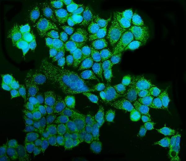
A07576-2 has real GRSF1 IHC images from human paraffin sections and an IF image from MCF-7 cells (catalog image captions).

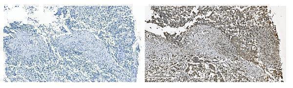
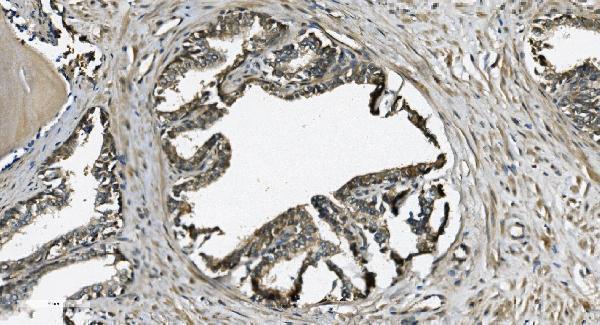
Real IHC data IHC analysis of GRSF1 using anti-GRSF1 antibody (A07576-2). GRSF1 was detected in paraffin-embedded section of human pancreatic cancer tissue. Heat mediated antigen retrieval was performed in EDTA buffer (pH8.0, epitope retrieval solution). The tissue section was blocked with 10% goat serum. The tissue section was then incubated with 2μg/ml rabbit anti-GRSF1 Antibody (A07576-2) overnight at 4°C. Biotinylated goat anti-rabbit IgG was used as secondary antibody and incubated for 30 minutes at 37°C. The tissue section was developed using Strepavidin-Biotin-Complex (SABC) (Catalog # SA1022) with DAB as the chromogen.
Anti-GRSF1 Antibody ®
Cat # A07576-2

A07576-2 is the only card with supplied IHC/IF figures: its IHC captions show human pancreatic and prostate cancer paraffin sections (catalog IHC captions). Its IF caption shows MCF-7 cells (catalog IF caption).

Which to pick: Choose A07576-2 for human paraffin-section IHC; its captions document that preparation, but do not report the fixative (catalog IHC captions). Choose A07576-2 for IF/ICC when a captioned cell example matters: its IF image uses MCF-7 cells (catalog IF caption). For mouse or rat IF/ICC, M07576 lists those species and applications, but has no supplied image or IHC application; M07576-1 lists human IHC and IF/ICC without a supplied figure (catalog applications, reactivity and image fields).

Each figure is that product's own IHC / IF validation image from its datasheet.

References

  1. UniProt Consortium. UniProt entry Q12849 (GRSF1_HUMAN, G-rich sequence factor 1).
  2. Human Protein Atlas. GRSF1 tissue IHC expression (reliability: Enhanced).
  3. Human Protein Atlas. GRSF1 subcellular location (ICC-IF): Localized to the mitochondria..
  4. Human Protein Atlas. GRSF1 antibody validation summary (2 antibodies).
  5. The RNA-binding protein GRSF1 promotes hepatocarcinogenesis via competitively binding to YY1 mRNA with miR-30e-5p. Journal of experimental & clinical cancer research : CR 2022 — PMC8742353.
  6. Network-based approach to identify prognosis-related genes in tamoxifen-treated patients with estrogen receptor-positive breast cancer. Bioscience reports 2021 — PMC8485391.
  7. Metabolic Heterogeneity of Tumor Cells and its Impact on Colon Cancer Metastasis: Insights from Single-Cell and Bulk Transcriptome Analyses. Journal of Cancer 2024 — PMC11212087.
  8. Guanine-Rich RNA Sequence Binding Factor 1 Deficiency Promotes Colorectal Cancer Progression by Regulating PI3K/AKT Signaling Pathway. Cancer management and research 2024 — PMC11179673.
  9. PubMed PMID:8036161 — UniProt-cited evidence.
  10. PubMed PMID:14702039 — UniProt-cited evidence.
  11. PubMed PMID:15815621 — UniProt-cited evidence.