This website uses cookies to ensure you get the best experience on our website.


- Table of Contents
Plan chromogenic GSPT1 IHC on paraffin sections with the IHC-validated antibody at 1:100–1:300 (datasheet: A07761). Expect broad cytoplasmic staining, with high staining in several glandular cell populations and no detected staining in adipocytes (HPA tissue IHC).

Expected localisation, validated protocols, controls and antibodies — the at-a-glance facts below, then the full design guide.
| Expected localisation | Cytoplasmic staining in tissue sections (HPA tissue IHC) | |
| Staining pattern | Broad cytoplasmic staining, often high in glandular cells (HPA tissue IHC) | |
| Antigen retrieval | Citrate pH 6.0 HIER, 95–98 °C, 20 min (rule: cytoplasmic / membrane antigen) | |
| Positive control | Appendix+4 more · see all | |
| Negative control | Adipose tissue |
| Fixation | Keep tissue fixation consistent across sections (standard IHC practice; not target-specific) | |
| Caveat | Adipocytes may be unstained despite broad tissue expression (HPA tissue IHC) | |
| Regulation | Expression regulation is not documented (UniProt) | |
| Isoform / epitope | 3 isoforms; antibody epitope coverage is unspecified (UniProt; datasheet) |
Compare the catalog antibody's IHC-P protocol with four published GSPT1 IHC methods (PMC9640916; PMC9937881; PMC8064227; PMC8798158).
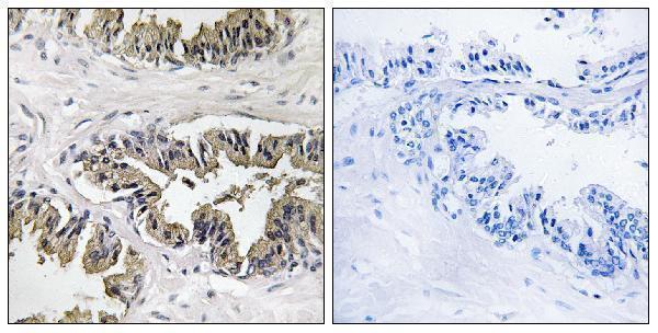
| Sample | Paraffin-embedded human prostate carcinoma tissue; fixative not specified (datasheet A07761) |
| Fixation | Image fixative and duration unreported (datasheet A07761); verify before use. |
| Sectioning | 4–5 µm sections on charged slides (standard) |
| Deparaffinisation | Xylene, graded ethanol series to water (standard) |
| Antigen retrieval | Heat-induced epitope retrieval in citrate buffer, pH 6.0, 20 min at 95–98 °C (standard rule: cytoplasmic / membrane antigen) |
| Peroxidase block | 3% H2O2, 10 min, room temperature (standard) |
| Blocking | 10% normal serum of the secondary host, 30 min, room temperature (standard) |
| Primary antibody | Rabbit anti-GSPT1, 1:100-1:300 (datasheet A07761) |
| Primary incubation | Overnight at 4 °C (standard) |
| Detection | HRP-polymer secondary, DAB chromogen 5–10 min (standard) |
| Counterstain | Hematoxylin, blue, dehydrate and mount (standard) |
| Expected result | GSPT1-positive staining in glandular cells of appendix (HPA tissue IHC: High). HPA tissue profile: Ubiquitous cytoplasmic expression. No signal in the no-primary control. |
GSPT1 should show cytoplasmic staining across many cell types, with high signal in several glandular and epithelial populations and bone marrow hematopoietic cells (HPA tissue IHC: ubiquitous cytoplasmic expression; listed high staining). Its lack of a transmembrane segment supports a nonmembranous pattern (UniProt P15170 topology). HPA rates the tissue IHC profile Approved, with medium consistency between antibody staining and RNA expression (HPA tissue IHC).
| Cytoplasmic staining in glandular, respiratory epithelial, or hematopoietic cells. | This fits the reported pattern. HPA lists high staining in appendix, breast, colon, duodenum, endometrium, and epididymis glandular cells, bronchial respiratory epithelial cells, and bone marrow hematopoietic cells (HPA tissue IHC). Compare intensity within the cell type being assessed; a high result in one listed tissue does not set a universal threshold. |
| Predominantly nuclear staining, or a crisp membrane outline with little cytoplasmic signal. | Treat this as discordant with the reported cytoplasmic IHC pattern (HPA tissue IHC) and cytosolic ICC-IF location (HPA subcellular). A membrane outline also lacks support from the sequence topology (UniProt P15170 topology). Review the negative control and detection conditions before assigning the signal to GSPT1. |
| Strong staining in adipocytes while nearby expected positive cells stain weakly. | HPA reports adipocytes in adipose tissue as not detected (HPA tissue IHC). Consider cross-reactivity or endogenous chromogen activity, particularly if the negative control also develops color; those are possible IHC artifacts, not established explanations for this specimen. Recheck cell identification and controls. |
| Diffuse color over stroma, empty spaces, or most cells without clear cytoplasmic boundaries. | This distribution does not resolve the reported cellular pattern (HPA tissue IHC: ubiquitous cytoplasmic expression). General IHC causes include nonspecific binding, incomplete blocking, or excess detection background. Interpret intensity only after background is low enough to distinguish stained cytoplasm from the surrounding section. |
| No staining in a section containing a listed high-staining cell population. | A failed positive control or insufficient assay sensitivity is possible when glandular, respiratory epithelial, or hematopoietic cells expected to stain high remain blank (HPA tissue IHC: listed high staining). Check the complete staining run before calling GSPT1 absent; HPA's medium RNA–protein consistency limits certainty for any individual specimen (HPA tissue IHC). |
| Tissue and cell selection | HPA reports low tissue specificity overall, yet staining varies by cell population: listed glandular, bronchial epithelial, and marrow hematopoietic cells are high; adipocytes are not detected (HPA tissue IHC). Select a control by the reported cell type, not by an assumption that every cell in a positive organ stains equally. |
| Strength of IHC evidence | The tissue profile is Approved, with medium consistency between antibody staining and RNA expression (HPA tissue IHC). HPA052488 is IHC Approved, whereas the supplied record gives no IHC status for HPA074588 (HPA antibodies). These labels support a reference pattern, not proof that every positive signal is specific. |
| Topology and processing | The protein has no transmembrane segment, signal peptide, or propeptide, and its recorded chain spans residues 1–499 (UniProt P15170 topology and processing). A sharp surface-only pattern therefore needs independent support. The record does not identify an IHC epitope or establish how retrieval affects it. |
| Isoform coverage | UniProt lists 3 isoforms (UniProt P15170 isoforms). The supplied evidence does not map the IHC antibody epitope across them, so staining cannot be assigned to a particular isoform. Check documented epitope coverage if an isoform-specific interpretation is required. |
| IF/ICC: what localization should I expect? | Mainly cytosolic signal, with additional vesicular localization, is the approved ICC-IF observation (HPA subcellular). This is a separate cellular readout; the paraffin IHC reference is ubiquitous cytoplasmic staining (HPA tissue IHC). The supplied evidence provides no IF/ICC protocol to recommend. |
| Situation | Likely cause | Next action |
|---|---|---|
| A known positive cell population is blank. | The run may lack detectable signal; HPA lists high staining in several glandular, bronchial epithelial, and marrow hematopoietic populations (HPA tissue IHC). | Inspect the positive control and reagent steps, then review retrieval, antibody incubation, and chromogen development as general IHC workflow checks. Do not infer a GSPT1-specific fixation effect; none is supplied. |
| Nuclear or membrane-rim color dominates. | The pattern conflicts with HPA cytoplasmic IHC and approved cytosolic ICC-IF localization (HPA tissue IHC; HPA subcellular). GSPT1 has no annotated transmembrane segment (UniProt P15170 topology). | Compare the same compartment in a listed high-staining control and its negative control. Review cell boundaries and detection background before scoring the unusual compartment as target signal. |
| Adipocytes show strong color. | Adipocytes are reported not detected (HPA tissue IHC); cross-reactivity or endogenous detection activity is possible under general IHC practice. | Confirm the stained cells are adipocytes, inspect the negative control for matching color, and reassess blocking and detection chemistry. Keep the result provisional until the discordance is resolved. |
| The entire section looks hazy or uniformly brown. | Diffuse background can obscure the expected cytoplasmic distribution (HPA tissue IHC: ubiquitous cytoplasmic expression). Excess reagent or inadequate washing are general IHC possibilities. | Compare background with the negative control, then review antibody concentration, wash steps, blocking, and chromogen time as general workflow variables. Score cells only where their cytoplasm remains distinguishable. |
| One listed positive tissue stains much less than another. | HPA reports high staining for specific cell populations, but its tissue IHC profile has only medium consistency with RNA expression (HPA tissue IHC). Different cell composition can also change the apparent field intensity. | Compare like cell types at the same scoring threshold and include a high-staining reference population from the same run (HPA tissue IHC). Confirm cellular localization before interpreting a weak field as absent expression. |


Comprehensive Human Protein Atlas IHC scoring per tissue (reliability: Approved — Medium consistency between antibody staining and RNA expression data.). Rows are taken directly from the HPA tissue chart — click any row's HPA link to view the source.
| Tissue | Cell type | Level | Evidence | Source |
|---|---|---|---|---|
| Appendix | Glandular cells | High | Protein (IHC) | HPA → |
| Bone marrow | Hematopoietic cells | High | Protein (IHC) | HPA → |
| Breast | Glandular cells | High | Protein (IHC) | HPA → |
| Bronchus | Respiratory epithelial cells | High | Protein (IHC) | HPA → |
| Colon | Glandular cells | High | Protein (IHC) | HPA → |
| Tissue | Cell type | Level | Evidence | Source |
|---|---|---|---|---|
| Adipose tissue | Adipocytes | Not detected | Protein (IHC) | HPA → |
Troubleshoot chromogenic IHC for GSPT1 in paraffin sections using the catalog antibody, with a separate IF/ICC question for multiplex experiments.
Both anti-GSPT1 antibodies have IHC images from paraffin-embedded human carcinoma tissue (catalog image captions) and list human, mouse, and rat reactivity (catalog reactivity); only A07761 lists IF (catalog applications).
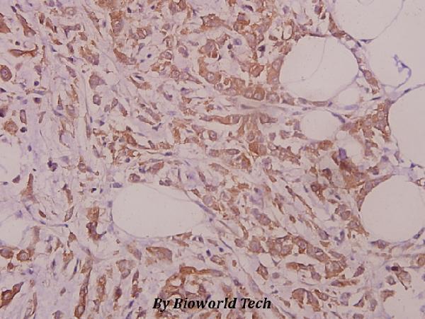
A07761 will render with IHC of paraffin-embedded human prostate carcinoma and a peptide-blocked comparison (A07761 image caption); its application list includes IF (A07761 catalog applications). A07761-1 will render with IHC of paraffin-embedded human colorectal carcinoma at 1:50 (A07761-1 image caption).
Which to pick: For tissue IHC, choose either SKU according to the pictured sample: A07761 shows paraffin-embedded human prostate carcinoma, while A07761-1 shows paraffin-embedded human colorectal carcinoma (respective image captions); neither caption reports the fixative (respective image captions). For IF, choose rabbit polyclonal A07761 because IF is listed (A07761 catalog applications and dilution record); ICC validation and an IF image are unreported (A07761 catalog applications and image alts). For mouse or rat work, both list reactivity in those species (catalog reactivity), although their IHC images show human tissue only (respective image captions).