This website uses cookies to ensure you get the best experience on our website.


- Table of Contents
Plan GSR paraffin IHC with high-staining epididymal or fallopian tube glandular cells as reference tissue (HPA tissue IHC). This guide covers the 2–5 μg/mL antibody range (datasheet A01479-1) and interpretation of cytoplasmic and nuclear staining alongside UniProt's mitochondrial and cytoplasmic annotation (HPA tissue IHC; UniProt).

Expected localisation, validated protocols, controls and antibodies — the at-a-glance facts below, then the full design guide.
| Expected localisation | Cytoplasmic and nuclear IHC; mitochondrial annotation (HPA tissue IHC; UniProt) | |
| Staining pattern | Widespread cytoplasmic and nuclear staining (HPA tissue IHC) | |
| Antigen retrieval | EDTA pH 8.0 HIER, heat-mediated (datasheet A01479-1) | |
| Positive control | Epididymis+4 more · see all | |
| Negative control | Adipose tissue+4 more · see all |
| Fixation | Keep fixation consistent across paraffin sections (standard IHC practice; not target-specific) | |
| Caveat | Nuclear IHC conflicts with UniProt localization (HPA tissue IHC; UniProt) | |
| Regulation | Regulation not annotated (UniProt) | |
| Isoform / epitope | Five isoforms; check the epitope against the mature chain (44–522) (UniProt) |
The catalog antibody’s IHC-P protocol is accompanied by two published GSR staining protocols for mouse lung sections (PMC12133717; PMC2634824).
| Sample | Paraffin-embedded human lung cancer tissue; fixative not specified (datasheet A01479-1) |
| Fixation | Image fixative and duration unreported (datasheet A01479-1); verify before use. |
| Sectioning | 4–5 µm sections on charged slides (standard) |
| Deparaffinisation | Xylene, graded ethanol series to water (standard) |
| Antigen retrieval | Heat retrieval: EDTA pH 8.0 (datasheet A01479-1); 20 min, 95–100 °C (standard) |
| Peroxidase block | 3% H2O2, 10 min, room temperature (standard) |
| Blocking | 10% goat serum (datasheet A01479-1) |
| Primary antibody | Rabbit anti-GSR, 2-5μg/ml (datasheet A01479-1) |
| Primary incubation | Overnight at 4 °C (datasheet A01479-1) |
| Detection | HRP-conjugated secondary, DAB chromogen (datasheet A01479-1) |
| Counterstain | Hematoxylin, blue, dehydrate and mount (standard) |
| Expected result | GSR-positive staining in glandular cells of epididymis (HPA tissue IHC: High). HPA tissue profile: Ubiquitous cytoplasmic and nuclear expression. No signal in the no-primary control. |
GSR is a soluble cytoplasmic and mitochondrial protein with no transmembrane segment (UniProt P00390: location and topology). In paraffin-section IHC, expect staining in glandular cells, especially in epididymis and fallopian tube, where HPA reports high staining (HPA: tissue IHC). HPA rates its tissue staining Approved, while noting medium agreement with RNA data and a subcellular location contradicted by external data (HPA: reliability).
| Glandular cells show cytoplasmic staining, strongest in epididymis or fallopian tube. | This fits HPA's high staining in those cells and UniProt's cytoplasmic and mitochondrial locations (HPA: tissue IHC; UniProt P00390: location). A granular accent can be compatible with mitochondria, but chromogenic IHC alone cannot assign individual organelles (general IHC practice). |
| Staining is confined to nuclei or sharply outlines cell membranes. | Recheck specificity and detection conditions: UniProt places GSR in cytoplasm and mitochondria and lists no transmembrane segment (UniProt P00390: location and topology). HPA describes cytoplasmic and nuclear tissue staining but flags a location conflict, so nuclear signal alone is inconclusive rather than proof of an artefact (HPA: tissue IHC and reliability). |
| Adipocytes or cardiomyocytes stain strongly while nearby expected cells do not. | HPA reports GSR as not detected in adipocytes and cardiomyocytes in its sampled tissues (HPA: tissue IHC). This reversal raises concern for off-target binding or endogenous detection activity; it does not prove either cause (general IHC practice). Check controls before treating those cells as GSR positive. |
| Brown colour spreads across stroma, section edges, or many unrelated cells. | A diffuse deposit that obscures cell boundaries is difficult to score as GSR (general IHC practice). Compare a section processed without primary antibody and inspect blocking, washing, and chromogen development; HPA's tissue pattern does not establish a cause for background in this specimen (HPA: tissue IHC). |
| No staining appears in epididymal or fallopian-tube glandular cells. | These are useful positive reference cells because HPA reports high staining in both tissues (HPA: tissue IHC). First confirm that cells are present and the detection control worked; then review antibody dilution, retrieval, and development as general IHC variables, without assuming GSR-specific fixation sensitivity (general IHC practice). |
| Compartment and interpretation | UniProt lists cytoplasm and mitochondrion, with no transmembrane segment (UniProt P00390: location and topology). HPA tissue IHC also describes nuclear staining, while its ICC-IF summary supports cytosol and approves an additional ER location (HPA: tissue IHC; HPA: subcellular). Treat compartment discrepancies as findings to investigate, not as interchangeable localisation claims. |
| Choice of reference cells | HPA reports high glandular-cell staining in epididymis and fallopian tube, medium staining in several other glandular epithelia, and no detected staining in sampled adipocytes or cardiomyocytes (HPA: tissue IHC). Compare the named cells within each tissue; HPA's low tissue RNA specificity does not make every cell equally useful as a control (HPA: RNA specificity). |
| Antibody evidence | HPA lists IHC as Approved for HPA001538 and CAB008632, while HPA064806 has no IHC status in the supplied record (HPA: antibodies). The tissue profile itself is Approved with medium RNA agreement and pending external verification (HPA: reliability). An Approved designation supports use as a reference, but does not settle every disputed compartment. |
| Isoforms and processing | UniProt lists five isoforms, including mitochondrial and cytoplasmic forms, and a mitochondrial chain spanning residues 44–522 (UniProt P00390: isoforms and processing). These facts help explain why cellular distribution can vary; without an epitope map for the antibody, they cannot predict which isoforms the IHC stain detects. |
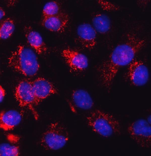
| IF/ICC Q&A | Q: Should IF/ICC show the same pattern as paraffin IHC? A: HPA's ICC-IF summary places GSR mainly in supported cytosol, with approved additional ER staining; its tissue IHC profile also describes nuclear staining (HPA: subcellular; HPA: tissue IHC). Interpret each assay against its own controls and keep IF/ICC optimisation on its separate guide page. |
| Situation | Likely cause | Next action |
|---|---|---|
| A high-staining reference tissue gives little or no signal. | The section may lack the relevant glandular cells, or a general IHC detection step may have failed; HPA reports high staining specifically in epididymal and fallopian-tube glandular cells (HPA: tissue IHC). | Identify the expected cells on the counterstain, verify a working detection control, then optimise retrieval, antibody dilution, and chromogen time using the antibody's IHC instructions (general IHC practice). Do not infer a GSR-specific fixation effect from this result. |
| Most cells show uniform brown colour, including a no-primary control. | Background can arise from endogenous enzyme activity or the detection system rather than specific antibody binding (general IHC practice). | Review the no-primary control, apply the detection system's appropriate endogenous-activity block, and check wash and development steps (general IHC practice). Judge any remaining signal against HPA's cell-level tissue pattern (HPA: tissue IHC). |
| Nuclei are prominent but cytoplasm is faint. | HPA reports nuclear tissue staining yet flags a subcellular conflict; UniProt places GSR in cytoplasm and mitochondria (HPA: tissue IHC and reliability; UniProt P00390: location). The slide alone cannot resolve that conflict. | Record nuclear and cytoplasmic scores separately, inspect no-primary and tissue controls, and seek independent antibody agreement before assigning nuclear localisation (general IHC practice; HPA: reliability). |
| A membrane rim or extracellular deposit dominates. | That distribution conflicts with GSR's reported intracellular locations and lack of a transmembrane segment (UniProt P00390: location and topology); edge deposits can also reflect nonspecific IHC background (general IHC practice). | Compare intact cells away from section edges, review the no-primary control and washing, and require reproducible intracellular staining in reference cells before scoring GSR (general IHC practice; HPA: tissue IHC). |
| A supposedly negative cell population stains. | HPA's “not detected” calls apply to the sampled cells and assay, including adipocytes and cardiomyocytes; they are not proof that every specimen of that cell type lacks GSR (HPA: tissue IHC). | Check cell identity and matched controls, compare staining with an expected glandular-cell reference, and describe the finding as discordant until specificity is supported (HPA: tissue IHC; general IHC practice). |
| Two antibodies give different compartment patterns. | HPA's supplied antibody statuses are assay-specific, and its tissue reliability note flags a subcellular discrepancy (HPA: antibodies and reliability). Isoforms exist, but an epitope-based explanation cannot be assigned without antibody epitope data (UniProt P00390: isoforms). | Compare like-for-like IHC controls and cell types, report each antibody's pattern separately, and avoid combining nuclear, ER, and mitochondrial labels into one confirmed result (general IHC practice; HPA: subcellular; UniProt P00390: location). |
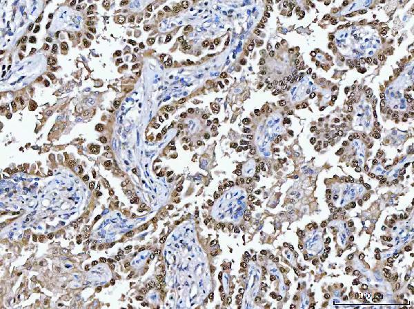
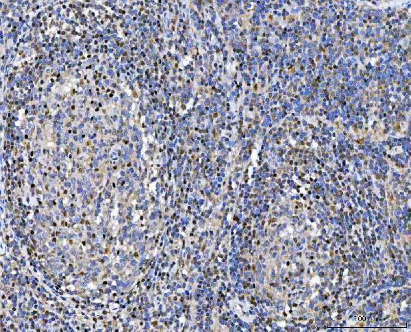


Comprehensive Human Protein Atlas IHC scoring per tissue (reliability: Approved — Medium consistency between antibody staining and RNA expression data. Subcellular location is contradicted by external data. Pending external verification.). Rows are taken directly from the HPA tissue chart — click any row's HPA link to view the source.
| Tissue | Cell type | Level | Evidence | Source |
|---|---|---|---|---|
| Epididymis | Glandular cells | High | Protein (IHC) | HPA → |
| Fallopian tube | Glandular cells | High | Protein (IHC) | HPA → |
| Adrenal gland | Glandular cells | Medium | Protein (IHC) | HPA → |
| Appendix | Glandular cells | Medium | Protein (IHC) | HPA → |
| Breast | Glandular cells | Medium | Protein (IHC) | HPA → |
| Tissue | Cell type | Level | Evidence | Source |
|---|---|---|---|---|
| Adipose tissue | Adipocytes | Not detected | Protein (IHC) | HPA → |
| Cerebellum | Cells in granular layer | Not detected | Protein (IHC) | HPA → |
| Heart muscle | Cardiomyocytes | Not detected | Protein (IHC) | HPA → |
| Hippocampus | Glial cells | Not detected | Protein (IHC) | HPA → |
| Smooth muscle | Smooth muscle cells | Not detected | Protein (IHC) | HPA → |
Troubleshoot GSR staining in paraffin sections by checking retrieval, staining controls and compartment patterns before comparing samples.
The IHC-validated antibody has paraffin-section data from human tissues and IF/ICC data from A549 cells (catalog image captions); listed reactivity covers human, mouse and rat (catalog applications/reactivity).
A01479-1 has IHC-P images from human lung cancer, tonsil, rectal cancer and thyroid cancer paraffin sections (A01479-1 IHC image captions). It also has an IF/ICC image from A549 cells and listed reactivity with human, mouse and rat (A01479-1 IF image caption; catalog applications/reactivity).
Which to pick: Choose A01479-1 for paraffin-section IHC: its tissue captions report EDTA retrieval at pH 8.0 and primary antibody at 2 μg/ml (A01479-1 IHC image captions); the fixative is unreported (A01479-1 IHC image captions). Choose A01479-1 for IF/ICC because its A549 image documents that application (A01479-1 IF image caption). For cross-species work, A01479-1 lists mouse and rat reactivity, although the supplied IHC and IF images show human samples (catalog reactivity; A01479-1 image captions).