GSTA2 / Glutathione S-transferase A2 · IHC design guide

Design Immunohistochemistry for GSTA2

Plan GSTA2 IHC-P with liver hepatocytes as a strong positive control and cytoplasmic staining as the expected pattern (HPA tissue IHC). Start the catalog antibody at 1:50–1:200 (datasheet), and assess unexpected staining in light of reported off-target binding (HPA tissue IHC).

Evidence assembled Oct 2026 · For research use; verify linked source records and product datasheet before use
Immunohistochemistry protocol sheet for GSTA2 (IHC for GSTA2): expected localisation Cytoplasmic staining in tissue (HPA tissue IHC), antibody A04171, validated IHC image, and IHC protocol steps
Printable GSTA2 IHC protocol sheet — expected localisation Cytoplasmic staining in tissue (HPA tissue IHC), antibody A04171, controls and protocol steps. Open the full GSTA2 IHC guide →

GSTA2 Immunohistochemistry Experimental Design Guide

Expected localisation, validated protocols, controls and antibodies — the at-a-glance facts below, then the full design guide.

Must know before staining
Expected localisation Cytoplasmic staining in tissue (HPA tissue IHC)
Staining pattern Hepatocytes show the most abundant cytoplasmic staining (HPA tissue IHC)
Antigen retrieval Citrate pH 6.0 HIER, 95–98 °C, 20 min (rule: cytoplasmic / membrane antigen)
Positive control ⓘ Duodenum+4 more · see all
Negative control ⓘ Adipose tissue+4 more · see all
Important caveats
Reasons your staining may differ from the expected pattern.
Fixation Keep fixation consistent across sections (standard IHC practice; not target-specific)
Caveat Presumed off-target binding may affect interpretation (HPA tissue IHC)
Regulation No expression regulator reported (UniProt)
Isoform / epitope No isoforms reported; mature chain spans residues 2–222 (UniProt)
Section 1

Recommended GSTA2 IHC & IF Protocols

Compare the catalog antibody’s IHC-P protocol with published GSTA2 IHC methods for colorectal tissue, paraffin sections, and FFPE epithelial specimens (PMC9008320; PMC6771233; PMC8295304; PMC13577711).

Recommended immunohistochemistry (IHC-P) protocol parameters
SampleParaffin-embedded human prostate cancer tissue; fixative not specified (datasheet A04171)
FixationImage fixative and duration unreported (datasheet A04171); verify before use.
Sectioning4–5 µm sections on charged slides (standard)
DeparaffinisationXylene, graded ethanol series to water (standard)
Antigen retrievalHeat-induced epitope retrieval in citrate buffer, pH 6.0, 20 min at 95–98 °C (standard rule: cytoplasmic / membrane antigen)
Peroxidase block3% H2O2, 10 min, room temperature (standard)
Blocking10% normal serum of the secondary host, 30 min, room temperature (standard)
Primary antibodyRabbit anti-GSTA2, 1:50-1:200 (datasheet A04171)
Primary incubationOvernight at 4 °C (standard)
DetectionHRP-polymer secondary, DAB chromogen 5–10 min (standard)
CounterstainHematoxylin, blue, dehydrate and mount (standard)
Expected resultGSTA2-positive staining in endocrine cells of duodenum (HPA tissue IHC: High). HPA tissue profile: Cytoplasmic expression in several tissues, most abundant in hepatocytes. No signal in the no-primary control.
💡Decision noteStart with citrate pH 6.0 HIER at 95–98 °C for 20 min (page retrieval rule); two published methods also report citrate retrieval (PMC6771233; PMC8295304).
Section 2

What Is the Expected GSTA2 Staining Pattern?

GSTA2 is cytoplasmic and lacks a transmembrane segment (UniProt P09210). In paraffin section IHC, expect the strongest cytoplasmic staining in hepatocytes, with high staining also reported in duodenal endocrine and testicular Leydig cells (HPA tissue IHC). HPA rates its tissue pattern Supported, while cautioning that the antibodies may target proteins from more than one gene and that presumed off target staining was disregarded (HPA tissue IHC).

What am I looking at on my slide?
Strong cytoplasmic staining in hepatocytes; little nuclear or membrane emphasis.This matches the most abundant reported tissue pattern and the expected compartment (HPA tissue IHC: High in hepatocytes; UniProt P09210: Cytoplasm). Compare cellular distribution as well as intensity, since a strong chromogenic signal alone cannot establish target identity (HPA tissue IHC: antibody specificity caution).
Predominantly nuclear or sharply membrane outlined staining.Treat this as a pattern requiring investigation: UniProt places GSTA2 in the cytoplasm and reports no transmembrane segment (UniProt P09210). Check morphology, counterstain and detection controls before interpreting it as GSTA2; the supplied sources do not establish a nuclear or membrane pattern.
Strong staining in cells reported as undetected, such as adipocytes in adipose tissue.This conflicts with the cell specific HPA observation (HPA tissue IHC: Not detected in adipose adipocytes). Consider antibody cross reactivity or chromogenic detection activity as possible causes, and assess an appropriate negative control (HPA tissue IHC: off target caution; standard IHC practice).
Diffuse color across cells and surrounding tissue, without a clear cellular pattern.Diffuse background cannot be scored as the expected cytoplasmic result (HPA tissue IHC: cytoplasmic profile). Review no primary controls, blocking, washes and detection conditions for nonspecific signal (standard IHC practice). HPA tissue observations alone do not identify which workflow step caused it.
No convincing signal in hepatocytes on a liver control section.This conflicts with the reported high hepatocyte staining (HPA tissue IHC: High in hepatocytes). First confirm section quality and that detection worked; then review antibody conditions and the laboratory's retrieval procedure (standard IHC practice). The supplied sources specify no GSTA2 specific retrieval condition or fixation sensitivity.
💡Expected GSTA2 appearanceCall a result consistent with GSTA2 when hepatocytes show strong cytoplasmic chromogen (HPA tissue IHC: High in hepatocytes; UniProt P09210: Cytoplasm); isolated nuclear, membrane outlined or cell discordant staining warrants investigation (HPA tissue IHC: specificity caution).
How each factor affects the staining
Tissue and cell selectionLiver hepatocytes provide a high staining reference; kidney tubular cells are reported at medium intensity, while adipose adipocytes are not detected (HPA tissue IHC). Score the named cell population rather than treating every cell in a tissue as equivalent.
Compartment and protein topologyA cytoplasmic pattern fits the reported location; no transmembrane segment or signal peptide is reported (UniProt P09210). These features guide interpretation of a membrane outlined pattern but do not, by themselves, validate antibody specificity.
Antibody specificityHPA calls the tissue pattern Supported but cautions that antibodies may target proteins from more than one gene; presumed off target binding was disregarded (HPA tissue IHC). Confirm unexpected staining against cell specific references and controls before assigning it to GSTA2.
Chromogenic detection backgroundEndogenous enzyme activity can contribute color when an enzyme based detection system is used; a no primary control helps reveal detection background (standard IHC practice). This is general workflow guidance, not a documented GSTA2 tissue or fixation effect.
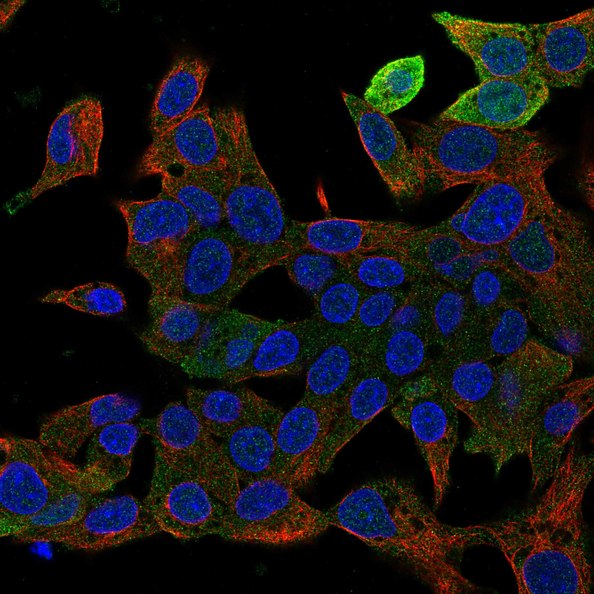
IF/ICC cross check?HPA reports cytosol as the supported ICC IF location and lists Hep-G2 and U2OS images, with a multi gene antibody caution (HPA subcellular). That supports a compartment cross check; IF/ICC setup and interpretation belong in its separate guide.
Why is my staining missing, weak or wrong?
SituationLikely causeNext action
Liver control is blank.A failed detection run or unsuitable assay conditions are possibilities (standard IHC practice); high hepatocyte staining is expected (HPA tissue IHC).Check a known working detection control, section integrity and the antibody's documented IHC conditions. Review retrieval settings without assuming a GSTA2 specific fixation effect (standard IHC practice).
Color appears throughout the section.Nonspecific detection signal, inadequate washing or endogenous activity may contribute (standard IHC practice).Compare a no primary control; review blocking, washes and detector controls. Score GSTA2 only where cellular cytoplasmic staining is distinguishable (standard IHC practice; UniProt P09210: Cytoplasm).
Nuclei dominate the signal.The location conflicts with cytoplasmic GSTA2 (UniProt P09210; HPA tissue IHC). The sources do not identify its cause.Check counterstain and control sections, then compare the signal with the expected hepatocyte cytoplasm (standard IHC practice; HPA tissue IHC: High in hepatocytes).
Adipose adipocytes stain strongly.The observation conflicts with HPA's undetected adipocytes; off target binding or detection background is possible (HPA tissue IHC; standard IHC practice).Run a no primary control and compare staining in a liver section. Do not assign the adipocyte signal to GSTA2 from intensity alone (standard IHC practice; HPA tissue IHC: specificity caution).
Kidney tubules are weaker than liver hepatocytes.This may reflect the reported tissue distribution: medium in kidney tubular cells versus high in hepatocytes (HPA tissue IHC).Evaluate the correct cell populations and control quality before changing assay conditions; these HPA levels are observations, not a quantitative acceptance threshold (HPA tissue IHC; standard IHC practice).
A second antibody gives a different tissue pattern.Antibody dependent off target staining is plausible given HPA's multi gene targeting caution (HPA tissue IHC).Compare cell type and cytoplasmic location in the same reference tissues, and review each antibody's validation and controls before resolving the discrepancy (HPA tissue IHC; standard IHC practice).

Sample controls for GSTA2 IHC & IF

🧪Start with liver: hepatocytes should show cytoplasmic staining (HPA: High in hepatocytes; UniProt P09210: cytoplasm). Use adipose tissue adipocytes as a negative comparison (HPA: Not detected in adipocytes); cells selected as internal negatives on the liver slide should show only background, but their GSTA2 status must be verified because the supplied HPA row specifies hepatocytes only.
Positive control tissue: Duodenum (Endocrine cells, HPA High)
Negative control tissue: Adipose tissue (HPA Not detected)
ICC-IF cell lines (HPA subcellular resource): HPA ICC-IF images show GSTA2 in Hep-G2, U2OS, with annotated localisation: Cytosol (supported) (HPA subcellular).
Technical controls: Include a no-primary, secondary-only control and an isotype control matched to the primary antibody’s host species and class; assess specificity with GSTA2 knockout material or immunizing-peptide competition when available (standard IHC practice). For liver chromogenic IHC, quench endogenous peroxidase and, if using avidin–biotin detection, block endogenous biotin (standard IHC practice).
⚠️Feasibility: A target-specific fixation window or fixation effect is unreported, and the selected A04171 paraffin-section caption does not state a fixative (A04171 tissue-IHC caption). Antigen-retrieval dependence is unreported; the supplied evidence does not establish whether frozen sections or IF/ICC are easier (supplied application evidence). Liver pigment and endogenous peroxidase can complicate chromogenic scoring (standard IHC practice).

HPA tissue IHC evidence for GSTA2

Comprehensive Human Protein Atlas IHC scoring per tissue (reliability: Supported — Caution, targets protein from more than one gene. Presumed off target binding observed and disregarded.). Rows are taken directly from the HPA tissue chart — click any row's HPA link to view the source.

Positive expression · recommended positive controls

TissueCell typeLevelEvidenceSource
Duodenum Endocrine cells High Protein (IHC) HPA →
Liver Hepatocytes High Protein (IHC) HPA →
Testis Leydig cells High Protein (IHC) HPA →
Adrenal gland Glandular cells Medium Protein (IHC) HPA →
Fallopian tube Glandular cells Medium Protein (IHC) HPA →

Undetected expression · recommended negative controls

TissueCell typeLevelEvidenceSource
Adipose tissue Adipocytes Not detected Protein (IHC) HPA →
Appendix Endocrine cells Not detected Protein (IHC) HPA →
Bone marrow Hematopoietic cells Not detected Protein (IHC) HPA →
Breast Adipocytes Not detected Protein (IHC) HPA →
Caudate Glial cells Not detected Protein (IHC) HPA →
Section 3

Advanced GSTA2 IHC Tips

Troubleshoot GSTA2 chromogenic IHC in paraffin sections by checking cytoplasmic localisation, cell identity and antibody specificity alongside each staining step.

What retrieval should I try when GSTA2 staining is weak in paraffin sections?
Start with citrate buffer at pH 6.0, heated to 95–98 °C for 20 min (page retrieval rule). Let sections cool before washing, then compare the result with a known positive hepatocyte section processed in the same run (HPA: High in hepatocytes; standard IHC practice). If staining remains weak, test a second retrieval condition on adjacent sections as a fallback, while holding antibody dilution and detection constant (standard IHC practice). Excessive retrieval can damage morphology or raise background, so judge the cytoplasmic signal together with tissue preservation (UniProt P09210: Cytoplasm; standard IHC practice). The paraffin-section caption for A04171 reports staining at 1:100 but does not report retrieval conditions (caption: A04171).
How should I troubleshoot fixation-related loss of GSTA2 staining?
Target-specific sensitivity to fixation is unknown from the supplied evidence; the A04171 caption identifies paraffin sections but does not state a fixative (caption: A04171). Record the fixative, fixation interval and processing history for each specimen before attributing a weak stain to GSTA2 biology (standard IHC practice). Compare similarly processed sections from a positive control and the test specimen in the same staining run, keeping retrieval and antibody conditions constant (HPA: High in hepatocytes; standard IHC practice). If signal varies with processing history, repeat the comparison using sections with matched handling and inspect morphology for damage (standard IHC practice). Do not infer fixation tolerance from GSTA2 localisation or its listed modifications (UniProt P09210: Cytoplasm, N-acetylalanine and N6-succinyllysine).
Where should convincing GSTA2 staining appear within a section?
Expect predominantly cytoplasmic staining, with hepatocytes providing a useful cell-level reference (UniProt P09210: Cytoplasm; HPA: most abundant in hepatocytes). GSTA2 has no transmembrane segment or signal peptide, so a crisp membrane rim alone does not fit its annotated topology (UniProt P09210: topology and processing). Assess localisation at sufficient magnification to distinguish cytoplasm from nuclei, luminal deposits and adjacent cells (standard IHC practice). Compare the staining pattern with a no-primary control and with neighbouring cells on the same section before calling an unexpected compartment positive (standard IHC practice). HPA also reports cytoplasmic expression across several tissues, but cautions that its antibodies may recognise proteins from more than one gene (HPA: tissue IHC reliability).
Could epitope recognition or related GST proteins explain an unexpected GSTA2 pattern?
The supplied UniProt record lists 0 isoforms, so an unexpected IHC pattern cannot be assigned to a documented GSTA2 splice isoform (UniProt P09210: isoforms). GSTA2 contains GST N-terminal residues 3–83 and GST C-terminal residues 85–207; epitope accessibility after processing may depend on where this antibody binds (UniProt P09210: domains; standard IHC practice). The epitope for A04171 is not supplied, so do not claim that a particular retrieval condition exposes it (caption: A04171). HPA cautions that its tissue antibody targets proteins from more than one gene, making cell pattern and orthogonal specificity evidence important when interpreting a stain (HPA: reliability description). GSTA2 can pair with GSTA1, which does not establish that this antibody cross-reacts with GSTA1 (UniProt P09210: subunit).
How can I use IF to investigate a disputed chromogenic GSTA2 signal?
Use IF as a separate check of the cell and compartment assigned by chromogenic IHC, while recognising that the supplied A04171 image documents paraffin-section IHC only (caption: A04171). Multiplex GSTA2 with an independently validated marker for the cell type under examination, and assess whether cytosolic signal falls within those cells (UniProt P09210: Cytoplasm; standard IF practice). Choose a fluorophore channel after checking tissue autofluorescence with an unstained section, especially when judging weak signal (standard IF practice). Because GSTA2 is cytosolic and lacks a transmembrane segment, include a mild permeabilisation comparison to allow antibody access, then check that cell morphology remains intact (UniProt P09210: topology; standard IF practice). Keep single-label and secondary-only controls for bleed-through and nonspecific fluorescence (standard IF practice).
What should I check when GSTA2 chromogenic staining looks diffuse or widespread?
First compare the section with a no-primary control to identify detection-reagent staining, and check whether the background follows tissue edges or damaged areas (standard IHC practice). Apply an appropriate peroxidase block before chromogenic detection and inspect a control section for residual endogenous enzyme activity (standard IHC practice). If diffuse stain persists, titrate the primary antibody and strengthen washing or protein blocking while keeping the positive control in the same run (standard IHC practice). The A04171 caption reports 1:100 in paraffin-embedded human prostate cancer, which is a documented starting dilution for that image, not a universal optimum (caption: A04171). Interpret widespread staining cautiously because HPA reports presumed off-target binding in its tissue IHC evidence (HPA: reliability description).
How should I score GSTA2 IHC across specimens with different tissue composition? ⚠ ANSWER MARKED FOR VERIFICATION
Score cytoplasmic signal in a defined cell population, recording the percentage of positive cells and intensity on a consistent 0–3 scale (UniProt P09210: Cytoplasm; standard IHC scoring practice). If an H-score is useful, multiply each intensity category by its cell percentage and sum the products, giving a 0–300 range (standard IHC scoring practice). Normalise comparisons to the number of eligible, viable cells in the annotated compartment, and report excluded necrotic or damaged regions (standard IHC practice). Keep section thickness, staining run, illumination and threshold rules consistent, with blinded review where practical (standard IHC practice). Report cell type because HPA describes high hepatocyte staining and medium staining in kidney tubular cells, among other patterns (HPA: tissue IHC).
How do I distinguish true GSTA2 staining from an artefact?
Give greatest weight to reproducible cytoplasmic staining in plausible cells, such as hepatocytes, alongside a clean no-primary control (UniProt P09210: Cytoplasm; HPA: High in hepatocytes; standard IHC practice). A membrane-only or nuclear-only pattern conflicts with the annotated cytoplasmic localisation and absence of a transmembrane segment (UniProt P09210: subcellular location and topology). Check isolated edge staining, necrotic regions and residual endogenous peroxidase signal against morphology and detection controls before scoring them as positive (standard IHC practice). Cell identity matters: HPA reports medium staining in kidney tubular cells but no detection in adipocytes from adipose tissue (HPA: tissue IHC). Treat an unexpected pattern as provisional because HPA flags presumed off-target binding and recognition of proteins from more than one gene (HPA: reliability description).
Boster reagents

Best GSTA2 / Glutathione S-transferase A2 IHC Antibodies

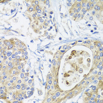
A04171 has real IHC data from a paraffin-embedded human prostate cancer section (IHC image caption); the catalog lists Human, Mouse, and Rat reactivity (catalog: A04171).

Real IHC data Immunohistochemistry of paraffin-embedded human prostate cancer using GSTA2 antibody at dilution of 1:100 (x40 lens).
Anti-Glutathione S-transferase A2 GSTA2 Antibody
Cat # A04171

A04171 will render with an IHC figure from a paraffin-embedded human prostate cancer section at 1:100 (IHC image caption). Its listed applications are IHC and WB, and its stated reactivity is Human, Mouse, and Rat (catalog: A04171).

Which to pick: For tissue IHC, choose A04171: its own image shows staining of a paraffin-embedded human prostate cancer section, but the fixative is unreported (IHC image caption). For IF/ICC, A04171 has no listed IF application or IF image (catalog: A04171). For cross-species IHC, A04171 lists Mouse and Rat alongside Human reactivity, although its supplied IHC image shows only human tissue; it is rabbit-derived, and clonality is unreported (catalog: A04171; IHC image caption).

Each figure is that product's own IHC / IF validation image from its datasheet.

References

  1. UniProt Consortium. UniProt entry P09210 (GSTA2_HUMAN, Glutathione S-transferase A2).
  2. Human Protein Atlas. GSTA2 tissue IHC expression (reliability: Supported).
  3. Human Protein Atlas. GSTA2 subcellular location (ICC-IF): Localized to the cytosol. Caution: Based on antibodies targeting proteins from multiple genes..
  4. Human Protein Atlas. GSTA2 antibody validation summary (3 antibodies).
  5. Chemotherapeutic Risk lncRNA-PVT1 SNP Sensitizes Metastatic Colorectal Cancer to FOLFOX Regimen. Frontiers in oncology 2022 — PMC9008320.
  6. Generation of hepatic spheroids using human hepatocyte-derived liver progenitor-like cells for hepatotoxicity screening. Theranostics 2019 — PMC6771233.
  7. Glutathione S-transferase A2 promotes hepatocellular carcinoma recurrence after liver transplantation through modulating reactive oxygen species metabolism. Cell death discovery 2021 — PMC8295304.
  8. Mutant p53 disrupts antioxidant defense in fallopian tube epithelium via GSTAs suppression: A pathway to serous tubal carcinogenesis. Free radical biology & medicine 2026 — PMC13577711.
  9. PubMed PMID:3036131 — UniProt-cited evidence.
  10. PubMed PMID:1329668 — UniProt-cited evidence.
  11. PubMed PMID:1497629 — UniProt-cited evidence.