This website uses cookies to ensure you get the best experience on our website.


- Table of Contents
This GUCY1B1 IHC-P guide identifies cytoplasmic staining and high-staining neuronal and glandular cells for tissue controls (HPA tissue IHC). Start M32396 at 1:50 (datasheet M32396), then compare staining within the relevant cell types.

Expected localisation, validated protocols, controls and antibodies — the at-a-glance facts below, then the full design guide.
| Expected localisation | Cytoplasmic staining in tissue (HPA tissue IHC) | |
| Staining pattern | Cytoplasmic staining across most tissues (HPA tissue IHC) | |
| Antigen retrieval | EDTA pH 8.0 HIER, heat-mediated (datasheet M32396) | |
| Positive control | Caudate+4 more · see all | |
| Negative control | Cervix+4 more · see all |
| Fixation | Keep fixation consistent across sections (standard IHC practice; not target-specific) | |
| Caveat | Some squamous epithelial cells are unstained (HPA tissue IHC) | |
| Regulation | Expression regulation unreported (UniProt) | |
| Isoform / epitope | 3 isoforms; epitope coverage unreported (UniProt) |
The catalog antibody’s IHC-P protocol (datasheet M32396) is accompanied by one published paraffin-section GUCY1B1 protocol (PMC12229189).
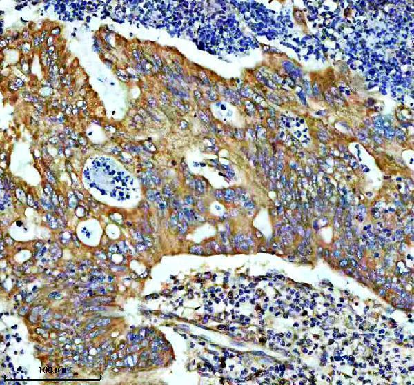
| Sample | Paraffin-embedded human colon cancer tissue; fixative not specified (datasheet M32396) |
| Fixation | Image fixative and duration unreported (datasheet M32396); verify before use. |
| Sectioning | 4–5 µm sections on charged slides (standard) |
| Deparaffinisation | Xylene, graded ethanol series to water (standard) |
| Antigen retrieval | Heat retrieval: EDTA pH 8.0 (datasheet M32396); 20 min, 95–100 °C (standard) |
| Peroxidase block | 3% H2O2, 10 min, room temperature (standard) |
| Blocking | 10% goat serum (datasheet M32396) |
| Primary antibody | Rabbit monoclonal (clone 20G57) anti-GUCY1B1, 1:50 (datasheet M32396) |
| Primary incubation | Overnight at 4 °C (datasheet M32396) |
| Detection | HRP-conjugated secondary, DAB chromogen (datasheet M32396) |
| Counterstain | Hematoxylin, blue, dehydrate and mount (standard) |
| Expected result | GUCY1B1-positive staining in neuronal cells of caudate (HPA tissue IHC: High). HPA tissue profile: Cytoplasmic expression in most tissues. No signal in the no-primary control. |
GUCY1B1 is a cytoplasmic protein with no transmembrane segment (UniProt Q02153). In paraffin-section IHC, expect cytoplasmic staining in neuronal cells of the caudate and cerebral cortex, cells of the cerebellar molecular layer, and glandular cells of the gallbladder and parathyroid gland (HPA: High in these cell populations). HPA rates the tissue IHC pattern Approved, with medium consistency between staining and RNA expression (HPA: tissue IHC).
| Cytoplasmic chromogen in caudate or cerebral cortex neuronal cells, or cerebellar molecular-layer cells. | This matches the reported High tissue IHC pattern and cytoplasmic profile (HPA: tissue IHC). Judge the named cells against nearby structures; staining somewhere else on the section does not establish the expected cell-specific pattern (general IHC practice). |
| Predominantly nuclear or sharply membrane-restricted staining, with little cytoplasmic signal. | That compartment conflicts with cytoplasmic tissue staining (HPA: tissue IHC), approved cytosolic localization (HPA: ICC-IF), and the absence of a transmembrane segment (UniProt Q02153). Treat it as a possible staining artefact and check morphology and controls before scoring it as GUCY1B1 (general IHC practice). |
| Strong staining in cells reported as undetected, such as lymph-node germinal-center cells. | HPA reports GUCY1B1 as Not detected in those cells (HPA: tissue IHC). Unexpected chromogen may reflect cross-reactivity or endogenous detection activity (general IHC practice); the HPA result is a comparison point, not proof that every specimen must be negative. |
| Diffuse chromogen across cells and extracellular spaces, obscuring cell boundaries. | An uninterpretable wash of color does not reproduce the reported cellular cytoplasmic pattern (HPA: tissue IHC). Excess detection background or nonspecific binding are possible explanations (general IHC practice); assess the no-primary control before assigning any positive cells. |
| No discernible signal in caudate neuronal cells or gallbladder glandular cells. | Both are reported High in tissue IHC (HPA: tissue IHC), so absence warrants an assay check. It does not alone show that the specimen lacks GUCY1B1: tissue handling, antibody performance, and detection can also affect an IHC result (general IHC practice). |
| Cell and tissue selection | HPA reports High staining in caudate and cortical neuronal cells, cerebellar molecular-layer cells, and gallbladder and parathyroid glandular cells, but Not detected in several other named cell populations (HPA: tissue IHC). Compare the same cell type when evaluating controls. |
| Antibody evidence | The tissue profile is Approved with medium staining–RNA consistency (HPA: tissue IHC). HPA020870 and CAB010890 each have Approved IHC status (HPA: antibodies). These ratings support comparison with the reported pattern but do not establish specificity in every preparation. |
| Compartment and protein form | UniProt places GUCY1B1 in the cytoplasm, reports no transmembrane segment, and lists a chain spanning residues 1–619 with no signal peptide or propeptide (UniProt Q02153). Those annotations support cytoplasmic scoring; they do not identify the catalog antibody's epitope. |
| Isoform coverage | UniProt lists 3 isoforms (UniProt Q02153). The supplied records do not specify which isoforms an IHC antibody recognizes, so a difference between samples cannot be assigned to isoform expression from this staining pattern alone. |
| Situation | Likely cause | Next action |
|---|---|---|
| No stain in an expected positive cell population. | The result differs from HPA's High staining in caudate neuronal or gallbladder glandular cells (HPA: tissue IHC); the technical cause is undetermined. | Confirm the cell type and slide controls, then review the IHC-validated antibody's supplied dilution and antigen-retrieval directions and the detection run (general IHC practice). No GUCY1B1-specific retrieval condition is supplied here. |
| Broad, hazy chromogen masks the cells. | Nonspecific binding or excessive detection background may obscure the cytoplasmic pattern (general IHC practice; HPA: tissue IHC pattern). | Compare a no-primary control, then review blocking, antibody dilution, washing, and chromogen development under the established IHC workflow (general IHC practice). |
| Predominantly nuclear or membrane-like stain. | The location disagrees with cytoplasmic and cytosolic annotations (UniProt Q02153; HPA: tissue IHC and ICC-IF). | Recheck cellular morphology and counterstain, inspect controls, and repeat with an IHC-approved antibody if the compartment remains discordant (HPA: antibodies; general IHC practice). |
| Strong color in an HPA-undetected cell population. | Cross-reactivity or endogenous detection activity is possible (general IHC practice); HPA reports germinal-center cells as Not detected (HPA: tissue IHC). | Compare the named cells with a positive tissue, inspect a no-primary control, and assess whether the signal follows cell boundaries before interpreting it (general IHC practice). |
| Uneven staining within a section. | This may reflect section or detection variability (general IHC practice); HPA's cell-specific levels do not establish the cause of patchiness in a particular specimen. | Check tissue integrity and whether positive and negative regions track recognizable cell types, then review coverage during reagent application (general IHC practice). |
| Q: Can ICC-IF help assess the expected compartment? | HPA reports approved cytosolic localization in ICC-IF (HPA: subcellular) and Supported ICC status for HPA077706 and CAB010890 (HPA: antibodies). | A: Use that evidence as a localization cross-check; follow the separate IF/ICC guide for assay design. An ICC-IF image does not replace cell-specific interpretation of paraffin-section IHC (HPA: tissue IHC; general IHC practice). |


Comprehensive Human Protein Atlas IHC scoring per tissue (reliability: Approved — Medium consistency between antibody staining and RNA expression data.). Rows are taken directly from the HPA tissue chart — click any row's HPA link to view the source.
| Tissue | Cell type | Level | Evidence | Source |
|---|---|---|---|---|
| Caudate | Neuronal cells | High | Protein (IHC) | HPA → |
| Cerebellum | Cells in molecular layer | High | Protein (IHC) | HPA → |
| Cerebral cortex | Neuronal cells | High | Protein (IHC) | HPA → |
| Gallbladder | Glandular cells | High | Protein (IHC) | HPA → |
| Parathyroid gland | Glandular cells | High | Protein (IHC) | HPA → |
| Tissue | Cell type | Level | Evidence | Source |
|---|---|---|---|---|
| Cervix | Squamous epithelial cells | Not detected | Protein (IHC) | HPA → |
| Lymph node | Germinal center cells | Not detected | Protein (IHC) | HPA → |
| Oral mucosa | Squamous epithelial cells | Not detected | Protein (IHC) | HPA → |
| Ovary | Ovarian stroma cells | Not detected | Protein (IHC) | HPA → |
| Seminal vesicle | Glandular cells | Not detected | Protein (IHC) | HPA → |
Troubleshoot chromogenic IHC for GUCY1B1 in paraffin sections using the catalog image conditions and independent tissue and localisation evidence.
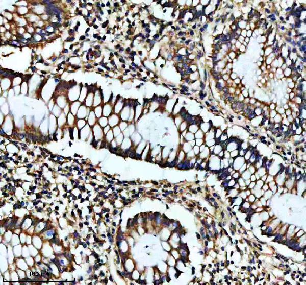
The catalog includes IHC images of human colon, colon cancer, ovarian cancer and mouse brain sections (M32396 image captions); IF/ICC is listed for human, mouse and rat (A32396-1 applications/reactivity).
M32396 is the SKU with a rendered card and its own IHC figure, showing a paraffin section of human colon cancer (card payload: M32396). Its additional IHC captions cover human colon, human ovarian cancer and mouse brain paraffin sections; rat reactivity is listed without a supplied IHC image (catalog: M32396 image captions/reactivity).
Which to pick: Choose monoclonal M32396 for tissue IHC when an illustrated paraffin-section procedure is useful (catalog: M32396 clone 20G57; M32396 IHC image captions). Choose polyclonal A32396-1 for IF/ICC because those applications are listed, although no IF image caption is supplied (catalog: A32396-1 applications/clone/IF image alts). Both list human, mouse and rat reactivity; M32396 has illustrated IHC in human and mouse, and its paraffin-section captions do not report the fixative (catalog: M32396 and A32396-1 reactivity; M32396 IHC image captions).