This website uses cookies to ensure you get the best experience on our website.


- Table of Contents
Plan paraffin-section IHC for GZMH using its cytolytic-granule location (UniProt) and staining in a subset of immune cells (HPA tissue IHC). The IHC-validated antibody has a 1:50–1:200 dilution range (datasheet); include a negative control (standard IHC practice).

Expected localisation, validated protocols, controls and antibodies — the at-a-glance facts below, then the full design guide.
| Expected localisation | Cytolytic granules expected (UniProt); tissue compartment unreported (HPA tissue IHC) | |
| Staining pattern | Staining in a subset of immune cells; compartment unreported (HPA tissue IHC) | |
| Antigen retrieval | Citrate pH 6.0 HIER, 95–98 °C, 20 min (rule: cytoplasmic / membrane antigen) | |
| Positive control | Skin+3 more · see all | |
| Negative control | Adipose tissue+4 more · see all |
| Fixation | Keep fixation consistent across sections (standard IHC practice; not target-specific) | |
| Caveat | Presumed off-target staining can mislead (HPA tissue IHC) | |
| Regulation | Constitutively expressed in NK cells (UniProt) | |
| Isoform / epitope | 3 isoforms; chain 21–246; epitope map unknown (UniProt) |
Compare the catalog antibody’s IHC-P protocol (datasheet) with the published GZMH tumor-tissue protocol (PMC13098063).
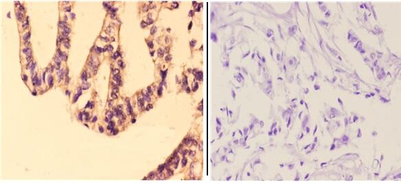
| Sample | Paraffin-embedded human breast carcinoma tissue; fixative not specified (datasheet A08559-1) |
| Fixation | Image fixative and duration unreported (datasheet A08559-1); verify before use. |
| Sectioning | 4–5 µm sections on charged slides (standard) |
| Deparaffinisation | Xylene, graded ethanol series to water (standard) |
| Antigen retrieval | Heat-induced epitope retrieval in citrate buffer, pH 6.0, 20 min at 95–98 °C (standard rule: cytoplasmic / membrane antigen) |
| Peroxidase block | 3% H2O2, 10 min, room temperature (standard) |
| Blocking | 10% normal serum of the secondary host, 30 min, room temperature (standard) |
| Primary antibody | Rabbit anti-GZMH, 1:50-1:200 (datasheet A08559-1) |
| Primary incubation | Overnight at 4 °C (standard) |
| Detection | HRP-polymer secondary, DAB chromogen 5–10 min (standard) |
| Counterstain | Hematoxylin, blue, dehydrate and mount (standard) |
| Expected result | GZMH-positive staining in cells in granular layer of skin (HPA tissue IHC: High). HPA tissue profile: Expression in a subset of immune cells. No signal in the no-primary control. |
In paraffin-section IHC, expect GZMH in a subset of immune cells, with a cytoplasmic pattern compatible with cytolytic granules (HPA: tissue IHC; UniProt P20718: cytolytic granule). UniProt reports constitutive expression in NK cells and no transmembrane segment (UniProt P20718). HPA rates its tissue IHC evidence Enhanced, while noting medium agreement with RNA and presumed off-target staining that was disregarded (HPA: tissue IHC).
| Discrete cytoplasmic staining in scattered cells of lymph node or tonsil outside germinal centers, or in splenic red pulp. | This fits the reported distribution and medium staining in those sites (HPA: tissue IHC). Granule-compatible cytoplasmic signal is consistent with GZMH location (UniProt P20718). Morphology alone does not identify each stained cell as an NK cell; HPA calls the tissue pattern a subset of immune cells (HPA: tissue IHC). |
| Predominantly nuclear staining, or a continuous membrane outline, across many cells. | That pattern conflicts with the reported cytolytic-granule location and absence of a transmembrane segment (UniProt P20718). Treat it as suspect staining and compare with the expected tissue distribution and a reagent control (HPA: tissue IHC; general IHC practice). A secreted protein may appear beyond its producing cell, so location alone cannot prove an artefact (HPA: subcellular summary). |
| Broad staining of adipocytes or respiratory epithelium, instead of a restricted cell subset. | HPA reports GZMH as undetected in adipocytes of adipose tissue and respiratory epithelial cells of bronchus (HPA: tissue IHC). Check antibody cross-reactivity and detection chemistry before scoring such staining as GZMH (general IHC practice). HPA also reports that presumed off-target staining was disregarded in its assessment (HPA: tissue IHC). |
| A diffuse haze spans the section or obscures individual stained cells. | That cannot be scored reliably as the reported subset-of-immune-cells pattern (HPA: tissue IHC). Diffuse chromogenic background can arise from nonspecific reagent binding or detection activity (general IHC practice). Because HPA summarizes GZMH as secreted, extracellular signal is possible; a uniform haze still needs control-based evaluation (HPA: subcellular summary; general IHC practice). |
| No discernible staining in lymph node, spleen, or tonsil. | HPA reports medium staining in specified cells of these tissues, making them useful reference sections when the relevant regions are present (HPA: tissue IHC). Check tissue representation and IHC controls before calling the assay negative (general IHC practice). A whole-section negative result cannot establish absent expression in every immune cell (HPA: subset-of-immune-cells profile). |
| Granule location and secretion | UniProt places GZMH in cytolytic granules; HPA summarizes it as secreted (UniProt P20718; HPA: subcellular summary). Interpret cell-associated cytoplasmic staining alongside morphology, while assessing any extracellular signal with controls (general IHC practice). |
| Tissue distribution and evidence strength | HPA reports medium staining in lymph node, spleen, and tonsil, and high staining in skin granular-layer cells (HPA: tissue IHC). Its Enhanced rating coexists with medium RNA agreement and disregarded presumed off-target staining; do not treat every stained cell as verified GZMH (HPA: tissue IHC). |
| Protein processing | UniProt annotates a signal peptide at residues 1–18, propeptide at 19–20, and mature chain at 21–246 (UniProt P20718). These boundaries matter when assessing a disclosed antibody epitope; no epitope is supplied here, so this record cannot predict which forms the antibody detects. |
| Isoforms and glycosylation | UniProt lists three isoforms and glycosylation sites at residues 71, 104, and 179 (UniProt P20718). Without an antibody epitope or isoform-specific validation, these annotations do not establish differences in IHC staining, antigen-retrieval needs, or fixation sensitivity. |
| Situation | Likely cause | Next action |
|---|---|---|
| Reference lymphoid tissue shows no signal. | The relevant cell subset may be absent from the sampled field, or assay sensitivity may be inadequate (HPA: tissue IHC; general IHC practice). | Inspect non-germinal-center areas in lymph node or tonsil, or splenic red pulp; verify a working IHC control and review antigen retrieval and antibody dilution using validated assay information (HPA: tissue IHC; general IHC practice). |
| Most nuclei or cell borders stain. | The distribution conflicts with granule localisation and lack of a transmembrane segment (UniProt P20718); nonspecific staining is possible (general IHC practice). | Compare a no-primary control and the reported restricted tissue pattern; review primary-antibody dilution and detection reagents before interpreting the stain (HPA: tissue IHC; general IHC practice). |
| Brown signal appears in cells HPA lists as undetected. | Antibody cross-reactivity or endogenous chromogenic detection activity may contribute (general IHC practice); HPA notes presumed off-target staining in its assessment (HPA: tissue IHC). | Check a no-primary control and the applicable endogenous-enzyme blocking step; compare cell identity and tissue distribution before assigning GZMH positivity (general IHC practice; HPA: tissue IHC). |
| Background masks scattered candidate-positive cells. | Nonspecific binding or detection background can obscure the restricted pattern reported by HPA (general IHC practice; HPA: tissue IHC). | Review blocking, washes, antibody dilution, and detection exposure against controls; score only cells whose signal remains distinguishable from background (general IHC practice). |
| Skin granular-layer staining dominates the interpretation. | HPA reports high staining there, but also cautions about medium RNA agreement and disregarded presumed off-target staining (HPA: tissue IHC). | Record the skin observation separately from the lymphoid-cell pattern and examine controls before assigning cell identity or target specificity (HPA: tissue IHC; general IHC practice). |
| Can this IHC pattern validate an IF/ICC result? | HPA provides no main subcellular location or ICC/IF image-bearing cell lines for this record (HPA: subcellular ICC/IF). | Use the separate IF/ICC guide for that application; assess fluorescence localisation and controls on their own terms, without treating this tissue IHC pattern as ICC/IF validation (HPA: subcellular ICC/IF; general IF practice). |
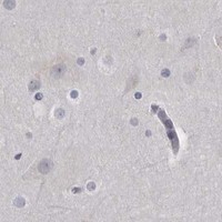


Comprehensive Human Protein Atlas IHC scoring per tissue (reliability: Enhanced — Medium consistency between antibody staining and RNA expression data. Secreted protein, tissue location of RNA and protein is expected to differ. Presumed off target binding observed and disregarded.). Rows are taken directly from the HPA tissue chart — click any row's HPA link to view the source.
| Tissue | Cell type | Level | Evidence | Source |
|---|---|---|---|---|
| Skin | Cells in granular layer | High | Protein (IHC) | HPA → |
| Lymph node | Non-germinal center cells | Medium | Protein (IHC) | HPA → |
| Spleen | Cells in red pulp | Medium | Protein (IHC) | HPA → |
| Tonsil | Non-germinal center cells | Medium | Protein (IHC) | HPA → |
| Tissue | Cell type | Level | Evidence | Source |
|---|---|---|---|---|
| Adipose tissue | Adipocytes | Not detected | Protein (IHC) | HPA → |
| Adrenal gland | Glandular cells | Not detected | Protein (IHC) | HPA → |
| Appendix | Glandular cells | Not detected | Protein (IHC) | HPA → |
| Bone marrow | Hematopoietic cells | Not detected | Protein (IHC) | HPA → |
| Breast | Adipocytes | Not detected | Protein (IHC) | HPA → |
Troubleshoot GZMH staining in paraffin sections by checking retrieval, controls, cellular pattern, and scoring before assigning biological meaning.
A08559-1 is listed for IHC and IF in human, mouse, and rat (catalog: applications/reactivity); its pictured IHC uses paraffin-embedded human breast carcinoma (IHC image caption).
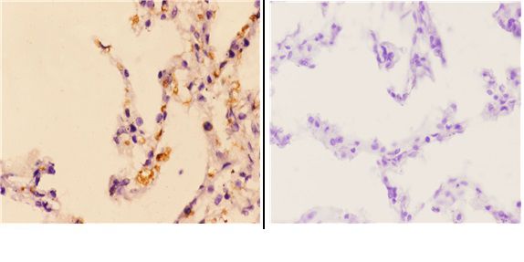
A08559-1 is listed for IHC and IF in human, mouse, and rat (catalog: applications/reactivity). Its IHC captions show paraffin-embedded human breast and lung carcinoma at 1:50; no IF image is supplied (IHC image captions; catalog: IF images).
Which to pick: For tissue IHC, choose A08559-1, a rabbit pAb pictured on paraffin-embedded human breast and lung carcinoma; the captions do not report the fixative (catalog: host; IHC image captions). For IF, A08559-1 is listed at 1:50–1:200, but ICC is unlisted and no IF image is supplied (catalog: applications, IF dilution, IF images). For mouse or rat samples, A08559-1 lists reactivity with both species, while its supplied IHC images show human tissue only (catalog: reactivity; IHC image captions).