This website uses cookies to ensure you get the best experience on our website.


- Table of Contents
Use 2 µg/ml of the catalog antibody for chromogenic IHC on paraffin sections (datasheet A06281-2). Compare cytoplasmic immune-cell staining with the tissue IHC profile and consider secretion when interpreting its distribution (HPA tissue IHC; UniProt).

Expected localisation, validated protocols, controls and antibodies — the at-a-glance facts below, then the full design guide.
| Expected localisation | Cytoplasm in immune cells (HPA tissue IHC) | |
| Staining pattern | Immune-cell cytoplasmic staining; texture unspecified (HPA tissue IHC) | |
| Antigen retrieval | EDTA pH 8.0 HIER, heat-mediated (datasheet A06281-2) | |
| Positive control | Appendix+3 more · see all | |
| Negative control | Adipose tissue+4 more · see all |
| Fixation | Keep fixation consistent across sections (standard IHC practice; not target-specific) | |
| Caveat | Secretion may separate tissue RNA and protein patterns (HPA tissue IHC) | |
| Regulation | Constitutive secretion by CD8+ T cells (UniProt) | |
| Isoform / epitope | No isoforms; cleavage removes 1–26 from mature GZMK (UniProt) |
The catalog antibody’s IHC-P protocol (datasheet: A06281-2) is followed by published chromogenic GZMK IHC protocols for breast tissue (PMC11150209) and lung tissue (PMC13618230).
| Sample | Paraffin-embedded human spleen tissue; fixative not specified (datasheet A06281-2) |
| Fixation | Image fixative and duration unreported (datasheet A06281-2); verify before use. |
| Sectioning | 4–5 µm sections on charged slides (standard) |
| Deparaffinisation | Xylene, graded ethanol series to water (standard) |
| Antigen retrieval | Heat retrieval: EDTA pH 8.0 (datasheet A06281-2); 20 min, 95–100 °C (standard) |
| Peroxidase block | 3% H2O2, 10 min, room temperature (standard) |
| Blocking | 10% goat serum (datasheet A06281-2) |
| Primary antibody | Rabbit anti-GZMK, 2 μg/ml (datasheet A06281-2) |
| Primary incubation | Overnight at 4 °C (datasheet A06281-2) |
| Detection | HRP-conjugated secondary, DAB chromogen (datasheet A06281-2) |
| Counterstain | Hematoxylin, blue, dehydrate and mount (standard) |
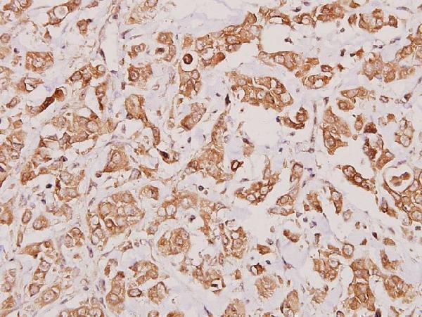
| Expected result | GZMK-positive staining in lymphoid tissue of appendix (HPA tissue IHC: Medium). HPA tissue profile: Cytoplasmic expression in immune cells. No signal in the no-primary control. |
In paraffin-section IHC, expect mainly cytoplasmic staining in immune cells (HPA tissue IHC: cytoplasmic immune-cell profile). Granule-associated staining is plausible, and extracellular signal may reflect secretion (UniProt P49863: cytoplasmic granule; secreted). GZMK has no transmembrane segment (UniProt P49863 topology). HPA rates tissue staining Enhanced, with medium consistency between staining and RNA and an explicit secretion-related location caveat (HPA tissue IHC: reliability description).
| Discrete cytoplasmic staining in immune cells within appendix lymphoid tissue, lymph-node or tonsil germinal centers. | This fits the tissue profile; HPA reports medium staining at these sites (HPA tissue IHC: positive tissues). Morphology alone does not identify stained cells as CD8-positive T cells (UniProt P49863: CD8-positive T-cell specificity). |
| Predominantly nuclear staining, especially without a cytoplasmic immune-cell pattern. | Treat this as discordant with the reported tissue profile, then check counterstain, detection and a control section before interpreting it as GZMK (HPA tissue IHC: cytoplasmic immune-cell profile; standard IHC practice). |
| Strong staining of adipocytes or respiratory epithelium without the expected immune-cell pattern. | Investigate nonspecific binding or endogenous detection activity: these cell types were reported as not detected in the sampled tissues (HPA tissue IHC: adipose adipocytes; bronchus respiratory epithelium). |
| Diffuse color across tissue, including areas without distinct stained cells. | Secreted GZMK can complicate assignment of extracellular signal to its producing cell (UniProt P49863: secreted; HPA tissue IHC: secretion caveat). Widespread color also calls for checks of blocking, washing and detection controls (standard IHC practice). |
| No convincing signal in appendix lymphoid tissue or lymph-node germinal-center cells. | These are reported medium staining sites, so review section quality, retrieval, antibody conditions and detection with controls before calling the assay negative (HPA tissue IHC: positive tissues; standard IHC practice). |
| Tissue choice and scoring | Appendix lymphoid tissue, lymph-node and tonsil germinal-center cells have reported medium staining; spleen red-pulp cells are reported low (HPA tissue IHC). Compare like cell populations rather than whole-section darkness (standard IHC practice). |
| Secretion and cell assignment | GZMK is granule-associated and secreted, including constitutive secretion by CD8-positive T cells without TCR stimulation (UniProt P49863: location note). An extracellular deposit need not mark the cell that made it (HPA tissue IHC: secretion caveat). |
| Topology and maturation | The precursor has a signal peptide at 1–24, a propeptide at 25–26 and a mature chain at 27–264; no transmembrane segment is annotated (UniProt P49863). The supplied record does not locate the antibody epitope. |
| Antibody evidence | HPA labels tissue IHC reliability Enhanced, while describing medium staining–RNA consistency and external characterization support (HPA tissue IHC: reliability description). Its IHC Enhanced entries are HPA063181 and HPA065895 (HPA antibodies). |
| Negative-cell comparison | HPA reports adipocytes in adipose tissue and respiratory epithelial cells in bronchus as not detected (HPA tissue IHC: negative tissues). Use their staining as a specificity warning, subject to section and detection controls (standard IHC practice). |
| IF/ICC Q: Should its reported Golgi pattern define the paraffin IHC readout? | A: No. HPA approves a Golgi location in ICC-IF, with images from JURKAT and REH; tissue IHC reports cytoplasmic immune-cell expression (HPA subcellular ICC-IF; HPA tissue IHC). Interpret each application against its own evidence. |
| Situation | Likely cause | Next action |
|---|---|---|
| Positive-control tissue shows little or no staining. | Target-specific fixation effects are not established by the supplied assay evidence. Verify with a matched IHC source before attributing a result to fixation. | Check section integrity, retrieval, antibody application and detection with appropriate run controls (standard IHC practice). |
| Most cells stain uniformly, obscuring immune-cell boundaries. | Nonspecific antibody binding, incomplete washing or detection background may be contributing (standard IHC practice). | Inspect a negative reagent control and review blocking, antibody concentration, washes and chromogen development (standard IHC practice). |
| Brown signal appears in nuclei but little appears in cytoplasm. | The compartment conflicts with the HPA tissue IHC profile; counterstain or detection artifact may be involved (HPA tissue IHC: cytoplasmic immune-cell profile; standard IHC practice). | Compare with the counterstain and negative control, then reassess localization in a reported positive tissue (HPA tissue IHC: positive tissues; standard IHC practice). |
| Adipocytes or bronchial respiratory epithelial cells stain strongly. | Those sampled cell types were not detected by HPA; cross-reactivity or endogenous detection activity is possible (HPA tissue IHC: negative tissues; standard IHC practice). | Check negative reagent controls and the detection-system blocking step before assigning the color to GZMK (standard IHC practice). |
| An extracellular rim is scored as a GZMK-positive cell. | GZMK can be secreted, so deposited protein may be spatially separated from its source (UniProt P49863: secreted; HPA tissue IHC: secretion caveat). | Score cellular staining separately from extracellular staining and record the compartment seen (standard IHC practice). |
| Only faint red-pulp staining is seen in spleen. | HPA reports low staining in spleen red-pulp cells; weak signal there alone does not establish assay failure (HPA tissue IHC: low tissue). | Check an HPA-reported medium site in the same run before changing assay conditions (HPA tissue IHC: positive tissues; standard IHC practice). |

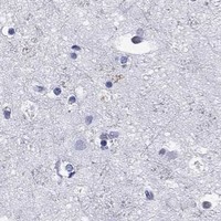
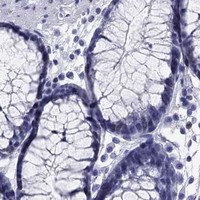
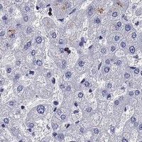


Comprehensive Human Protein Atlas IHC scoring per tissue (reliability: Enhanced — Medium consistency between antibody staining and RNA expression data. Secreted protein, tissue location of RNA and protein is expected to differ. External characterization data supports antibody staining.). Rows are taken directly from the HPA tissue chart — click any row's HPA link to view the source.
| Tissue | Cell type | Level | Evidence | Source |
|---|---|---|---|---|
| Appendix | Lymphoid tissue | Medium | Protein (IHC) | HPA → |
| Lung | Alveolar cells type I | Medium | Protein (IHC) | HPA → |
| Lymph node | Germinal center cells | Medium | Protein (IHC) | HPA → |
| Tonsil | Germinal center cells | Medium | Protein (IHC) | HPA → |
| Tissue | Cell type | Level | Evidence | Source |
|---|---|---|---|---|
| Adipose tissue | Adipocytes | Not detected | Protein (IHC) | HPA → |
| Adrenal gland | Glandular cells | Not detected | Protein (IHC) | HPA → |
| Bone marrow | Hematopoietic cells | Not detected | Protein (IHC) | HPA → |
| Breast | Adipocytes | Not detected | Protein (IHC) | HPA → |
| Bronchus | Respiratory epithelial cells | Not detected | Protein (IHC) | HPA → |
Troubleshoot GZMK staining in paraffin sections by checking retrieval, cell identity, subcellular pattern, and controls before scoring signal.
Anti-GZMK antibodies have IHC images from paraffin sections of human spleen and breast carcinoma (image captions); one also lists IF for Human, Mouse, and Rat (catalog: applications/reactivity).
A06281-2 has an IHC image from a paraffin-embedded human spleen section (A06281-2 image caption). A06281-1 has an IHC image from paraffin-embedded human breast carcinoma and lists IF for Human, Mouse, and Rat (A06281-1 image caption; catalog: applications/reactivity).
Which to pick: For human tissue IHC, choose A06281-2 when its documented spleen workflow fits: EDTA retrieval, 2 μg/ml primary antibody, and DAB detection; the fixative is unreported (A06281-2 image caption). For IF or work across Human, Mouse, and Rat, choose A06281-1, which lists IF and IHC at 1:50–1:200 and has its own paraffin-section IHC image at 1:100 (catalog: applications/reactivity/dilutions; A06281-1 image caption). Both are described as polyclonal, while ICC validation is unreported (A06281-2 datasheet: Polyclonal; A06281-1 image caption: pAb; catalog: applications).