This website uses cookies to ensure you get the best experience on our website.


- Table of Contents
Plan GZMM paraffin IHC with the catalog antibody at 2–5 μg/ml (datasheet A08286-2). Bone marrow hematopoietic cells and spleen red pulp cells show high staining, while secretion may complicate interpretation of tissue location (HPA tissue IHC).

Expected localisation, validated protocols, controls and antibodies — the at-a-glance facts below, then the full design guide.
| Expected localisation | Cytoplasmic granules or secreted; tissue location unconfirmed (UniProt) | |
| Staining pattern | High in a subset of immune cells; compartment unreported (HPA tissue IHC) | |
| Antigen retrieval | EDTA pH 8.0 HIER, heat-mediated (datasheet A08286-2) | |
| Positive control | Bone marrow+4 more · see all | |
| Negative control | Adipose tissue+4 more · see all |
| Fixation | Matched tissue-IHC evidence does not establish the fixation claim. Validate the specimen-specific method before use. (selected-SKU IHC image A08286-2) | |
| Caveat | Secretion may separate tissue staining from RNA expression (HPA tissue IHC) | |
| Regulation | Constitutive in activated NK cells (UniProt) | |
| Isoform / epitope | No isoforms annotated; epitopes in aa 1–25 may be lost (UniProt) |
Compare the catalog antibody’s IHC-P protocol (datasheet A08286-2) with published GZMM staining in endometrial cancer (PMC10792215) and colon carcinoma (PMC4467404).
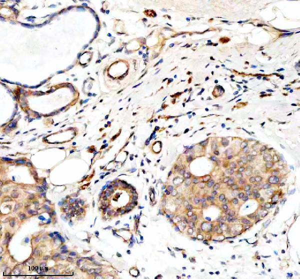
| Sample | Paraffin-embedded human breast cancer tissue; fixative not specified (datasheet A08286-2) |
| Fixation | Image fixative and duration unreported (datasheet A08286-2); verify before use. |
| Sectioning | 4–5 µm sections on charged slides (standard) |
| Deparaffinisation | Xylene, graded ethanol series to water (standard) |
| Antigen retrieval | Heat retrieval: EDTA pH 8.0 (datasheet A08286-2); 20 min, 95–100 °C (standard) |
| Peroxidase block | 3% H2O2, 10 min, room temperature (standard) |
| Blocking | 10% goat serum (datasheet A08286-2) |
| Primary antibody | Rabbit anti-GZMM, 2-5 μg/ml (datasheet A08286-2) |
| Primary incubation | Overnight at 4 °C (datasheet A08286-2) |
| Detection | HRP-conjugated secondary, DAB chromogen (datasheet A08286-2) |
| Counterstain | Hematoxylin, blue, dehydrate and mount (standard) |
| Expected result | GZMM-positive staining in hematopoietic cells of bone marrow (HPA tissue IHC: High). HPA tissue profile: High expression in subset of immune cells. No signal in the no-primary control. |
GZMM is a secreted protein associated with cytoplasmic granules of large granular lymphocytes and has no transmembrane segment (UniProt P51124). Expect staining in a subset of immune cells, consistent with its expression in activated NK cells (HPA tissue IHC: Enhanced; UniProt P51124). Tissue location can be difficult to interpret for a secreted protein, and HPA disregarded presumed off-target staining in its assessment (HPA tissue IHC: reliability description).
| Distinct cytoplasmic granules in scattered immune cells, with strong staining among bone marrow hematopoietic cells or splenic red-pulp cells. | This fits the reported granule location and tissue pattern (UniProt P51124; HPA tissue IHC: High in bone marrow hematopoietic cells and splenic red-pulp cells). Cell shape alone does not establish NK identity; use an appropriate cell marker if that distinction matters (general IHC practice). |
| Strong, uniform nuclear or sharply membrane-bound staining dominates the section. | That pattern is inconsistent with the annotated granule and secreted locations and absence of a transmembrane segment (UniProt P51124). Treat it as suspect until controls and a second detection approach support it (general IHC practice). |
| Strong staining appears in adipocytes or bronchial respiratory epithelium. | HPA reports GZMM as not detected in those cells, so investigate nonspecific binding or endogenous detection activity (HPA tissue IHC: adipocytes and bronchial respiratory epithelial cells not detected; general IHC practice). Do not apply this rule to every nonimmune cell: HPA reports medium staining in alveolar type I and tonsillar squamous epithelial cells (HPA tissue IHC). |
| Diffuse color covers many cell types or the section background, obscuring individual positive cells. | The result cannot be scored as the reported subset-of-immune-cells pattern (HPA tissue IHC: profile). Check the reagent control, detection system and wash steps for background, and compare with a known-positive tissue processed in the same run (general IHC practice). |
| No staining is visible in bone marrow hematopoietic cells or splenic red-pulp cells. | These are reported high-staining locations, so an entirely blank known-positive control calls the assay result into question (HPA tissue IHC: High). Check antibody and detection performance before interpreting an unstained study specimen as GZMM-negative (general IHC practice). |
| Compartment and secretion | Granule-associated cytoplasmic signal is expected, but extracellular signal may require cautious interpretation because GZMM is also secreted (UniProt P51124). HPA warns that RNA and protein tissue locations may differ for a secreted variant (HPA tissue IHC: reliability description). |
| Protein processing and epitope | UniProt annotates a signal peptide at residues 1–23, propeptide at 24–25 and mature chain at 26–257 (UniProt P51124). If an antibody's epitope is known, compare it with these regions when interpreting staining; no epitope is supplied here (general IHC practice). |
| Antibody validation and tissue comparison | The listed antibody HPA015624 has Enhanced IHC validation (HPA antibodies: HPA015624). Use the reported High bone marrow or spleen locations alongside a reported not-detected cell population to judge whether the observed pattern is selective (HPA tissue IHC). |
| IF/ICC: Is a cellular staining pattern established? | HPA lists GZMM as secreted but provides no main subcellular location or ICC-IF cell-line images (HPA subcellular). Granule localisation is annotated by UniProt, so an IF/ICC result needs independent validation before assigning a precise cellular pattern (UniProt P51124; general IF practice). |
| Situation | Likely cause | Next action |
|---|---|---|
| Known-positive bone marrow or spleen control is blank. | The run may have insufficient primary-antibody or detection signal; those locations are reported High by HPA (HPA tissue IHC; general IHC practice). | Verify the control section, primary-antibody step and detection reagents; review antigen-retrieval settings as a general IHC workflow check (general IHC practice). No GZMM-specific retrieval condition is supplied. |
| Study specimen is blank while the positive control stains. | The specimen may contain few expressing cells; GZMM staining is reported in a subset of immune cells (HPA tissue IHC: profile). | Review an appropriate immune-rich region and score the relevant cells rather than the entire section; document the positive control in the same run (HPA tissue IHC: profile; general IHC practice). |
| Nuclear or membrane signal overwhelms cytoplasmic staining. | The dominant compartment conflicts with the granule and secreted annotations and lack of a transmembrane segment (UniProt P51124). | Inspect the reagent control and repeat with a validated detection setup; avoid calling the compartment GZMM-specific without corroboration (general IHC practice). |
| Color appears broadly in cells HPA reports as not detected. | Nonspecific binding or endogenous detection activity is possible (general IHC practice); HPA reports no staining in adipocytes and bronchial respiratory epithelial cells (HPA tissue IHC). | Compare reagent controls and examine the detection-blocking step; assess the named cell population, since HPA also reports medium staining in some nonimmune cells (general IHC practice; HPA tissue IHC). |
| Background obscures scattered positive cells. | Broad reagent or detection background can prevent assessment of the reported subset pattern (general IHC practice; HPA tissue IHC: profile). | Compare a no-primary control, review blocking and washes, and rescore only when individual stained cells can be distinguished (general IHC practice). |
| A second tissue gives a different staining intensity. | HPA reports High staining in bone marrow hematopoietic and splenic red-pulp cells, but Medium staining in lymph-node non-germinal-center cells (HPA tissue IHC). | Compare the same annotated cell populations and run controls before treating an intensity difference as assay failure (HPA tissue IHC; general IHC practice). |


Comprehensive Human Protein Atlas IHC scoring per tissue (reliability: Enhanced — At least one protein variant secreted, tissue location of RNA and protein might differ and correlation is complex. Presumed off target binding observed and disregarded. Antibody staining in cells/structures not annotated, view images.). Rows are taken directly from the HPA tissue chart — click any row's HPA link to view the source.
| Tissue | Cell type | Level | Evidence | Source |
|---|---|---|---|---|
| Bone marrow | Hematopoietic cells | High | Protein (IHC) | HPA → |
| Spleen | Cells in red pulp | High | Protein (IHC) | HPA → |
| Lung | Alveolar cells type I | Medium | Protein (IHC) | HPA → |
| Lymph node | Non-germinal center cells | Medium | Protein (IHC) | HPA → |
| Tonsil | Squamous epithelial cells | Medium | Protein (IHC) | HPA → |
| Tissue | Cell type | Level | Evidence | Source |
|---|---|---|---|---|
| Adipose tissue | Adipocytes | Not detected | Protein (IHC) | HPA → |
| Adrenal gland | Glandular cells | Not detected | Protein (IHC) | HPA → |
| Appendix | Glandular cells | Not detected | Protein (IHC) | HPA → |
| Breast | Adipocytes | Not detected | Protein (IHC) | HPA → |
| Bronchus | Respiratory epithelial cells | Not detected | Protein (IHC) | HPA → |
Troubleshoot GZMM chromogenic IHC by checking retrieval, tissue processing, cellular localisation, controls and scoring before interpreting weak or unexpected staining.
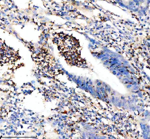
One anti-GZMM antibody has real IHC data from human paraffin-embedded breast and colon cancer sections; no IF figure is supplied (A08286-2 image captions; catalog: IF images).
A08286-2 is listed for human IHC at 2–5 μg/ml (catalog: applications, reactivity, IHC dilution). Its own IHC captions show paraffin-embedded human breast and colon cancer sections stained at 2 μg/ml (A08286-2 image captions).
Which to pick: Choose A08286-2 for human paraffin-section IHC: it is a rabbit polyclonal antibody listed for human IHC, with breast and colon cancer section images (catalog: host, clonality, applications, reactivity; A08286-2 image captions). No SKU in the supplied catalog is listed for IF/ICC or species beyond human (catalog: applications, reactivity). The A08286-2 captions report paraffin embedding but leave the fixative unreported (A08286-2 image captions).