This website uses cookies to ensure you get the best experience on our website.


- Table of Contents
HAAO shows strong cytoplasmic staining in liver hepatocytes and kidney proximal tubules (HPA tissue IHC). For paraffin IHC, start antibody A10482 at 2.5 μg/mL (datasheet: A10482) and score staining by cell type (HPA tissue IHC).

Expected localisation, validated protocols, controls and antibodies — the at-a-glance facts below, then the full design guide.
| Expected localisation | Cytoplasmic in hepatocytes and renal tubules (HPA tissue IHC) | |
| Staining pattern | Strong cytoplasmic hepatocytes and proximal tubules (HPA tissue IHC) | |
| Antigen retrieval | Citrate pH 6.0 HIER, 95–98 °C, 20 min (rule: cytoplasmic / membrane antigen) | |
| Positive control | Kidney+2 more · see all | |
| Negative control | Adipose tissue+4 more · see all |
| Fixation | Keep paraffin-section fixation consistent (standard IHC practice; not target-specific) | |
| Caveat | Signal varies sharply by tissue and cell type (HPA tissue IHC) | |
| Regulation | Liver-enriched expression (HPA tissue IHC) | |
| Isoform / epitope | 2 isoforms; epitope coverage is unspecified (UniProt) |
The catalog antibody’s IHC-P protocol is accompanied by published HAAO IHC conditions for lung, cancer tissue microarrays, and mouse kidney (PMC4448130; PMC9951368; PMC12081153).
| Sample | Tissue sections; selected-image fixative not specified (standard IHC workflow) |
| Fixation | Image fixative and duration unreported (datasheet A10482); verify before use. |
| Sectioning | 4–5 µm sections on charged slides (standard) |
| Deparaffinisation | Xylene, graded ethanol series to water (standard) |
| Antigen retrieval | Heat-induced epitope retrieval in citrate buffer, pH 6.0, 20 min at 95–98 °C (standard rule: cytoplasmic / membrane antigen) |
| Peroxidase block | 3% H2O2, 10 min, room temperature (standard) |
| Blocking | 10% normal serum of the secondary host, 30 min, room temperature (standard) |
| Primary antibody | Rabbit anti-HAAO, 2.5 μg/mL (datasheet A10482) |
| Primary incubation | Overnight at 4 °C (standard) |
| Detection | HRP-polymer secondary, DAB chromogen 5–10 min (standard) |
| Counterstain | Hematoxylin, blue, dehydrate and mount (standard) |
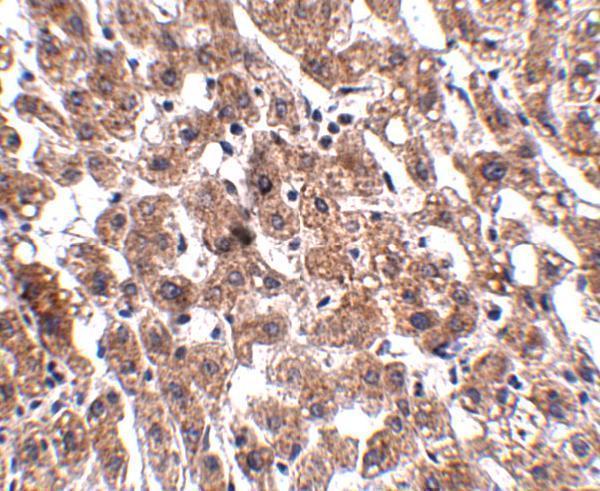
| Expected result | HAAO-positive staining in proximal tubules (cell body) of kidney (HPA tissue IHC: High). HPA tissue profile: Selective cytoplasmic expression most abundant in hepatocytes and renal tubules. No signal in the no-primary control. |
HAAO should show cytoplasmic staining, strongest in hepatocytes and kidney proximal tubule cell bodies, with medium staining in placental decidual cells (HPA tissue IHC: Enhanced; High in hepatocytes and proximal tubules; Medium in decidual cells). This distribution agrees with a cytosolic protein lacking a transmembrane segment (UniProt P46952: cytosol; no transmembrane segment).
| Strong cytoplasmic staining in hepatocytes and proximal tubule cell bodies (HPA tissue IHC: High in both). | This matches the reported selective pattern and expected compartment (HPA tissue IHC: selective cytoplasmic expression; UniProt P46952: cytosol). Compare intensity within the same staining run, because the HPA levels describe observed tissue patterns, not a required numerical threshold (HPA tissue IHC: High). |
| Predominantly nuclear or sharply membrane-localized staining in otherwise positive cells. | Treat this as a localization mismatch and investigate an artefact before calling it HAAO: the reported pattern is cytoplasmic, and HAAO has no annotated transmembrane segment (HPA tissue IHC: selective cytoplasmic expression; UniProt P46952: cytosol, no transmembrane segment). |
| Strong staining in adipocytes, adrenal glandular cells, or respiratory epithelial cells. | These cells are reported as not detected, so strong signal raises concern for cross-reactivity or endogenous detection activity (HPA tissue IHC: Not detected in these cells; general IHC practice). Check the corresponding tissue and detection controls before assigning HAAO positivity (general IHC practice). |
| Diffuse color covers cells and surrounding tissue without a clear cytoplasmic pattern. | This does not support a cell-specific HAAO call (HPA tissue IHC: selective cytoplasmic expression). General IHC causes to assess include excess antibody, incomplete blocking, inadequate washes, and chromogen development; the supplied HAAO sources do not identify a target-specific cause (general IHC practice). |
| No staining in hepatocytes or proximal tubule cell bodies. | An expected positive has failed: both cell types show High staining in the HPA tissue profile (HPA tissue IHC: High in hepatocytes and proximal tubules). Check tissue identity, antibody and detection controls, and the run conditions before interpreting a test specimen as negative (general IHC practice). |
| Cellular location and topology (UniProt P46952: cytosol; no transmembrane segment). | Score signal in the cell body cytoplasm rather than using a membrane outline or nuclear deposit as the expected pattern (HPA tissue IHC: selective cytoplasmic expression; UniProt P46952: cytosol). Topology alone does not specify an antigen-retrieval condition (UniProt P46952: topology; general IHC practice). |
| Tissue and cell distribution (HPA tissue IHC: High in hepatocytes and proximal tubules; Medium in decidual cells). | Use liver or kidney as an expected positive reference. Placental decidual cells provide a reported medium-level comparison; adipocytes and several other listed cells are not detected (HPA tissue IHC: cell-level observations). These levels are observations, not universal pass/fail intensity cutoffs (HPA tissue IHC: staining levels). |
| IHC validation (HPA antibodies: HPA036394 and HPA042024, both IHC Enhanced). | The HPA tissue profile has Enhanced reliability, described as high consistency between staining and RNA expression (HPA tissue IHC: Enhanced). This supports the reported IHC pattern; it does not establish how a different antibody or detection system will perform (HPA antibodies: validation status; general IHC practice). |
| Isoforms and processing (UniProt P46952: isoforms 1 and 2; chain 1–286; no signal peptide or propeptide). | Two isoforms are annotated, but the supplied record gives no antibody epitope or isoform-specific staining data (UniProt P46952: two isoforms; HPA antibodies: IHC validation only). Do not interpret a regional staining difference as isoform-specific from these sources alone. |
| IF evidence (HPA subcellular: main location unavailable; no ICC-IF images). | The HPA subcellular record provides no image-based ICC-IF localization for HAAO (HPA subcellular: no ICC-IF images). Its Hep-G2 value is an RNA expression measure, not an IF protein pattern (HPA subcellular: 42.5 nTPM); assess IF on its dedicated guide page. |
| Chromogenic detection background (general IHC practice). | Endogenous enzyme activity can produce color independent of primary-antibody binding in enzyme-based IHC (general IHC practice). Interpret unexpected staining against an appropriate no-primary detection control; HPA's HAAO tissue profile does not identify a sample-specific background mechanism (HPA tissue IHC: tissue observations). |
| Situation | Likely cause | Next action |
|---|---|---|
| The liver or kidney reference has no signal (HPA tissue IHC: High in hepatocytes and proximal tubules). | The run may have failed, or the sampled section may lack the expected cells; the source record does not identify a HAAO-specific failure mechanism (general IHC practice; HPA tissue IHC: positive cells). | Confirm tissue and cell identity, then inspect antibody application, retrieval, detection reagents, and the run control before scoring other sections (general IHC practice). Keep retrieval changes empirical because no HAAO-specific setting is supplied (HPA tissue IHC: no retrieval data). |
| Signal is mainly nuclear or outlines membranes (HPA tissue IHC: selective cytoplasmic expression). | The pattern conflicts with reported HAAO localization (UniProt P46952: cytosol, no transmembrane segment). Nonspecific binding or detection artefact is possible, but the image alone cannot distinguish causes (general IHC practice). | Review the cell morphology and no-primary control, then repeat with adjusted blocking or antibody conditions if the controls implicate background (general IHC practice). Require a cytoplasmic pattern in expected positive cells for interpretation (HPA tissue IHC: selective cytoplasmic expression). |
| Strong color appears in reported negative cell types (HPA tissue IHC: Not detected in adipocytes, adrenal glandular cells, and respiratory epithelial cells). | Cross-reactivity or endogenous detection activity may explain staining outside the reported HAAO distribution (general IHC practice; HPA tissue IHC: negative cell types). A single unexpected cell cannot establish which mechanism applies. | Compare a no-primary control and the expected positive tissue in the same run; review blocking, detection, and antibody concentration if unexpected color persists (general IHC practice). Do not reclassify the HPA-negative cells from color alone (HPA tissue IHC: Not detected). |
| Diffuse background obscures hepatocytes or proximal tubules (HPA tissue IHC: expected positive cells). | High background can conceal cell-level cytoplasmic staining (general IHC practice). Excess reagent, insufficient washing, or overdevelopment are possible general IHC causes; none is a documented HAAO-specific effect in the supplied sources (general IHC practice). | Use the run controls to locate the background source, then optimize blocking, washing, antibody concentration, or chromogen development one variable at a time (general IHC practice). Reassess whether signal is cytoplasmic and cell-selective (HPA tissue IHC: selective cytoplasmic expression). |
| The no-primary section develops color (general IHC practice). | Color without primary antibody points to the detection workflow or endogenous activity, rather than proving HAAO binding (general IHC practice). The HPA tissue observations do not establish a HAAO-specific source of this color (HPA tissue IHC: tissue observations). | Review the detection reagents and the block appropriate to the enzyme system, then repeat the control alongside the positive tissue (general IHC practice). Interpret test-section staining only after the control is acceptable (general IHC practice). |
| Can the HPA IHC pattern be used as an IF/ICC result? | The HPA subcellular record has no ICC-IF images or assigned main location (HPA subcellular: no ICC-IF images; main location unavailable). Its tissue IHC pattern establishes an IHC expectation, not an observed IF distribution (HPA tissue IHC: selective cytoplasmic expression). | Use the separate IF/ICC guide for that application. If assessing IF, require application-appropriate controls and evaluate the observed compartment directly; do not infer IF validation or an IF protocol from the IHC antibody status (HPA antibodies: IHC Enhanced, ICC unreported; general IF practice). |

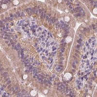


Comprehensive Human Protein Atlas IHC scoring per tissue (reliability: Enhanced — High consistency between antibody staining and RNA expression data.). Rows are taken directly from the HPA tissue chart — click any row's HPA link to view the source.
| Tissue | Cell type | Level | Evidence | Source |
|---|---|---|---|---|
| Kidney | Proximal tubules (cell body) | High | Protein (IHC) | HPA → |
| Liver | Hepatocytes | High | Protein (IHC) | HPA → |
| Placenta | Decidual cells | Medium | Protein (IHC) | HPA → |
| Tissue | Cell type | Level | Evidence | Source |
|---|---|---|---|---|
| Adipose tissue | Adipocytes | Not detected | Protein (IHC) | HPA → |
| Adrenal gland | Glandular cells | Not detected | Protein (IHC) | HPA → |
| Appendix | Glandular cells | Not detected | Protein (IHC) | HPA → |
| Bone marrow | Hematopoietic cells | Not detected | Protein (IHC) | HPA → |
| Breast | Adipocytes | Not detected | Protein (IHC) | HPA → |
Use cytoplasmic staining in hepatocytes and renal tubules as the main reference when optimising HAAO staining in paraffin sections (HPA tissue IHC).
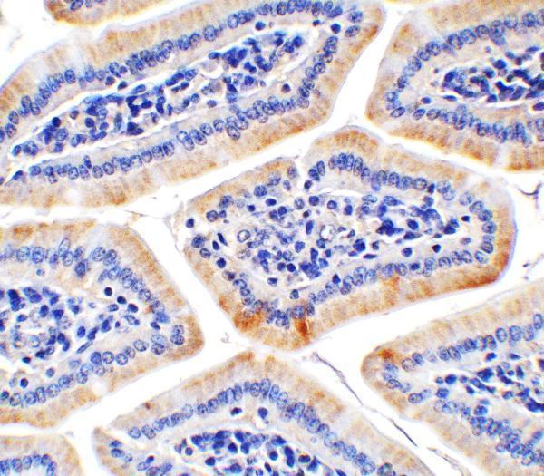
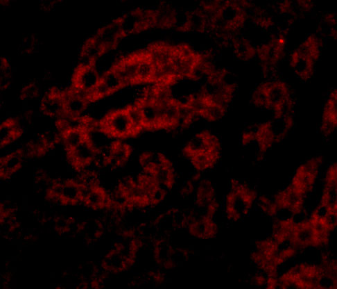
The IHC-validated antibody has images of human liver and mouse colon tissue; its IF image shows human liver cells (catalog image captions).
A10482 is the SKU that will render; its IHC images show human liver and mouse colon tissue (catalog image captions). Its IF image shows human liver cells, and its listed reactivity is Human, Mouse and Rat (catalog image caption; catalog reactivity).
Which to pick: Choose A10482 for tissue IHC: its own images show human liver at 2.5 μg/mL and mouse colon at 2 μg/mL (A10482 IHC image captions); the fixative is unreported. For IF, A10482 has a human liver cell image at 20 μg/mL and lists IF as an application; A10482-1 has an ICC image of Caco-2 cells but does not list IF as an application (catalog IF image captions; catalog applications). For cross-species work, A10482 lists Human, Mouse and Rat reactivity, whereas A10482-1 lists Human and Rat; both list a rabbit host, and neither provides a clone designation (catalog reactivity; catalog host and clone fields).