This website uses cookies to ensure you get the best experience on our website.


- Table of Contents
Plan HADH staining in paraffin sections using the catalog antibody’s IHC protocol. The guide covers granular cytoplasmic staining (HPA tissue IHC), mitochondrial matrix localisation (UniProt), and interpretation across broadly positive tissues (HPA tissue IHC).

Expected localisation, validated protocols, controls and antibodies — the at-a-glance facts below, then the full design guide.
| Expected localisation | Granular cytoplasm (HPA tissue IHC); mitochondrial matrix (UniProt) | |
| Staining pattern | Ubiquitous granular cytoplasmic staining (HPA tissue IHC) | |
| Antigen retrieval | EDTA pH 8.0 HIER, heat-mediated (datasheet A03650-1) | |
| Positive control | Adipose tissue+4 more · see all | |
| Negative control | None in HPA (detected in all 44 tissues); use no-primary + isotype controls |
| Fixation | Keep fixation consistent across paraffin sections. (standard IHC practice; not target-specific) | |
| Caveat | Broad expression limits tissue-specific interpretation (HPA tissue IHC) | |
| Regulation | No specific expression regulator reported (UniProt) | |
| Isoform / epitope | 3 isoforms; mature chain starts at residue 13; epitope map unknown (UniProt) |
The catalog antibody’s IHC-P protocol uses EDTA pH 8.0 retrieval (datasheet A03650-1). Three published HADH IHC protocols below provide tissue-specific comparisons (PMC12860641; PMC11402447; PMC13326317).
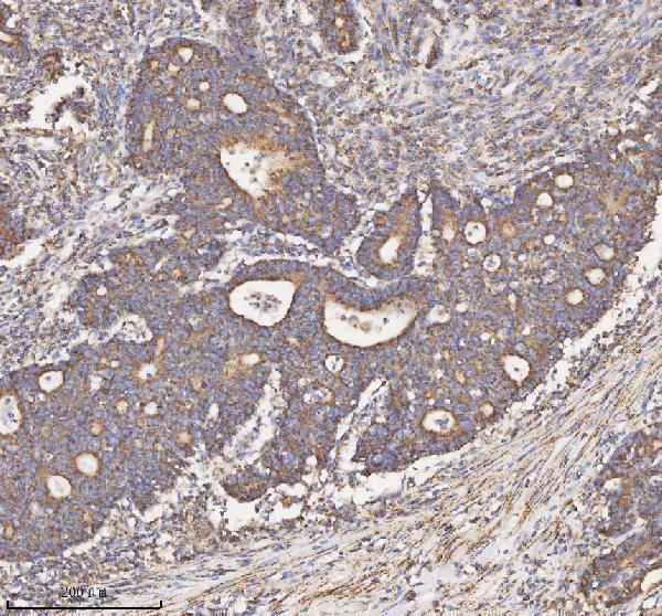
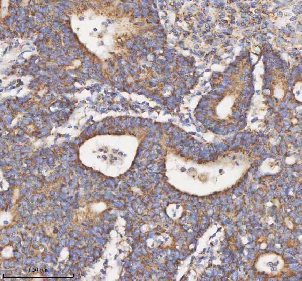
| Sample | Paraffin-embedded human colon adenocarcinoma tissue; fixative not specified (datasheet A03650-1) |
| Fixation | Image fixative and duration unreported (datasheet A03650-1); verify before use. |
| Sectioning | 4–5 µm sections on charged slides (standard) |
| Deparaffinisation | Xylene, graded ethanol series to water (standard) |
| Antigen retrieval | Heat retrieval: EDTA pH 8.0 (datasheet A03650-1); 20 min, 95–100 °C (standard) |
| Peroxidase block | 3% H2O2, 10 min, room temperature (standard) |
| Blocking | 10% goat serum (datasheet A03650-1) |
| Primary antibody | Rabbit anti-HADH, 2-5 μg/ml (datasheet A03650-1) |
| Primary incubation | Overnight at 4 °C (datasheet A03650-1) |
| Detection | HRP-conjugated secondary, DAB chromogen (datasheet A03650-1) |
| Counterstain | Hematoxylin, blue, dehydrate and mount (standard) |
| Expected result | HADH-positive staining in adipocytes of adipose tissue (HPA tissue IHC: High). HPA tissue profile: Ubiquitous cytoplasmic expression with a granular expression. No signal in the no-primary control. |
HADH is a mitochondrial matrix protein with no transmembrane segment (UniProt Q16836 localisation and topology). In paraffin-section IHC, expect granular cytoplasmic staining across multiple cell types (HPA: ubiquitous granular cytoplasmic expression; Enhanced tissue reliability). HPA reports high staining in adipocytes, glandular cells, endocrine cells, hematopoietic cells and glial cells in the listed tissues (HPA: tissue IHC).
| Granular cytoplasmic staining in adipocytes, with nuclei relatively clear. | This fits the reported IHC pattern and a mitochondrial matrix location (HPA: High in adipocytes; HPA: granular cytoplasmic expression; UniProt Q16836 localisation). Routine chromogenic IHC can support this compartment-level interpretation without resolving individual mitochondria (standard IHC practice). Compare staining with nearby tissue and detection controls before judging intensity (standard IHC practice). |
| Strong nuclear or sharply outlined surface staining dominates the section. | That distribution conflicts with the expected mitochondrial, granular cytoplasmic pattern (UniProt Q16836 localisation and topology; HPA: tissue IHC profile). Treat it as a localisation mismatch and investigate background or antibody specificity; the image alone cannot establish which mechanism caused it (standard IHC interpretation). |
| An unexpected cell population stains strongly while an HPA-listed high population is blank. | Cross-reactivity or endogenous detection activity is possible (standard IHC practice). HPA describes broad HADH expression and supplies no negative cell list, so an unlisted positive cell type is not, by itself, evidence of a false positive (HPA: ubiquitous expression; negative list empty). Confirm cell identity and inspect detection controls (standard IHC practice). |
| Uniform colour obscures cell boundaries and granular cytoplasmic detail. | Diffuse deposition prevents a confident localisation call, even where the tissue contains an HPA-listed positive population (HPA: tissue IHC; standard IHC interpretation). Background from detection chemistry or insufficient washing is a general possibility; neither HPA nor UniProt identifies a HADH-specific cause here (standard IHC practice). |
| No staining appears in adipocytes in adipose tissue. | This conflicts with an HPA high-staining example, but one blank section does not establish absence of HADH (HPA: High in adipose-tissue adipocytes). Review tissue quality, retrieval, antibody application and detection controls as general IHC checks (standard IHC practice). The supplied sources give no HADH-specific fixation sensitivity or retrieval condition. |
| Compartment and topology | HADH resides in the mitochondrial matrix and has no transmembrane segment (UniProt Q16836). Score granular cytoplasmic staining against that expectation; do not require visible mitochondrial outlines in routine chromogenic sections (HPA: granular cytoplasmic IHC; standard IHC interpretation). |
| Tissue breadth | HPA calls expression ubiquitous with low tissue RNA specificity and reports high IHC staining in several cell populations (HPA: tissue IHC). Use its named examples as positive references, while allowing other cell types to stain; the supplied HPA record lists no negative tissue or cell population. |
| Evidence strength | The tissue profile has Enhanced reliability because antibody staining is highly consistent with RNA expression data (HPA: tissue IHC reliability). HPA039588 is Supported for IHC and HPA043888 is Enhanced for IHC (HPA: antibody validation); these grades support interpretation but do not validate an unspecified catalog antibody. |
| Processing and isoforms | UniProt lists a mature chain at residues 13–314 and 3 isoforms (UniProt Q16836 processing and isoforms). The supplied record does not locate the antibody epitope or establish isoform coverage, so a staining difference cannot be assigned to processing or a particular isoform. |
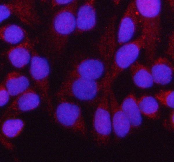
| IF/ICC Q: what should fluorescence show? | A: Mainly mitochondrial localisation, supported by HPA ICC-IF images; this agrees with the UniProt matrix annotation (HPA: mitochondria, supported; UniProt Q16836 localisation). IF/ICC images are a localisation cross-check for IHC interpretation, not a paraffin-IHC intensity standard (standard assay interpretation). |
| Situation | Likely cause | Next action |
|---|---|---|
| The expected granular pattern is absent in an HPA-listed high population. | The result conflicts with the reference pattern, but the source data do not identify a HADH-specific technical cause (HPA: tissue IHC). | Check tissue integrity, retrieval execution, antibody application and detection controls; repeat with a documented positive tissue if needed (standard IHC practice). |
| Nuclei stain more strongly than cytoplasm. | Predominantly nuclear signal conflicts with the mitochondrial matrix annotation and granular cytoplasmic IHC profile (UniProt Q16836; HPA: tissue IHC). | Review primary-omission and detection controls, then compare the pattern with an HPA-listed positive cell population (standard IHC practice; HPA: tissue IHC). |
| A crisp cell-surface rim dominates. | Surface localisation conflicts with HADH's matrix location and lack of a transmembrane segment (UniProt Q16836 localisation and topology). | Inspect background controls and whether the signal tracks tissue edges or other nonspecific deposition patterns (standard IHC practice). |
| Colour is diffuse across cells and extracellular areas. | The background masks the granular cytoplasmic pattern described by HPA; excess detection signal is one general possibility (HPA: tissue IHC; standard IHC practice). | Review blocking, washes, detection development and a primary-omission control before scoring positive cells (standard IHC practice). |
| Only an unlisted cell population stains. | Specificity or endogenous activity may need review, but HPA's ubiquitous profile does not exclude that cell type (HPA: tissue IHC; standard IHC practice). | Confirm cell identification, check detection controls and compare an HPA-listed high population on a suitable section (standard IHC practice; HPA: tissue IHC). |
| Two sections differ markedly in staining intensity. | The supplied sources establish a qualitative pattern, not a HADH-specific fixation effect or a quantitative intensity threshold (HPA: tissue IHC; UniProt Q16836). | Review section quality and keep retrieval, detection and scoring conditions comparable before interpreting the difference (standard IHC practice). |
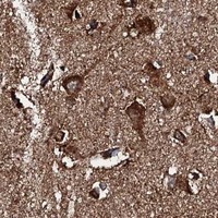
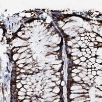
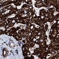
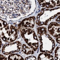


Comprehensive Human Protein Atlas IHC scoring per tissue (reliability: Enhanced — High consistency between antibody staining and RNA expression data.). Rows are taken directly from the HPA tissue chart — click any row's HPA link to view the source.
| Tissue | Cell type | Level | Evidence | Source |
|---|---|---|---|---|
| Adipose tissue | Adipocytes | High | Protein (IHC) | HPA → |
| Adrenal gland | Glandular cells | High | Protein (IHC) | HPA → |
| Appendix | Endocrine cells | High | Protein (IHC) | HPA → |
| Bone marrow | Hematopoietic cells | High | Protein (IHC) | HPA → |
| Breast | Adipocytes | High | Protein (IHC) | HPA → |
| Tissue | Cell type | Level | Evidence | Source |
|---|---|---|---|---|
| None in HPA: HADH is detected in all 44 scored tissues. Use a no-primary (secondary-only) and an isotype control instead. | ||||
Troubleshoot HADH staining in paraffin sections by checking retrieval, compartment, cell type and background before comparing chromogenic signal across samples.
A03650-1 has real IHC images from human paraffin sections and IF images from human cells and a paraffin section (catalog image captions). Its listed reactivity is human, mouse and rat (catalog applications and reactivity).
A03650-1 has IHC images from human colon adenocarcinoma and glioblastoma paraffin sections (catalog IHC image captions). The same SKU has IF images from HeLa cells and a human intestinal cancer paraffin section, and lists IHC, ICC and IF applications (catalog IF image captions; catalog applications).
Which to pick: Choose A03650-1 for tissue IHC: its own image captions show paraffin sections with EDTA retrieval at pH 8.0 and primary antibody at 2 μg/ml; the fixative is unreported (catalog IHC image captions). Choose the same SKU for IF/ICC because both applications are listed and its IF captions show human cells and a paraffin section (catalog applications; catalog IF image captions). For cross-species work, the catalog lists human, mouse and rat reactivity, but its IHC-specific dilution listing names human and rat, so mouse IHC requires separate validation (catalog reactivity; catalog dilution listing).