This website uses cookies to ensure you get the best experience on our website.


- Table of Contents
This HAND1 IHC-P guide covers the nuclear membranous tissue pattern (HPA tissue IHC) alongside the nucleoplasmic and nucleolar molecular location (UniProt). Start the catalog antibody at 1:100–1:300 (datasheet), and interpret staining with controls because tissue evidence has uncertain reliability (HPA tissue IHC).

Expected localisation, validated protocols, controls and antibodies — the at-a-glance facts below, then the full design guide.
| Expected localisation | Nuclear membranous tissue staining (HPA tissue IHC) | |
| Staining pattern | Nuclear membranous staining in most tissues, including CNS (HPA tissue IHC) | |
| Antigen retrieval | Tris-EDTA pH 9.0 HIER, 95–98 °C, 20 min (rule: nuclear antigen) | |
| Positive control | Caudate+4 more · see all | |
| Negative control | Adipose tissue+4 more · see all |
| Fixation | Matched tissue-IHC evidence does not establish the fixation claim. Validate the specimen-specific method before use. (selected-SKU IHC image A06496-1) | |
| Caveat | Presumed off-target staining and low RNA agreement (HPA tissue IHC) | |
| Regulation | PLK4 may release HAND1 from the nucleolus (UniProt) | |
| Isoform / epitope | No annotated isoforms; one 1–215 chain (UniProt) |
The catalog antibody has its own IHC-P protocol. Published HAND1 IHC methods add paraffin bone preparation and chromogenic detection details (PMC4769249), plus antibody dilution and incubation details (PMC9760445).
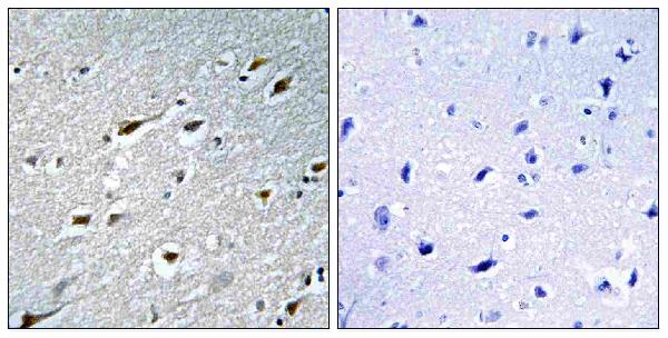
| Sample | Paraffin-embedded human brain tissue; fixative not specified (datasheet A06496-1) |
| Fixation | Image fixative and duration unreported (datasheet A06496-1); verify before use. |
| Sectioning | 4–5 µm sections on charged slides (standard) |
| Deparaffinisation | Xylene, graded ethanol series to water (standard) |
| Antigen retrieval | Heat-induced epitope retrieval in Tris-EDTA buffer, pH 9.0, 20 min at 95–98 °C (standard rule: nuclear antigen) |
| Peroxidase block | 3% H2O2, 10 min, room temperature (standard) |
| Blocking | 10% normal serum of the secondary host, 30 min, room temperature (standard) |
| Primary antibody | Rabbit anti-HAND1, 1:100 - 1:300 (datasheet A06496-1) |
| Primary incubation | Overnight at 4 °C (standard) |
| Detection | HRP-polymer secondary, DAB chromogen 5–10 min (standard) |
| Counterstain | Hematoxylin, blue, dehydrate and mount (standard) |
| Expected result | HAND1-positive staining in glial cells of caudate (HPA tissue IHC: High). HPA tissue profile: Nuclear membranous expression in most tissues including CNS. No signal in the no-primary control. |
HAND1 is a nuclear transcription factor found in the nucleoplasm and, under some conditions, the nucleolus (UniProt O96004). In paraffin-section IHC, assess staining within cells of the tissue being examined; HPA reports high staining in selected glial cells and cerebellar molecular-layer cells, but rates its tissue IHC profile Uncertain because staining and RNA expression show low consistency and presumed off-target binding was observed (HPA: tissue IHC). HAND1 has no transmembrane segment (UniProt O96004).
| Distinct nuclear staining in a subset of cells, with visible nuclear detail. | This fits HAND1's reported nuclear location (UniProt O96004). Record the stained cell type and compartment; nuclear location alone cannot establish specificity because HPA rates tissue IHC Uncertain (HPA: tissue IHC). |
| Predominantly cytoplasmic stain, with little or no nuclear signal. | This conflicts with HAND1's nucleoplasmic and nucleolar locations (UniProt O96004). Review the staining controls and antibody specificity before scoring these cells as positive (general IHC practice). |
| Strong signal across many cell types, especially as a nuclear rim without clear nucleoplasmic staining. | HPA describes nuclear-membranous staining across most tissues but flags presumed off-target binding and low consistency with RNA data (HPA: tissue IHC). A nuclear rim alone is therefore insufficient evidence of HAND1 in tissue IHC. |
| Diffuse color over nuclei, cytoplasm and surrounding tissue. | Diffuse deposition prevents reliable compartment scoring (general IHC practice). Compare a matched negative detection control and inspect whether the color follows tissue structures rather than individual nuclei (general IHC practice). |
| No stain in a section selected because HPA reports high staining there. | The absence needs a technical check, but the HPA-high designation is not an independently validated positive control: the tissue IHC profile is Uncertain (HPA: tissue IHC). |
| Nuclear staining restricted to a cell population reported as not detected by HPA. | For example, HPA reports adipocytes and bone-marrow hematopoietic cells as not detected (HPA: tissue IHC). Treat a result in those cells as a specificity question requiring controls, while recognizing that the HPA tissue profile itself is uncertain (HPA: tissue IHC). |
| Tissue IHC evidence | HPA reports high staining in caudate and cerebral-cortex glial cells and cerebellar molecular-layer cells, yet assigns Uncertain reliability because staining and RNA data have low consistency (HPA: tissue IHC). |
| Tissue choice | UniProt lists heart tissue specificity; HPA reports RNA enhancement in heart muscle and intestine (UniProt O96004; HPA: tissue IHC). Neither statement establishes a validated heart-section IHC positive control (HPA: tissue IHC). |
| Nuclear distribution | HAND1 is reported in nucleoplasm and nucleolus; MDFIC interaction can sequester it in the nucleolus, with PLK4 phosphorylation linked to release in a by-similarity annotation (UniProt O96004). Do not require identical nuclear distribution in every cell. |
| Topology and processing | HAND1 has no transmembrane segment or signal peptide, and UniProt lists a single chain spanning residues 1–215 (UniProt O96004). The record supplies no antibody epitope or target-specific antigen-retrieval response. |
| Antibody validation | HPA040925 is Uncertain for IHC; HPA040925 and HPA075313 are Approved for ICC (HPA: antibody validation). ICC approval does not resolve the uncertainty of paraffin-section IHC. |
| Situation | Likely cause | Next action |
|---|---|---|
| Nuclear stain is weak or absent throughout the run. | A run-level detection or staining problem is possible (general IHC practice). | Check the run controls, section integrity and detection reagents; interpret a negative HAND1 result only after the controls perform as expected (general IHC practice). |
| An HPA-high comparison section is negative. | HPA's tissue IHC profile is Uncertain, so its high-staining images do not establish a validated positive control (HPA: tissue IHC). | Check run controls and review tissue and cell identification before concluding that the test section lacks HAND1 (general IHC practice). |
| Most tissues show a strong nuclear rim. | HPA reports a widespread nuclear-membranous pattern and presumed off-target binding (HPA: tissue IHC). | Compare the rim with nucleoplasmic staining and negative controls; avoid scoring rim-only signal as confirmed HAND1 (UniProt O96004; general IHC practice). |
| Color appears in places where no primary-antibody signal is expected. | Endogenous detection activity or nonspecific detection can produce color in chromogenic IHC (general IHC practice). | Review the negative detection control and apply blocking appropriate to the detection system, then reassess the cellular pattern (general IHC practice). |
| Cytoplasmic staining dominates the section. | The pattern disagrees with the reported nuclear localization of HAND1 (UniProt O96004). | Check controls and reassess localization before calling cells positive; record the cytoplasmic signal as discordant if it persists (general IHC practice). |
| IF/ICC: where should HAND1 appear? | HPA's ICC-IF localization evidence is separate from its Uncertain tissue IHC assessment (HPA: subcellular; HPA: tissue IHC). | Expect mainly nucleoplasmic signal, with additional nuclear-body or nuclear-membrane signal reported in ICC-IF; use the separate IF/ICC guide for that assay (HPA: subcellular). |
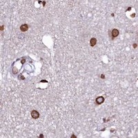


Comprehensive Human Protein Atlas IHC scoring per tissue (reliability: Uncertain — Low consistency between antibody staining and RNA expression data. Presumed off target binding observed and disregarded.). Rows are taken directly from the HPA tissue chart — click any row's HPA link to view the source.
| Tissue | Cell type | Level | Evidence | Source |
|---|---|---|---|---|
| Caudate | Glial cells | High | Protein (IHC) | HPA → |
| Cerebellum | Cells in molecular layer | High | Protein (IHC) | HPA → |
| Cerebral cortex | Glial cells | High | Protein (IHC) | HPA → |
| Adrenal gland | Glandular cells | Medium | Protein (IHC) | HPA → |
| Duodenum | Glandular cells | Medium | Protein (IHC) | HPA → |
| Tissue | Cell type | Level | Evidence | Source |
|---|---|---|---|---|
| Adipose tissue | Adipocytes | Not detected | Protein (IHC) | HPA → |
| Bone marrow | Hematopoietic cells | Not detected | Protein (IHC) | HPA → |
| Cervix | Glandular cells | Not detected | Protein (IHC) | HPA → |
| Endometrium | Cells in endometrial stroma | Not detected | Protein (IHC) | HPA → |
| Esophagus | Squamous epithelial cells | Not detected | Protein (IHC) | HPA → |
Troubleshoot HAND1 staining by checking retrieval, nuclear localisation, controls, and cell-level scoring while treating uncertain tissue staining cautiously (HPA tissue IHC).
The catalog antibody has a real IHC image from paraffin-embedded human brain and lists IF/ICC use; listed reactivity covers human, mouse, and rat (catalog image caption; catalog applications/reactivity).
A06496-1 will render with an IHC image of paraffin-embedded human brain, including a peptide-blocked companion image (catalog IHC caption). It is listed for IHC, IF, ICC, and ELISA in human, mouse, and rat (catalog applications/reactivity).
Which to pick: For tissue IHC, choose A06496-1: its own caption documents paraffin-embedded human brain, and the listed IHC dilution is 1:100–1:300 (catalog IHC caption; datasheet: 1:100–1:300). The caption does not report the fixative (catalog IHC caption). For IF/ICC or work across the listed human, mouse, and rat species, A06496-1 is the available rabbit polyclonal option; IF/ICC are listed applications, though no IF image is supplied (catalog host/clonality, applications, reactivity, and image alts).