This website uses cookies to ensure you get the best experience on our website.


- Table of Contents
Plan chromogenic HAP1 IHC on paraffin sections with hippocampal neurons as a positive reference and adipocytes as a negative reference (HPA tissue IHC). Assess cytoplasmic staining using the IHC-validated antibody starting at 2.5 μg/mL (datasheet: A01658).

Expected localisation, validated protocols, controls and antibodies — the at-a-glance facts below, then the full design guide.
| Expected localisation | Cytoplasmic tissue staining (HPA tissue IHC) | |
| Staining pattern | Cytoplasmic in most tissues; high in hippocampal neurons (HPA tissue IHC) | |
| Antigen retrieval | Tris-EDTA pH 9.0 HIER, 95–98 °C, 20 min (rule: nuclear antigen) | |
| Positive control | Hippocampus+4 more · see all | |
| Negative control | Adipose tissue+4 more · see all |
| Fixation | Matched tissue-IHC evidence does not establish the fixation claim. Validate the specimen-specific method before use. (selected-SKU IHC image A01658) | |
| Caveat | Staining has medium consistency with RNA expression (HPA tissue IHC) | |
| Regulation | No expression regulator specified (UniProt) | |
| Isoform / epitope | 4 isoforms; verify antibody epitope coverage (UniProt) |
The catalog antibody’s IHC-P protocol is accompanied by four published HAP1 immunoperoxidase protocols covering mouse neural tissues and human gastrointestinal tissue (PMC7560692; PMC12321798; PMC9954002; PMC6525053).
| Sample | Tissue sections; selected-image fixative not specified (standard IHC workflow) |
| Fixation | Image fixative and duration unreported (datasheet A01658); verify before use. |
| Sectioning | 4–5 µm sections on charged slides (standard) |
| Deparaffinisation | Xylene, graded ethanol series to water (standard) |
| Antigen retrieval | Heat-induced epitope retrieval in Tris-EDTA buffer, pH 9.0, 20 min at 95–98 °C (standard rule: nuclear antigen) |
| Peroxidase block | 3% H2O2, 10 min, room temperature (standard) |
| Blocking | 10% normal serum of the secondary host, 30 min, room temperature (standard) |
| Primary antibody | Rabbit anti-HAP1, 2.5 μg/mL (datasheet A01658) |
| Primary incubation | Overnight at 4 °C (standard) |
| Detection | HRP-polymer secondary, DAB chromogen 5–10 min (standard) |
| Counterstain | Hematoxylin, blue, dehydrate and mount (standard) |
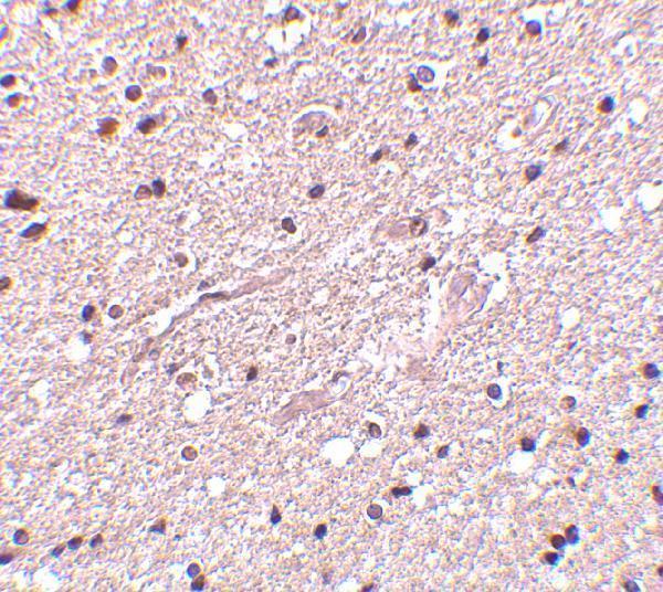
| Expected result | HAP1-positive staining in neuronal cells of hippocampus (HPA tissue IHC: High). HPA tissue profile: Cytoplasmic expression in most tissues, most abundant in CNS. No signal in the no-primary control. |
HAP1 is predominantly neuronal and brain enriched (UniProt P54257 tissue specificity); expect chiefly cytoplasmic staining in neuronal cells, especially hippocampus, with possible signal in neuronal processes (HPA: hippocampal neuronal cells High; UniProt P54257 localisation). It has no transmembrane segment, so an apparent membrane edge alone does not establish membrane insertion (UniProt P54257 topology). HPA rates tissue IHC reliability Enhanced but reports only medium consistency between staining and RNA expression (HPA: tissue IHC reliability).
| Neuronal cell bodies stain in hippocampus, with some staining extending into processes. | This fits high hippocampal neuronal staining and HAP1 localisation in cytoplasm, axons and dendrites (HPA: hippocampal neuronal cells High; UniProt P54257 localisation). Score cells and compartments together; process staining may be harder to assign to a single cell in a section (general IHC practice). |
| Strong staining is predominantly nuclear, with little neuronal cytoplasmic signal. | Recheck the result against the expected cytoplasmic pattern (HPA: cytoplasmic expression in most tissues). Nuclear staining alone is not proof of an artefact: UniProt also lists the nucleus and nuclear rods (UniProt P54257 localisation). Compare an IHC positive control and a detection control before calling the pattern specific (general IHC practice). |
| Staining is strongest in a cell population expected to be unstained in the chosen control tissue. | Consider cross-reactivity or endogenous chromogenic activity, and check cell identity and control staining (general IHC practice). HPA lists adipocytes in adipose tissue and hematopoietic cells in bone marrow as Not detected; that designation is specific to those sampled cells and is not an absolute absence claim (HPA: tissue IHC). |
| Diffuse colour covers cells and surrounding tissue without a discernible cellular pattern. | Treat this as background until a clear compartment and cell population can be scored (general IHC practice). Inspect the no-primary control, blocking and detection steps; diffuse colour does not match HPA's reported cytoplasmic cellular expression (general IHC practice; HPA: tissue IHC profile). |
| No signal appears in hippocampal neuronal cells. | This conflicts with HPA's High hippocampal neuronal staining, but does not by itself show that HAP1 is absent (HPA: tissue IHC). Confirm tissue identity and assay controls, then review the antibody's IHC validation and the run conditions (general IHC practice; HPA: HPA076992 IHC Enhanced). |
| Tissue and cell selection | HPA reports High staining in hippocampal neuronal cells and syncytiotrophoblast microvilli, Medium in several other listed tissues, and Not detected in selected cell populations (HPA: tissue IHC). The placental microvillus observation identifies a staining site; it does not establish that HAP1 spans a membrane (HPA: placental syncytiotrophoblasts High; UniProt P54257 topology). |
| Antibody validation | HPA076992 has Enhanced IHC validation, while HPA023394 has no IHC status in the supplied record (HPA: antibody validation). Use the IHC-validated antibody's documented conditions when comparing a new stain with HPA tissue observations (HPA: antibody validation; general IHC practice). |
| Isoforms and epitope | UniProt lists four HAP1 isoforms, but the supplied record gives no antibody epitope (UniProt P54257 isoforms; HPA: antibody validation). Do not infer which isoforms an antibody detects or explain a discrepant tissue pattern by isoform selection without epitope evidence (general IHC practice). |
| Processing and topology | UniProt lists one chain spanning residues 1–671, with no signal peptide, propeptide, transmembrane segment or recorded glycosylation sites (UniProt P54257 processing, topology and glycosylation). These annotations do not establish fixation sensitivity, antigen retrieval conditions or shedding in a paraffin section (UniProt P54257 annotations). |
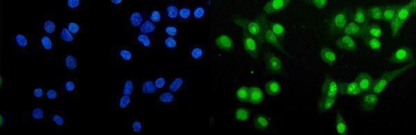
| Q: What should IF/ICC show? | A: HPA reports mainly cytosolic signal; nucleolar localisation is additional but uncertain (HPA: ICC-IF subcellular summary). Interpret that evidence on the separate IF/ICC guide page; the HPA ICC-IF observation does not supply an IHC-P detection or retrieval protocol (HPA: ICC-IF subcellular summary). |
| Situation | Likely cause | Next action |
|---|---|---|
| Hippocampal neurons are unstained. | A failed staining run or unsuitable assay conditions are possibilities; the slide alone cannot distinguish them from specimen variation (general IHC practice). | Verify the neuronal region, inspect run controls, and review the IHC-validated antibody's documented conditions (HPA: hippocampal neuronal cells High; HPA: HPA076992 IHC Enhanced; general IHC practice). |
| Colour appears on the no-primary control. | The detection system or endogenous tissue activity can produce chromogenic signal without primary antibody (general IHC practice). | Check the detection reagents and apply an appropriate endogenous enzyme block for the chosen chromogen system; repeat the control (general IHC practice). |
| Most of the section is uniformly coloured. | Diffuse background can obscure the cellular pattern needed to interpret HAP1 staining (general IHC practice; HPA: tissue IHC profile). | Compare the no-primary control, then review blocking, washing and antibody concentration against the antibody documentation (general IHC practice). |
| The stain is almost entirely nuclear. | A dominant nuclear pattern diverges from HPA's chiefly cytoplasmic tissue profile, although UniProt includes nuclear localisation (HPA: tissue IHC profile; UniProt P54257 localisation). | Compare cytoplasmic signal in hippocampal neurons and inspect detection controls before attributing the nuclear colour to HAP1 (HPA: hippocampal neuronal cells High; general IHC practice). |
| An expected low or Not detected cell population stains strongly. | Cross-reactivity, endogenous detection activity or cell misidentification may account for the discrepancy (general IHC practice). | Recheck morphology and controls; compare the exact cell population with its HPA entry rather than treating a whole tissue as negative (HPA: tissue IHC; general IHC practice). |
| A researcher reads placental microvillus staining as proof of a membrane protein. | The staining site is being interpreted as transmembrane topology (HPA: placental syncytiotrophoblasts High; UniProt P54257 topology). | Report the observed microvillus staining, while keeping localisation separate from topology: UniProt records no HAP1 transmembrane segment (HPA: placental syncytiotrophoblasts High; UniProt P54257 topology). |
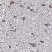
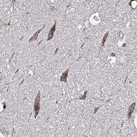
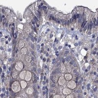


Comprehensive Human Protein Atlas IHC scoring per tissue (reliability: Enhanced — Medium consistency between antibody staining and RNA expression data.). Rows are taken directly from the HPA tissue chart — click any row's HPA link to view the source.
| Tissue | Cell type | Level | Evidence | Source |
|---|---|---|---|---|
| Hippocampus | Neuronal cells | High | Protein (IHC) | HPA → |
| Placenta | Syncytiotrophoblasts - microvilli | High | Protein (IHC) | HPA → |
| Adrenal gland | Glandular cells | Medium | Protein (IHC) | HPA → |
| Caudate | Neuronal cells | Medium | Protein (IHC) | HPA → |
| Cerebral cortex | Neuronal cells | Medium | Protein (IHC) | HPA → |
| Tissue | Cell type | Level | Evidence | Source |
|---|---|---|---|---|
| Adipose tissue | Adipocytes | Not detected | Protein (IHC) | HPA → |
| Bone marrow | Hematopoietic cells | Not detected | Protein (IHC) | HPA → |
| Cervix | Glandular cells | Not detected | Protein (IHC) | HPA → |
| Endometrium | Cells in endometrial stroma | Not detected | Protein (IHC) | HPA → |
| Epididymis | Glandular cells | Not detected | Protein (IHC) | HPA → |
Troubleshoot HAP1 staining in paraffin sections by checking retrieval, cellular distribution, controls, and scoring before interpreting chromogenic signal.
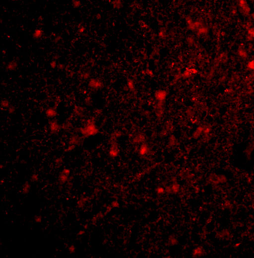
Anti-HAP1 catalog antibodies have IHC images from human brain tissue and IF images from human brain cells and A549 cells (image captions). Both list Human, Mouse and Rat reactivity (catalog reactivity).
A01658 will render with an IHC image of human brain tissue at 2.5 μg/mL; its catalog also lists IHC-P and IF (A01658 image caption; catalog applications). A01658-3 will render with an IF/ICC image of A549 cells at 5 μg/mL; its catalog lists IF and ICC but no IHC-P (A01658-3 image caption; catalog applications).
Which to pick: Choose A01658 for paraffin-section IHC: its IHC-P listing and own human brain tissue image support that application, while the image caption does not report the fixative (catalog applications; A01658 image caption). Choose A01658-3 for IF/ICC of A549 cells, as shown in its own image (A01658-3 image caption). Both list Human, Mouse and Rat reactivity, but the supplied images show human samples; neither catalog entry specifies a clone (catalog reactivity; image captions; catalog clone fields).