This website uses cookies to ensure you get the best experience on our website.


- Table of Contents
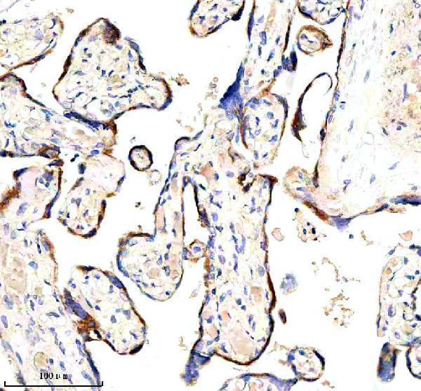
Plan chromogenic HAPLN1 IHC in paraffin sections using the human placenta example at 2 μg/ml (datasheet A05980-3). Interpret selective cytoplasmic tissue staining (HPA tissue IHC) alongside its secreted extracellular location (UniProt).

Expected localisation, validated protocols, controls and antibodies — the at-a-glance facts below, then the full design guide.
| Expected localisation | Observed cytoplasmic staining (HPA tissue IHC); expected extracellular matrix location (UniProt) | |
| Staining pattern | Selective cytoplasmic staining in cartilage, placenta and CNS, including decidual cells (HPA tissue IHC) | |
| Antigen retrieval | EDTA pH 8.0 HIER, heat-mediated (datasheet A05980-3) | |
| Positive control | Placenta+1 more · see all | |
| Negative control | Adipose tissue+4 more · see all |
| Fixation | Matched tissue-IHC evidence does not establish the fixation claim. Validate the specimen-specific method before use. (selected-SKU IHC image A05980-3) | |
| Caveat | Secreted protein may stain away from cells expressing its RNA (HPA tissue IHC) | |
| Regulation | Regulation not reported (UniProt) | |
| Isoform / epitope | No isoforms; propeptide cleaved; no cytoplasmic region (UniProt) |
Compare the catalog antibody’s IHC-P protocol (datasheet: A05980-3) with two published chromogenic HAPLN1 protocols (PMC6328344; PMC3761224).
| Sample | Paraffin-embedded human placenta tissue; fixative not specified (datasheet A05980-3) |
| Fixation | Image fixative and duration unreported (datasheet A05980-3); verify before use. |
| Sectioning | 4–5 µm sections on charged slides (standard) |
| Deparaffinisation | Xylene, graded ethanol series to water (standard) |
| Antigen retrieval | Heat retrieval: EDTA pH 8.0 (datasheet A05980-3); 20 min, 95–100 °C (standard) |
| Peroxidase block | 3% H2O2, 10 min, room temperature (standard) |
| Blocking | 10% goat serum (datasheet A05980-3) |
| Primary antibody | Rabbit anti-HAPLN1, 2-5 μg/ml (datasheet A05980-3) |
| Primary incubation | Overnight at 4 °C (datasheet A05980-3) |
| Detection | HRP-conjugated secondary, DAB chromogen (datasheet A05980-3) |
| Counterstain | Hematoxylin, blue, dehydrate and mount (standard) |
| Expected result | HAPLN1-positive staining in decidual cells of placenta (HPA tissue IHC: Medium). HPA tissue profile: Selective cytoplasmic expression in cartilage, placenta and CNS. No signal in the no-primary control. |
HAPLN1 is secreted into the extracellular matrix and has no transmembrane segment (UniProt P10915). In paraffin IHC, assess cartilage-associated staining and selective cytoplasmic staining in placenta and CNS (HPA tissue IHC: Enhanced; medium consistency with RNA). Decidual cells and elongated or late spermatids show medium staining, while the listed CNS cell populations show low staining (HPA tissue IHC). Protein location need not match the tissue where its RNA is detected (HPA tissue IHC).
| Cartilage-associated staining with selective cytoplasmic staining in placenta or CNS (HPA tissue IHC). | This fits the reported tissue profile (HPA tissue IHC). Extracellular staining is biologically plausible because HAPLN1 stabilizes proteoglycan aggregates in cartilage matrix (UniProt P10915). Score cellular and matrix signal separately; the record does not give a cartilage intensity threshold (HPA tissue IHC). |
| Medium staining in decidual cells or elongated or late spermatids; low staining in the listed CNS cells (HPA tissue IHC). | These are the reported cell-specific levels, including low neuronal staining in cerebral cortex, hippocampus and caudate (HPA tissue IHC). A weak CNS signal can therefore be consistent with HAPLN1; compare it with background and a stronger positive control before calling it absent (HPA tissue IHC; general IHC practice). |
| Predominantly nuclear staining without the reported cellular or cartilage-associated pattern (HPA tissue IHC; UniProt P10915). | A nuclear-only result conflicts with the reported cytoplasmic tissue profile and secreted extracellular localization (HPA tissue IHC; UniProt P10915). Review morphology and control slides, then assess whether nonspecific detection or misidentified structures explain the signal (general IHC practice). |
| Strong staining in adipocytes or other cells reported as not detected (HPA tissue IHC). | Adipocytes in adipose tissue and glandular cells in adrenal gland are reported as not detected (HPA tissue IHC). If staining is reproducible, assess antibody specificity and endogenous chromogenic activity with appropriate controls; a single unexpected stain does not establish HAPLN1 expression (general IHC practice). |
| Diffuse color over tissue, extracellular spaces and the negative control (general IHC practice). | This distribution cannot be assigned confidently to HAPLN1, even though HAPLN1 is extracellular (UniProt P10915). Compare a no-primary control and tissue morphology; excessive detection signal, incomplete blocking or inadequate washing are general IHC possibilities, not HAPLN1-specific findings (general IHC practice). |
| Secreted matrix protein (UniProt P10915) | HAPLN1 can be seen outside producing cells in cartilage matrix (UniProt P10915). Interpret extracellular color alongside the reported cytoplasmic tissue pattern; do not require a membrane outline, because no transmembrane segment is annotated (HPA tissue IHC; UniProt P10915). |
| Tissue and cell context (HPA tissue IHC) | Placental decidual cells and elongated or late spermatids are medium positive, whereas listed CNS populations are low and several surveyed cell types are not detected (HPA tissue IHC). Choose comparisons by annotated cell population, since an entire tissue is not uniformly positive or negative (HPA tissue IHC). |
| Protein processing and modification (UniProt P10915) | The annotated mature chain spans residues 16–354 after a 1–15 propeptide, and glycosylation sites occur at residues 21 and 56 (UniProt P10915). These annotations do not locate any antibody epitope or establish a retrieval or fixation effect; avoid predicting stain strength from them. |
| Antibody evidence (HPA antibodies; HPA tissue IHC) | HPA019105 and HPA019482 have Enhanced IHC status; HPA025238 is Approved (HPA antibodies). The tissue profile is labeled Enhanced but has medium RNA–protein consistency, with a stated caveat for secreted proteins (HPA tissue IHC). Interpret discordant sites with controls and tissue context. |
| Situation | Likely cause | Next action |
|---|---|---|
| No stain in decidual cells expected to be medium positive (HPA tissue IHC). | The run may lack sensitivity, or the selected section may lack identifiable decidual cells (general IHC practice). | Confirm cell identity and section quality, then check the IHC-validated antibody's stated retrieval and dilution conditions and the detection controls (general IHC practice). |
| Low CNS staining is scored as negative (HPA tissue IHC). | The expected signal is low in the listed CNS populations (HPA tissue IHC). | Compare cells with local background and a positive control; assess the reported cell population before making a tissue-wide call (HPA tissue IHC; general IHC practice). |
| Unexpected color appears in adipocytes reported as not detected (HPA tissue IHC). | Cross-reactivity or endogenous detection activity is possible, but the stain alone cannot distinguish them (general IHC practice). | Review a no-primary control and cellular morphology; if color persists, check blocking and the detection system before attributing it to HAPLN1 (general IHC practice). |
| Cartilage shows broad extracellular color (UniProt P10915). | Matrix localization is plausible for HAPLN1, but diffuse background can look similar (UniProt P10915; general IHC practice). | Check whether color follows cartilage structures and exceeds the no-primary control; score matrix and cellular compartments separately (HPA tissue IHC; general IHC practice). |
| Staining is mainly nuclear despite the reported cytoplasmic profile (HPA tissue IHC). | The compartment disagrees with secreted extracellular localization and the reported tissue pattern (UniProt P10915; HPA tissue IHC). | Inspect tissue morphology and controls, then verify antibody and detection specificity before interpreting nuclear color as HAPLN1 (general IHC practice). |
| IF/ICC: where should HAPLN1 appear? | HPA reports vesicles as the approved ICC–IF location; UniProt also describes secretion into extracellular space (HPA subcellular; UniProt P10915). | Interpret vesicular signal against the ICC–IF control and cell context; do not apply the paraffin IHC tissue pattern as an ICC–IF intensity threshold (HPA subcellular; HPA tissue IHC). |


Comprehensive Human Protein Atlas IHC scoring per tissue (reliability: Enhanced — Medium consistency between antibody staining and RNA expression data. Secreted protein, tissue location of RNA and protein is expected to differ. Antibody staining in cells/structures not annotated, view images.). Rows are taken directly from the HPA tissue chart — click any row's HPA link to view the source.
| Tissue | Cell type | Level | Evidence | Source |
|---|---|---|---|---|
| Placenta | Decidual cells | Medium | Protein (IHC) | HPA → |
| Testis | Elongated or late spermatids | Medium | Protein (IHC) | HPA → |
| Tissue | Cell type | Level | Evidence | Source |
|---|---|---|---|---|
| Adipose tissue | Adipocytes | Not detected | Protein (IHC) | HPA → |
| Adrenal gland | Glandular cells | Not detected | Protein (IHC) | HPA → |
| Appendix | Glandular cells | Not detected | Protein (IHC) | HPA → |
| Bone marrow | Hematopoietic cells | Not detected | Protein (IHC) | HPA → |
| Breast | Adipocytes | Not detected | Protein (IHC) | HPA → |
Troubleshoot HAPLN1 chromogenic IHC by anchoring retrieval to the catalog antibody’s placenta image and interpreting staining alongside its secreted extracellular role (datasheet A05980-3; UniProt P10915).
IHC images show human placenta (A05980-3 caption), plus rat liver, mouse skeletal muscle and heart, and human glioblastoma (M05980-1 captions); IF images have unspecified samples (M05980-1 IF captions).
A05980-3 has IHC data from a paraffin-embedded human placenta section (A05980-3 IHC caption). M05980-1 has IHC images from paraffin-embedded rat liver, mouse skeletal muscle and heart, and human glioblastoma, plus IF images with unspecified samples (M05980-1 image captions).
Which to pick: For human placenta tissue IHC, choose A05980-3: its polyclonal antibody was shown on a paraffin-embedded section with EDTA retrieval at pH 8.0 and 2 μg/ml primary antibody (A05980-3 catalog and IHC caption). For IF/ICC, choose monoclonal M05980-1 because those applications and IF images are listed, although the IF sample is unspecified (M05980-1 catalog and IF captions). For IHC across human, mouse and rat samples, M05980-1 has paraffin-section images from all three species; the fixative is unreported in both SKUs’ IHC captions (M05980-1 and A05980-3 IHC captions).