This website uses cookies to ensure you get the best experience on our website.


- Table of Contents
Plan HAX1 staining in paraffin sections using the IHC-validated antibody and its documented chromogenic protocol. Compare cytoplasmic staining in glandular and immune cells with matched controls, mindful that tissue staining has low consistency with RNA expression (HPA tissue IHC).

Expected localisation, validated protocols, controls and antibodies — the at-a-glance facts below, then the full design guide.
| Expected localisation | Cytoplasmic in most glandular and immune cells (HPA tissue IHC) | |
| Staining pattern | Glandular and immune cells show cytoplasmic staining (HPA tissue IHC) | |
| Antigen retrieval | EDTA pH 8.0 HIER, heat-mediated (datasheet A01495-2) | |
| Positive control | Appendix+4 more · see all | |
| Negative control | Adipose tissue+4 more · see all |
| Fixation | Keep fixation consistent across sections. (standard IHC practice; not target-specific) | |
| Caveat | Antibody staining has low consistency with RNA expression (HPA tissue IHC) | |
| Regulation | Up-regulated in oral cancers (UniProt) | |
| Isoform / epitope | 6 isoforms; confirm antibody epitope coverage (UniProt) |
The catalog antibody’s IHC-P protocol is paired with three published HAX1 IHC protocols covering NPC tumour tissues, a CRC tissue array, and paraffin-embedded tumour specimens (PMC9077140; PMC11217408; PMC8650040).
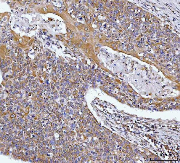
| Sample | Paraffin-embedded human esophageal squamous carcinoma tissue; fixative not specified (datasheet A01495-2) |
| Fixation | Image fixative and duration unreported (datasheet A01495-2); verify before use. |
| Sectioning | 4–5 µm sections on charged slides (standard) |
| Deparaffinisation | Xylene, graded ethanol series to water (standard) |
| Antigen retrieval | Heat retrieval: EDTA pH 8.0 (datasheet A01495-2); 20 min, 95–100 °C (standard) |
| Peroxidase block | 3% H2O2, 10 min, room temperature (standard) |
| Blocking | 10% goat serum (datasheet A01495-2) |
| Primary antibody | Rabbit anti-HAX1, 2-5 μg/ml (datasheet A01495-2) |
| Primary incubation | Overnight at 4 °C (datasheet A01495-2) |
| Detection | HRP-conjugated secondary, DAB chromogen (datasheet A01495-2) |
| Counterstain | Hematoxylin, blue, dehydrate and mount (standard) |
| Expected result | HAX1-positive staining in glandular cells of appendix (HPA tissue IHC: High). HPA tissue profile: Cytoplasmic expression in most glandular cells and in immune cells. No signal in the no-primary control. |
In paraffin section IHC, expect HAX1 mainly in the cytoplasm of glandular and immune cells (HPA: tissue IHC profile; UniProt O00165: predominantly cytoplasmic). Several glandular cell populations show high staining, while some other sampled cell populations show none (HPA: tissue IHC). HAX1 has no transmembrane segment (UniProt O00165: topology). HPA rates the tissue IHC pattern Approved but reports low consistency with RNA expression (HPA: reliability).
| Glandular cells show cytoplasmic chromogenic staining, with strong signal in an appendix, duodenum, or stomach section. | This fits the reported compartment and cell type; glandular cells in those tissues are rated High (HPA: tissue IHC). Judge the cell distribution as well as intensity. An Approved IHC rating supports use of the pattern, but low agreement with RNA expression limits how confidently staining alone identifies HAX1 (HPA: reliability). |
| Staining appears only in nuclei, or forms a continuous extracellular deposit without discernible cytoplasmic signal. | Treat this as discordant with the usual IHC pattern and inspect controls and detection background (HPA: cytoplasmic tissue IHC profile; standard IHC practice). Nuclear HAX1 can occur when export is inhibited or after arsenite stress in vitro, so an isolated nuclear finding needs context before biological interpretation (UniProt O00165: subcellular location). |
| Adipocytes or oral mucosal squamous epithelial cells stain as strongly as glandular cells in a positive control. | Those specific cell populations are reported Not detected, whereas several glandular populations are High (HPA: tissue IHC). Check nonspecific antibody binding and endogenous detection activity before calling the staining HAX1 (standard IHC practice). A negative call applies to the listed cell population, not every cell in its tissue (HPA: tissue IHC). |
| Weak, even color covers cells, stroma, and spaces between cells, obscuring cell boundaries. | Diffuse color lacks the cell restricted cytoplasmic distribution described for HAX1 (HPA: tissue IHC profile). Compare a no primary control, blocking, washes, and detection chemistry to locate background (standard IHC practice). Do not score uniform haze as positive merely because HAX1 is described as ubiquitous (UniProt O00165: tissue specificity). |
| Glandular cells in a designated positive tissue show no signal while the counterstain and tissue morphology remain visible. | Appendix, duodenum, gallbladder, pancreas exocrine glands, rectum, small intestine, stomach, and thyroid gland contain listed High populations (HPA: tissue IHC). Confirm the relevant cells are present, then review the IHC validated antibody, retrieval, dilution, and detection controls (standard IHC practice); the payload gives no HAX1 specific retrieval condition. |
| Tissue and cell choice (HPA: tissue IHC). | High staining is reported in appendix and duodenal glandular cells and pancreatic exocrine glandular cells; adipocytes and oral mucosal squamous epithelial cells are listed Not detected (HPA: tissue IHC). Compare the named cell populations, not whole tissues. |
| Compartment used for scoring (HPA: tissue IHC; UniProt O00165: subcellular location). | Predominantly cytoplasmic staining is the IHC expectation, despite additional organelle and membrane locations annotated by UniProt (HPA: tissue IHC profile; UniProt O00165: subcellular location). A fine organelle pattern need not be resolved by chromogenic IHC (standard IHC practice). |
| Antibody validation and interpretation limit (HPA: antibody validation; HPA: reliability). | HPA055141 is Approved for IHC, while the overall tissue pattern has low consistency with RNA expression (HPA: antibody validation; HPA: reliability). Use cell level morphology and controls when interpreting an unexpected signal (standard IHC practice). |
| Isoforms and antibody epitope (UniProt O00165: isoforms). | Six HAX1 isoforms are annotated (UniProt O00165: isoforms 1–6). Their recognition by the catalog antibody cannot be inferred because its epitope is absent from the supplied record; do not assign an unusual pattern to one isoform without separate evidence. |
| IF/ICC Q: What pattern is expected? (HPA: subcellular ICC-IF). | A: HPA reports a supported mitochondrial location in ICC-IF, with images from MCF-7, SK-MEL-30, and U2OS (HPA: subcellular ICC-IF). This is a cell imaging observation; score paraffin section IHC against its reported cytoplasmic tissue pattern (HPA: tissue IHC). |
| Situation | Likely cause | Next action |
|---|---|---|
| No staining in appendix or stomach glandular cells (HPA: High). | The positive cells may be absent from the examined area, or an IHC workflow step may have failed (HPA: tissue IHC; standard IHC practice). | Locate intact glands, check the positive control and detection reagents, then review retrieval and antibody dilution using the IHC validated antibody's instructions (standard IHC practice). |
| A section looks positive mainly in adipocytes (HPA: Not detected). | That cell level result conflicts with the HPA observation; nonspecific binding or detection background is possible (HPA: tissue IHC; standard IHC practice). | Inspect cell identity and a no primary control; assess blocking and washes before assigning HAX1 positivity (standard IHC practice). |
| Broad brown haze obscures cytoplasmic boundaries. | Background from antibody or chromogenic detection can prevent cell level scoring (standard IHC practice). | Review a no primary control, blocking, wash stringency, and detection time; rescore only where cells remain identifiable (standard IHC practice). |
| Only nuclei stain in an otherwise intact paraffin section. | This differs from the predominant cytoplasmic pattern, although conditional nuclear detection is annotated in vitro (HPA: tissue IHC; UniProt O00165: subcellular location). | Check a positive tissue control and nonspecific background; require experimental context before treating nuclear staining as a HAX1 localization change (standard IHC practice; UniProt O00165: subcellular location). |
| Glandular populations differ unexpectedly across tissues. | HPA reports both High and Not detected glandular populations, and its IHC pattern has low RNA consistency (HPA: tissue IHC; HPA: reliability). | Score each named cell population against its own HPA observation and review section quality and controls before interpreting the difference (HPA: tissue IHC; standard IHC practice). |
| A punctate ICC-IF image seems inconsistent with broad cytoplasmic IHC. | The two HPA observations use different imaging applications: mitochondrial ICC-IF and cytoplasmic tissue IHC (HPA: subcellular ICC-IF; HPA: tissue IHC). | Interpret each result with its application specific compartment and appropriate controls; avoid treating the ICC-IF image as a paraffin IHC scoring template (HPA: subcellular ICC-IF; HPA: tissue IHC). |


Comprehensive Human Protein Atlas IHC scoring per tissue (reliability: Approved — Low consistency between antibody staining and RNA expression data.). Rows are taken directly from the HPA tissue chart — click any row's HPA link to view the source.
| Tissue | Cell type | Level | Evidence | Source |
|---|---|---|---|---|
| Appendix | Glandular cells | High | Protein (IHC) | HPA → |
| Duodenum | Glandular cells | High | Protein (IHC) | HPA → |
| Gallbladder | Glandular cells | High | Protein (IHC) | HPA → |
| Pancreas | Exocrine glandular cells | High | Protein (IHC) | HPA → |
| Rectum | Glandular cells | High | Protein (IHC) | HPA → |
| Tissue | Cell type | Level | Evidence | Source |
|---|---|---|---|---|
| Adipose tissue | Adipocytes | Not detected | Protein (IHC) | HPA → |
| Caudate | Glial cells | Not detected | Protein (IHC) | HPA → |
| Cerebellum | Cells in granular layer | Not detected | Protein (IHC) | HPA → |
| Cerebral cortex | Endothelial cells | Not detected | Protein (IHC) | HPA → |
| Fallopian tube | Glandular cells | Not detected | Protein (IHC) | HPA → |
These questions address HAX1 staining in paraffin sections with chromogenic IHC, with one question on IF.
A01495-2 has real IHC data from human paraffin sections (A01495-2 image captions); the catalog provides no IF image or IF dilution (A01495-2 catalog).
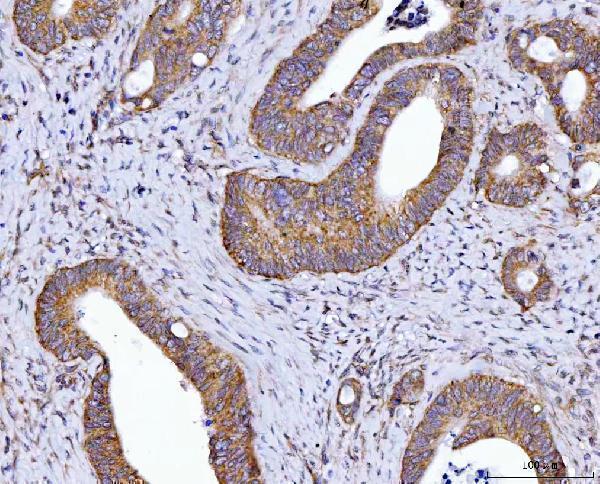
A01495-2 is listed for human IHC (A01495-2 applications and reactivity). Its captions document staining in paraffin sections of human esophageal squamous carcinoma, colorectal adenocarcinoma and lung cancer tissue (A01495-2 image captions).
Which to pick: Choose A01495-2 for human paraffin-section IHC; its rabbit antibody was used at 2 μg/ml after EDTA pH 8.0 heat retrieval in the documented images (A01495-2 image captions). No IF/ICC application or IF image is supplied for A01495-2, so there is no validated IF/ICC choice in this catalog (A01495-2 applications and image alts). No cross-species choice is documented because A01495-2 lists human reactivity only; its paraffin-section captions do not report the fixative (A01495-2 reactivity and image captions).