HBB / Hemoglobin subunit beta · IHC design guide

Design Immunohistochemistry for HBB

Plan chromogenic HBB IHC around erythrocyte staining (HPA tissue IHC). Use the catalog antibody’s paraffin-section example (datasheet A00147-1) and score erythrocytes separately from other cells (HPA tissue IHC).

Evidence assembled Oct 2026 · For research use; verify linked source records and product datasheet before use
Immunohistochemistry protocol sheet for HBB (IHC for HBB): expected localisation Erythrocytes; compartment not established (HPA tissue IHC), antibody A00147-1, validated IHC image, and IHC protocol steps
Printable HBB IHC protocol sheet — expected localisation Erythrocytes; compartment not established (HPA tissue IHC), antibody A00147-1, controls and protocol steps. Open the full HBB IHC guide →

HBB Immunohistochemistry Experimental Design Guide

Expected localisation, validated protocols, controls and antibodies — the at-a-glance facts below, then the full design guide.

Must know before staining
Expected localisation Erythrocytes; compartment not established (HPA tissue IHC)
Staining pattern Erythrocyte staining; subcellular pattern unreported (HPA tissue IHC)
Antigen retrieval EDTA pH 8.0 HIER, heat-mediated (datasheet A00147-1)
Positive control ⓘ Bone marrow+2 more · see all
Negative control ⓘ Adipose tissue+4 more · see all
Important caveats
Reasons your staining may differ from the expected pattern.
Fixation Keep fixation consistent across paraffin sections. (standard IHC practice; not target-specific)
Caveat Antibody may recognize products of more than one gene (HPA tissue IHC)
Regulation Signal tracks erythrocyte content (HPA tissue IHC)
Isoform / epitope No annotated isoforms; mature chain spans aa 2–147 (UniProt)
Section 1

Recommended HBB IHC & IF Protocols

The catalog antibody’s IHC-P protocol is accompanied by four published HBB IHC protocols covering renal, thyroid, and breast tissue sections (PMC10216096; PMC2361827; PMC12131617; PMC5625664).

Recommended immunohistochemistry (IHC-P) protocol parameters
SampleParaffin-embedded mouse liver tissue; fixative not specified (datasheet A00147-1)
FixationImage fixative and duration unreported (datasheet A00147-1); verify before use.
Sectioning4–5 µm sections on charged slides (standard)
DeparaffinisationXylene, graded ethanol series to water (standard)
Antigen retrievalHeat retrieval: EDTA pH 8.0 (datasheet A00147-1); 20 min, 95–100 °C (standard)
Peroxidase block3% H2O2, 10 min, room temperature (standard)
Blocking10% goat serum (datasheet A00147-1)
Primary antibodyRabbit anti-HBB, 1:50 recommended; image 1:100 (datasheet A00147-1)
Primary incubationOvernight at 4 °C (datasheet A00147-1)
DetectionHRP-conjugated secondary, DAB chromogen (datasheet A00147-1)
CounterstainHematoxylin, blue, dehydrate and mount (standard)
Expected resultHBB-positive staining in hematopoietic cells of bone marrow (HPA tissue IHC: Medium). HPA tissue profile: Expression in erythrocytes. No signal in the no-primary control.
💡Decision noteStart with the catalog antibody’s heat-mediated EDTA retrieval at pH 8.0 (datasheet A00147-1); published citrate conditions offer alternatives for optimization (PMC10216096; PMC5625664).
Section 2

What Is the Expected HBB Staining Pattern?

HBB is a globin without a transmembrane segment or signal peptide (UniProt P68871 topology and processing). On IHC sections, expect staining associated primarily with erythrocytes (HPA tissue IHC: expression in erythrocytes; UniProt P68871: red blood cell specificity). Treat apparent staining in other cells cautiously: HPA rates its tissue profile Enhanced but warns that antibody staining may reflect proteins from more than one gene (HPA tissue IHC).

What am I looking at on my slide?
Erythrocytes show clear chromogenic staining with limited staining elsewhere.This matches the principal tissue pattern (HPA tissue IHC: expression in erythrocytes; UniProt P68871: red blood cell specificity). Compare cellular staining with the slide's background before assigning intensity; HPA's Enhanced reliability does not make every stained structure HBB-specific (HPA tissue IHC: reliability caveat).
A sharp nuclear or cell-membrane outline is the dominant pattern.That compartment is unexpected for a globin lacking a transmembrane segment and signal peptide (UniProt P68871 topology and processing). Check whether the outline also appears in controls and whether erythrocytes show the expected pattern (HPA tissue IHC: expression in erythrocytes).
Cells other than erythrocytes stain prominently.Do not automatically score them as HBB-positive. HPA reports Medium staining in bone-marrow hematopoietic cells, lung alveolar type II cells and placental trophoblastic cells, while warning that staining can reflect proteins from more than one gene (HPA tissue IHC). Compare controls for cross-reactivity or endogenous detection activity.
Color is diffuse across tissue, including cells expected to be negative.A broadly distributed haze weakens the distinction between specific staining and background. For example, adipocytes and bronchial respiratory epithelial cells are Not detected in the HPA profile (HPA tissue IHC). Review the no-primary control and general IHC blocking and washing before interpreting weak color.
A known-positive section has no convincing erythrocyte staining.The result conflicts with the reported erythrocyte pattern (HPA tissue IHC; UniProt P68871: red blood cell specificity). First confirm that identifiable erythrocytes are present; then check the assay's positive control, antibody and detection steps. A blank slide alone cannot establish biological absence.
💡Expected HBB appearanceCall positive when clear erythrocyte-associated staining exceeds local background (HPA tissue IHC: erythrocyte expression); dominant nuclear or membrane-outline staining is suspect given HBB topology (UniProt P68871).
How each factor affects the staining
Cell and tissue distributionErythrocytes are the main expectation (HPA tissue IHC; UniProt P68871). HPA also records Medium staining in selected non-erythrocyte cells, so identify the stained cell before interpreting a whole-tissue result (HPA tissue IHC).
Antibody validationHPA lists CAB009526 as IHC Enhanced and HPA043234 as IHC Supported (HPA antibodies). Its tissue profile still cautions that staining may reflect proteins from more than one gene; validation status does not resolve each unexpected cell type (HPA tissue IHC).
Protein topology and processingHBB has no transmembrane segment or signal peptide, and its annotated chain spans residues 2–147 (UniProt P68871). These features support an intracellular, non-membrane-outline expectation; UniProt supplies no subcellular-location annotation (UniProt P68871).
Epitope interpretationUniProt records several modified residues and no isoforms (UniProt P68871). The supplied sources do not map either antibody's epitope, so those annotations cannot establish which modification changes staining or which retrieval condition works.
IF/ICC evidenceHPA provides no ICC-IF images or subcellular-location call for HBB (HPA subcellular). Tissue IHC therefore supports a cellular expectation, but does not validate a fluorescent subcellular pattern.
Why is my staining missing, weak or wrong?
SituationLikely causeNext action
Erythrocytes are present, but staining is absent.A failed assay step or unsuitable antibody condition is possible; erythrocyte staining is expected (HPA tissue IHC; UniProt P68871).Verify the positive control and detection reagents, then review the IHC-validated antibody's documented conditions. Any retrieval adjustment is general IHC optimization; the supplied sources establish no HBB-specific fixation or retrieval effect.
Erythrocytes and background are both strongly colored.The readout may include nonspecific chromogen deposition or endogenous detection activity; color alone cannot establish HBB identity.Inspect a no-primary control, blocking and washing, then compare erythrocytes with adjacent background. This is general chromogenic IHC troubleshooting, not an HBB-specific effect reported by HPA.
A nuclear or membrane-outline pattern dominates.That pattern does not follow from HBB's lack of a transmembrane segment or signal peptide (UniProt P68871).Check the no-primary control and reassess cell boundaries and counterstain. Require the expected erythrocyte-associated pattern before treating the compartment signal as HBB (HPA tissue IHC).
A non-erythrocyte population appears positive.HPA reports some such staining but cautions that antibodies may detect proteins from more than one gene (HPA tissue IHC).Name the cell type and compare its HPA entry, the antibody's IHC validation and a no-primary control (HPA tissue IHC; HPA antibodies). Report an unresolved signal as staining, without assigning HBB identity.
A presumed negative tissue has staining.A tissue label alone does not identify the stained structure; HPA's Not detected entries refer to specified cell types (HPA tissue IHC).Identify the colored cells. For example, distinguish adipocytes, listed Not detected, from erythrocytes within adipose tissue, where HBB staining is expected (HPA tissue IHC; UniProt P68871).
IF/ICC Q&A: can a fluorescent compartment pattern confirm HBB?HPA has no ICC-IF images or subcellular-location call for HBB (HPA subcellular).No compartment pattern is confirmed by that record. Interpret fluorescence with assay-specific controls and use the separate IF/ICC guide for method design; retain the erythrocyte expectation as tissue context (HPA tissue IHC).

Sample controls for HBB IHC & IF

🧪Run bone marrow first and score staining in hematopoietic cells (Medium; HPA: bone marrow), while nonhematopoietic cells on the same section serve as an internal negative reference. Use adipose tissue as the negative tissue and score adipocytes, where HBB is not detected (HPA: adipose tissue).
Positive control tissue: Bone marrow (Hematopoietic cells, HPA Medium)
Negative control tissue: Adipose tissue (HPA Not detected)
ICC-IF cell lines (HPA subcellular resource): HPA carries no ICC-IF cell line for HBB; derive a cell-line control from the positive tissue's cell type (Hematopoietic cells) and confirm it by RNA or western blot first.
Technical controls: Include a no-primary (secondary-only) control, a host- and clonality-matched rabbit IgG isotype control (caption: rabbit primary; clonality unreported), and HBB-knockout tissue as a biological negative control. Check endogenous peroxidase and background pigment in marrow erythrocytes before interpreting DAB staining (UniProt P68871: red blood cell specificity; caption: HRP/DAB detection).
⚠️Feasibility: The selected IHC-P caption demonstrates heat retrieval in EDTA, pH 8.0, followed by overnight incubation at 4°C, but does not establish that retrieval is required (caption: A00147-1). A target-specific fixation window and fixation effect are unreported, and the selected caption does not report a fixative (caption: fixative not stated). The supplied evidence does not establish whether frozen sections or IF are easier; erythrocytes can complicate marrow scoring through endogenous peroxidase and pigment (UniProt P68871: red blood cell specificity; caption: HRP/DAB detection).

HPA tissue IHC evidence for HBB

Comprehensive Human Protein Atlas IHC scoring per tissue (reliability: Enhanced — Caution, targets protein from more than one gene. Antibody staining in cells/structures not annotated, view images. Tissue location of RNA and protein might differ and correlation is complex.). Rows are taken directly from the HPA tissue chart — click any row's HPA link to view the source.

Positive expression · recommended positive controls

TissueCell typeLevelEvidenceSource
Bone marrow Hematopoietic cells Medium Protein (IHC) HPA →
Lung Alveolar cells type II Medium Protein (IHC) HPA →
Placenta Trophoblastic cells Medium Protein (IHC) HPA →

Undetected expression · recommended negative controls

TissueCell typeLevelEvidenceSource
Adipose tissue Adipocytes Not detected Protein (IHC) HPA →
Adrenal gland Glandular cells Not detected Protein (IHC) HPA →
Appendix Glandular cells Not detected Protein (IHC) HPA →
Breast Adipocytes Not detected Protein (IHC) HPA →
Bronchus Respiratory epithelial cells Not detected Protein (IHC) HPA →
Section 3

Advanced HBB IHC Tips

Use erythrocyte distribution and matched controls to assess HBB staining in paraffin sections (UniProt P68871: red blood cell specificity; HPA: expression in erythrocytes).

Which retrieval conditions should I try first for HBB in paraffin sections?
Start with heat-mediated retrieval in EDTA at pH 8.0 (datasheet A00147-1). The catalog antibody detected HBB in a paraffin-embedded mouse liver section after that retrieval, followed by 1:100 primary antibody overnight at 4°C (datasheet A00147-1). If staining is weak, compare retrieval duration on adjacent sections while holding antibody dilution and detection conditions constant (standard IHC practice). Include an erythrocyte-containing area as an internal reference, because HBB is associated with red blood cells and a weak tissue-cell signal alone is difficult to interpret (UniProt P68871: tissue specificity; HPA: expression in erythrocytes).
How should I troubleshoot weak HBB staining when fixation details are incomplete?
Target-specific fixation sensitivity is unknown from the supplied evidence, and the catalog image identifies a paraffin-embedded section without stating its fixative (datasheet A00147-1). Record the fixative and fixation duration for each specimen, then compare sections processed with the same pH 8.0 EDTA retrieval and 1:100 primary dilution (datasheet A00147-1; standard IHC practice). Assess erythrocytes in each section before attributing a weak result to fixation, since HBB is associated with red blood cells (UniProt P68871: tissue specificity). Use matched positive and reagent controls when comparing processing conditions, and report fixation history as unknown when it cannot be recovered (standard IHC practice).
Where should HBB signal appear, and how should unexpected staining be checked?
Expect the most readily interpretable signal in erythrocytes, consistent with HBB tissue specificity and the HPA tissue profile (UniProt P68871: red blood cells; HPA: expression in erythrocytes). HBB has no annotated transmembrane segment, while UniProt supplies no subcellular location annotation; a precise organelle pattern is therefore unsupported here (UniProt P68871: topology and subcellular record). HPA also reports medium staining in bone marrow hematopoietic cells, lung alveolar type II cells, and placental trophoblastic cells, with a warning about antibody cross-target staining (HPA: tissue IHC and reliability note). For unexpected cellular signal, inspect morphology and adjacent erythrocytes, then compare reagent controls and an independent antibody or method before assigning it to HBB (standard IHC practice).
Could isoforms or epitope changes explain discrepant HBB IHC staining?
The supplied record lists 0 HBB isoforms, so an isoform-specific explanation is unsupported by this evidence (UniProt P68871: isoforms). HBB contains a globin domain across residues 3–147 and reported modified residues, but the catalog antibody’s precise epitope is not supplied (UniProt P68871: domains and modifications; datasheet A00147-1). Consequently, do not assign loss of staining to a particular modification or residue without epitope mapping and a suitable comparison sample (standard IHC practice). Compare retrieval and primary-antibody conditions across adjacent sections first, and assess concordance with erythrocyte morphology before proposing an epitope-dependent biological difference (datasheet A00147-1: retrieval and dilution; UniProt P68871: tissue specificity).
How can I assess HBB by IF alongside an erythroid cell marker?
For multiplex IF, pair HBB with a validated erythroid cell marker and check that the signals align with cell morphology, since HBB is associated with red blood cells (UniProt P68871: tissue specificity; standard IF practice). Choose spectrally separated fluorophores and favor a far-red channel when the specimen has strong shorter-wavelength autofluorescence (standard IF practice). Because HBB has no transmembrane segment and its antibody epitope is unspecified, establish whether the epitope is intracellular before selecting permeabilisation, then compare mild permeabilisation with an untreated control (UniProt P68871: topology; standard IF practice). Set exposure and background thresholds using single-stain and secondary-only controls; the supplied tissue caption establishes chromogenic IHC conditions, not an IF dilution (datasheet A00147-1; standard IF practice).
What causes diffuse brown staining around HBB-positive areas?
First compare the suspect area with erythrocyte morphology and a section processed without primary antibody, because HBB is associated with red blood cells and DAB can reveal nonspecific background (UniProt P68871: tissue specificity; standard IHC practice). The catalog tissue image used 10% goat serum blocking, a peroxidase-linked secondary antibody, and DAB detection (datasheet A00147-1). If staining persists without primary antibody, check endogenous peroxidase blocking, washes, and DAB development time on matched sections (standard chromogenic IHC practice). If it appears only with primary antibody, titrate from the documented 1:100 dilution and examine whether staining follows erythrocytes rather than tissue edges or damaged regions (datasheet A00147-1; standard IHC practice).
How should I quantify HBB staining without conflating cells and blood content? ⚠ ANSWER MARKED FOR VERIFICATION
Define the region of interest and score erythrocyte-rich areas separately from identified tissue-cell compartments, because HBB is associated with red blood cells (UniProt P68871: tissue specificity; standard IHC practice). For discrete nucleated cells, report the percentage positive or an H-score from intensity categories 0–3; for distributed erythrocyte signal, report positive area or stained-cell density per mm² (standard IHC quantification practice). Normalise each measure to the corresponding cell count, tissue area, or erythrocyte area, as appropriate, and keep thresholds and imaging settings consistent across sections (standard IHC quantification practice). Report compartments separately when comparing HPA-listed hematopoietic, alveolar type II, or trophoblastic staining with erythrocyte signal (HPA: tissue IHC).
When is unexpected HBB-positive staining convincing rather than an artefact?
A convincing result should follow identifiable cells or erythrocytes across intact tissue and reproduce in comparable sections, with appropriate reagent controls (UniProt P68871: tissue specificity; standard IHC practice). Treat isolated nuclear or sharply membranous staining cautiously: HBB has no annotated transmembrane segment and no supplied subcellular location, so those patterns lack support from this record (UniProt P68871: topology and subcellular record). Inspect edges and necrotic regions for processing artefact, and use a primary-omission control plus endogenous peroxidase blocking to assess DAB signal unrelated to the antibody (standard chromogenic IHC practice). HPA warns that its antibody may detect protein from more than one gene, so unexpected cell-type staining needs independent confirmation before attribution to HBB (HPA: reliability note).
Boster reagents

Best HBB / Hemoglobin subunit beta IHC Antibodies

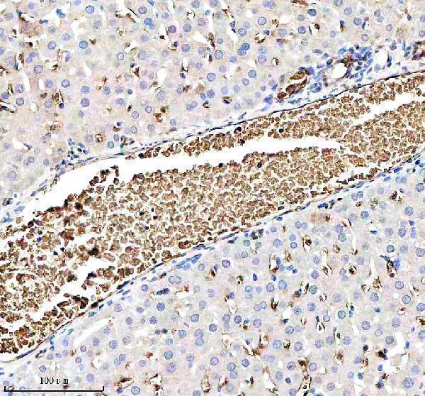
The IHC-validated anti-HBB antibody has paraffin-section images from mouse and rat liver (IHC image captions); its listed reactivity also includes human (catalog: reactivity). No IF data are supplied (catalog: IF images).

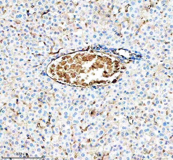
Real IHC data IHC analysis of HBB using anti-HBB antibody (A00147-1). HBB was detected in a paraffin-embedded section of mouse liver tissue. Heat mediated antigen retrieval was performed in EDTA buffer (pH 8.0, epitope retrieval solution). The tissue section was blocked with 10% goat serum. The tissue section was then incubated with 1:100 rabbit anti-HBB Antibody (A00147-1) overnight at 4°C. Peroxidase Conjugated Goat Anti-rabbit IgG was used as secondary antibody and incubated for 30 minutes at 37°C. The tissue section was developed using HRP Conjugated Rabbit IgG Super Vision Assay Kit (Catalog # SV0002) with DAB as the chromogen.
Anti-HBB Antibody
Cat # A00147-1

A00147-1 will render with its mouse liver paraffin-section IHC figure (IHC image caption). Its catalog also lists IHC and human, mouse, and rat reactivity, with a separate rat liver paraffin-section IHC image (catalog: applications and reactivity; IHC image captions).

Which to pick: Choose A00147-1 for paraffin-section tissue IHC, supported by mouse and rat liver images showing EDTA pH 8.0 retrieval; the fixative is unreported (IHC image captions). No SKU in this payload lists IF/ICC or supplies an IF image, so there is no IF/ICC pick here (catalog: applications and IF images). For work across human, mouse, and rat, A00147-1 is the listed polyclonal option, although the supplied IHC images show only mouse and rat tissue (catalog: dilution_raw and reactivity; IHC image captions).

Each figure is that product's own IHC / IF validation image from its datasheet.

References

  1. UniProt Consortium. UniProt entry P68871 (HBB_HUMAN, Hemoglobin subunit beta).
  2. Human Protein Atlas. HBB tissue IHC expression (reliability: Enhanced).
  3. Human Protein Atlas. HBB subcellular location (ICC-IF): Highest expression in K-562: 49.6 nTPM.
  4. Human Protein Atlas. HBB antibody validation summary (2 antibodies).
  5. Hemoglobin β Expression Is Associated with Poor Prognosis in Clear Cell Renal Cell Carcinoma. Biomedicines 2023 — PMC10216096.
  6. Decreased expression of haemoglobin beta (HBB) gene in anaplastic thyroid cancer and recovery of its expression inhibits cell growth. British journal of cancer 2005 — PMC2361827.
  7. PDE3B and HBB are key prognostic biomarkers driving cell proliferation and regulating immune microenvironment in breast cancer. Hereditas 2025 — PMC12131617.
  8. Non-conventional role of haemoglobin beta in breast malignancy. British journal of cancer 2017 — PMC5625664.
  9. PubMed PMID:1019344 — UniProt-cited evidence.
  10. PubMed PMID:6254664 — UniProt-cited evidence.
  11. PubMed PMID:16175509 — UniProt-cited evidence.