This website uses cookies to ensure you get the best experience on our website.


- Table of Contents
Plan HCAR2 IHC in paraffin sections using the reported cytoplasmic staining in immune cells as a tissue reference (HPA tissue IHC). Compare that pattern with HCAR2’s annotated cell membrane location (UniProt) and account for the antibody’s potential to detect proteins from more than one gene (HPA tissue IHC).

Expected localisation, validated protocols, controls and antibodies — the at-a-glance facts below, then the full design guide.
| Expected localisation | Cytoplasmic tissue staining (HPA tissue IHC); cell membrane location (UniProt) | |
| Staining pattern | Cytoplasmic staining in several tissues, including immune cells (HPA tissue IHC) | |
| Antigen retrieval | Citrate pH 6.0 HIER, heat-mediated (datasheet A02511) | |
| Positive control | Bone marrow+4 more · see all | |
| Negative control | Adipose tissue+4 more · see all |
| Fixation | Matched tissue-IHC evidence does not establish the fixation claim. Validate the specimen-specific method before use. (selected-SKU IHC image A02511) | |
| Caveat | Antibody may detect proteins from more than one gene (HPA tissue IHC) | |
| Regulation | Expression depends on neutrophil maturity (UniProt) | |
| Isoform / epitope | No isoforms annotated; check extracellular vs cytoplasmic epitope (UniProt) |
The catalog antibody uses citrate pH 6.0 retrieval (datasheet A02511). One published HCAR2 IHC protocol stains paraffin sections (PMC11561673).
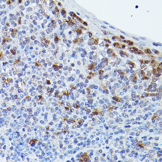
| Sample | Paraffin-embedded human tonsil tissue; fixative not specified (datasheet A02511) |
| Fixation | Image fixative and duration unreported (datasheet A02511); verify before use. |
| Sectioning | 4–5 µm sections on charged slides (standard) |
| Deparaffinisation | Xylene, graded ethanol series to water (standard) |
| Antigen retrieval | Heat retrieval: Citrate pH 6.0 (datasheet A02511); 20 min, 95–100 °C (standard) |
| Peroxidase block | 3% H2O2, 10 min, room temperature (standard) |
| Blocking | 10% normal serum of the secondary host, 30 min, room temperature (standard) |
| Primary antibody | Rabbit anti-HCAR2, 1:50-1:200 (datasheet A02511) |
| Primary incubation | Overnight at 4 °C (standard) |
| Detection | HRP-polymer secondary, DAB chromogen 5–10 min (standard) |
| Counterstain | Hematoxylin, blue, dehydrate and mount (standard) |
| Expected result | HCAR2-positive staining in hematopoietic cells of bone marrow (HPA tissue IHC: Medium). HPA tissue profile: Cytoplasmic expression in several tissues, including immune cells. No signal in the no-primary control. |
HCAR2 is a seven-pass cell-membrane receptor (UniProt Q8TDS4: topology and subcellular location). In paraffin-section IHC, assess staining in reported immune-cell populations, including splenic red-pulp cells and bone-marrow hematopoietic cells (HPA: Medium staining). HPA also describes cytoplasmic staining across several tissues; its IHC profile is Approved with medium consistency against RNA data and cautions that the antibody targets proteins from more than one gene (HPA: tissue IHC).
| Distinct staining in splenic red-pulp cells or bone-marrow hematopoietic cells. | These are plausible positive populations: HPA reports Medium IHC staining in both (HPA: tissue IHC). Evaluate the stained cells and their boundaries against nearby unstained cells. A membrane-associated component fits HCAR2's seven-pass topology (UniProt Q8TDS4: topology); cytoplasmic staining also appears in HPA's tissue profile (HPA: tissue IHC). |
| Predominantly nuclear staining, especially without staining at cell boundaries. | A nuclear pattern conflicts with the annotated cell-membrane location and seven transmembrane segments (UniProt Q8TDS4: subcellular location and topology). Treat it as suspect and check the counterstain, detection controls and antibody specificity before scoring it as HCAR2 (general IHC practice). |
| Strong staining in cells reported as unstained, such as adipocytes. | HPA reports adipocytes as Not detected by tissue IHC, despite UniProt's statement that expression is largely restricted to adipose tissue and spleen (HPA: tissue IHC; UniProt Q8TDS4: tissue specificity). Investigate cell identification, cross-reactivity and endogenous detection activity; this discrepancy prevents a simple tissue-wide positive or negative call. |
| Diffuse color across cells and surrounding tissue, with little distinction between structures. | That distribution is difficult to assign to the reported cell populations or receptor compartment (HPA: tissue IHC; UniProt Q8TDS4: subcellular location). Background from detection reagents or insufficient blocking is possible; compare a no-primary control and review washes and detection conditions (general IHC practice). |
| No visible signal in spleen red-pulp cells or bone-marrow hematopoietic cells. | These populations show Medium staining in HPA, so a blank result warrants a workflow check (HPA: tissue IHC). Confirm that the tissue and expected cells are present, then review primary-antibody conditions, antigen retrieval and detection with appropriate controls (general IHC practice). Medium staining is an expectation, not a guarantee for every section. |
| Membrane annotation versus observed tissue pattern. | UniProt places HCAR2 at the cell membrane and annotates seven membrane-spanning segments (UniProt Q8TDS4: subcellular location and topology). HPA describes cytoplasmic tissue staining (HPA: tissue IHC). Record the compartment actually seen rather than rejecting every cytoplasmic signal solely on topology. |
| Antibody-validation limit. | The listed rabbit polyclonal antibody, HPA028660, has Approved IHC status, while HPA flags medium agreement with RNA data and targeting of proteins from more than one gene (HPA: antibody validation and tissue IHC). A matching location and tissue pattern support interpretation but do not establish exclusive HCAR2 detection. |
| Choice of comparison tissue and cell population. | HPA reports Medium staining in splenic red-pulp, bone-marrow hematopoietic, lymph-node non-germinal-center, tonsillar germinal-center, seminal-vesicle glandular and bladder urothelial cells (HPA: tissue IHC). It reports adipocytes and several other sampled populations as Not detected (HPA: tissue IHC). Compare identified cells, since a whole-section label can hide the relevant population. |
| Epitope and preparation information. | UniProt identifies extracellular and cytoplasmic regions but supplies no antibody epitope in this record (UniProt Q8TDS4: topology). The supplied sources do not establish HCAR2-specific retrieval requirements or fixation sensitivity; choose and document retrieval and detection conditions as general IHC workflow decisions. |
| Situation | Likely cause | Next action |
|---|---|---|
| Spleen or bone-marrow section is blank. | Expected cells may be absent from the sampled field, or the IHC workflow may have failed; HPA reports Medium staining only in specified populations (HPA: tissue IHC). | Locate red-pulp or hematopoietic cells on the counterstain, then check retrieval, primary-antibody conditions and detection using a suitable control section (general IHC practice). |
| Adipose section is blank. | HPA reports adipocytes as Not detected by IHC, although UniProt describes adipose-tissue expression (HPA: tissue IHC; UniProt Q8TDS4: tissue specificity). | Do not use an adipocyte-negative slide alone to declare assay failure. Examine an HPA-reported Medium-staining population and document the adipose discrepancy when interpreting results. |
| Color appears mainly in nuclei. | Nuclear localization does not match the annotated cell-membrane receptor (UniProt Q8TDS4: subcellular location and topology). | Compare the counterstain and no-primary control; reassess detection background and specificity before counting nuclear signal as positive (general IHC practice). |
| Many unrelated cell types stain strongly. | HPA cautions that its antibody targets proteins from more than one gene (HPA: tissue IHC); nonspecific or endogenous detection signal is also possible (general IHC practice). | Map staining by cell type against HPA's reported pattern, run no-primary and detection controls, and report unexpected populations separately from supported positives. |
| Diffuse chromogen obscures cell boundaries. | Background can arise from blocking, washing or detection conditions (general IHC practice); such color cannot reliably establish the annotated membrane location (UniProt Q8TDS4: subcellular location). | Inspect a no-primary control, review blocking and washes, and score only distinguishable cellular staining after background is resolved (general IHC practice). |
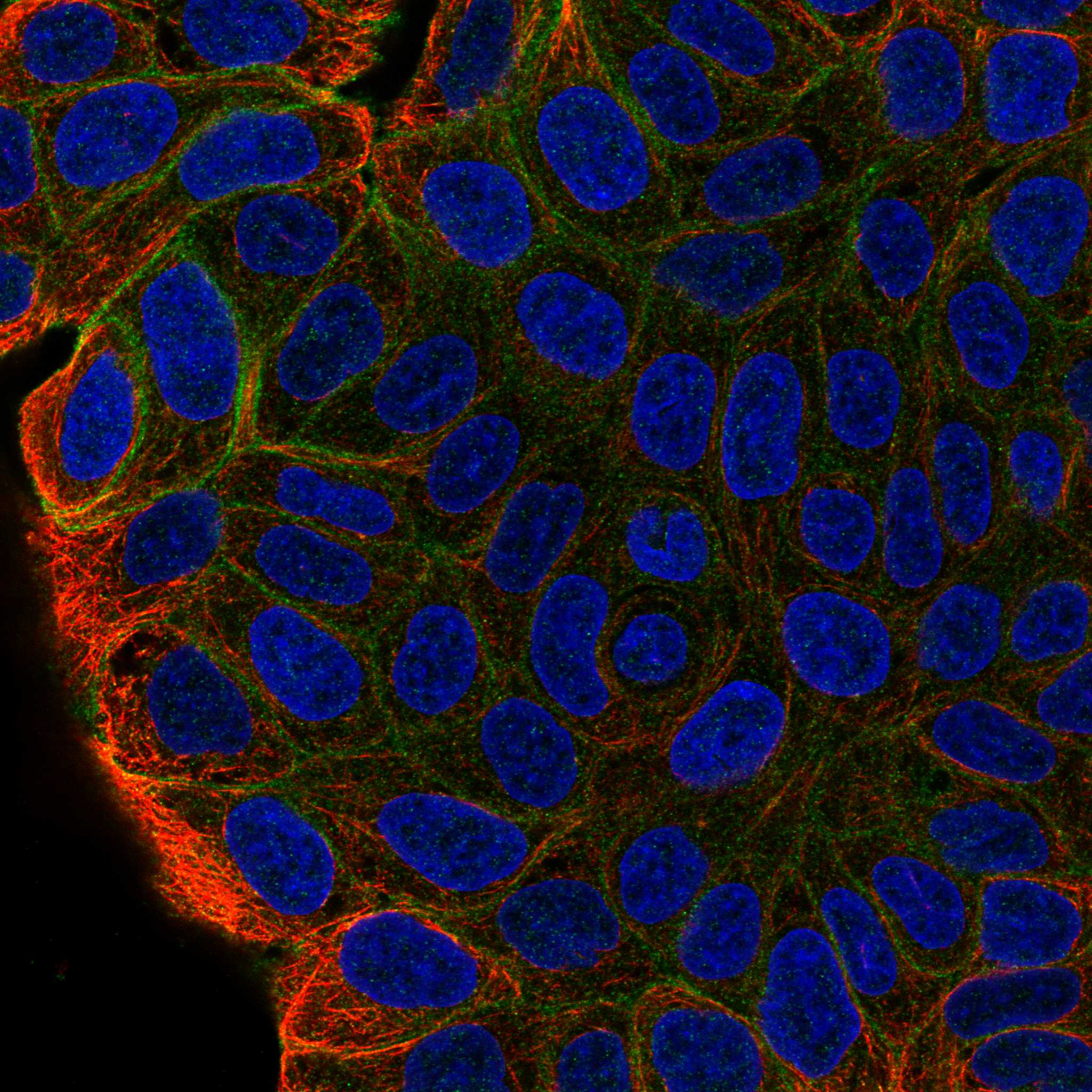
| Does an ICC-IF junction signal establish the expected paraffin IHC pattern? | HPA reports supported cell-junction localization from ICC-IF images in RT-4, with the same multi-gene-targeting caution (HPA: subcellular and antibody validation). That observation uses a different application. | Use the junction result as context for membrane-associated staining; interpret paraffin IHC against HPA's tissue-cell observations (HPA: subcellular and tissue IHC). Consult the separate IF/ICC guide for that application. |

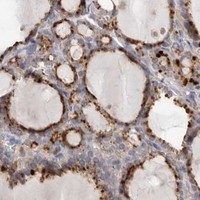



Comprehensive Human Protein Atlas IHC scoring per tissue (reliability: Approved — Medium consistency between antibody staining and RNA expression data. Caution, targets protein from more than one gene.). Rows are taken directly from the HPA tissue chart — click any row's HPA link to view the source.
| Tissue | Cell type | Level | Evidence | Source |
|---|---|---|---|---|
| Bone marrow | Hematopoietic cells | Medium | Protein (IHC) | HPA → |
| Lymph node | Non-germinal center cells | Medium | Protein (IHC) | HPA → |
| Seminal vesicle | Glandular cells | Medium | Protein (IHC) | HPA → |
| Spleen | Cells in red pulp | Medium | Protein (IHC) | HPA → |
| Tonsil | Germinal center cells | Medium | Protein (IHC) | HPA → |
| Tissue | Cell type | Level | Evidence | Source |
|---|---|---|---|---|
| Adipose tissue | Adipocytes | Not detected | Protein (IHC) | HPA → |
| Adrenal gland | Glandular cells | Not detected | Protein (IHC) | HPA → |
| Cerebellum | Cells in granular layer | Not detected | Protein (IHC) | HPA → |
| Endometrium | Cells in endometrial stroma | Not detected | Protein (IHC) | HPA → |
| Esophagus | Squamous epithelial cells | Not detected | Protein (IHC) | HPA → |
Troubleshoot HCAR2 staining in paraffin sections by checking retrieval, cell identity, and compartment before interpreting chromogenic signal.
A02511 is listed for IHC and IF/ICC in human, mouse, and rat (catalog applications/reactivity). Its supplied figure shows IHC in paraffin-embedded human tonsil; no IF figure is supplied (IHC image caption; catalog).
A02511 will render with an IHC figure from paraffin-embedded human tonsil (IHC image caption). It is a rabbit polyclonal antibody listed for IHC and IF/ICC with human, mouse, and rat reactivity; the payload supplies no IF image (catalog applications/reactivity; catalog host/clonality; catalog images).
Which to pick: For tissue IHC, choose A02511: its figure shows staining in paraffin-embedded human tonsil after high-pressure retrieval in citrate buffer at pH 6.0; the fixative is unreported (IHC image caption). For IF/ICC, A02511 is listed for those applications, but no IF figure is supplied (catalog applications; catalog images). For mouse or rat samples, A02511 lists reactivity in both species, while the supplied IHC figure documents human tissue only (catalog reactivity; IHC image caption).