This website uses cookies to ensure you get the best experience on our website.


- Table of Contents
Plan HCFC1 paraffin IHC with the catalog antibody at 2–5 μg/ml (datasheet A01729-2) and assess nuclear and cytoplasmic staining (HPA tissue IHC). Use highly stained nasopharyngeal ciliated cells as a positive reference (HPA tissue IHC), and consider HCFC1R1-dependent redistribution when interpreting compartment shifts (UniProt).

Expected localisation, validated protocols, controls and antibodies — the at-a-glance facts below, then the full design guide.
| Expected localisation | Nuclear and cytoplasmic tissue staining (HPA tissue IHC) | |
| Staining pattern | Variable nuclear and cytoplasmic staining in most tissues (HPA tissue IHC) | |
| Antigen retrieval | EDTA pH 8.0 HIER, heat-mediated (datasheet A01729-2) | |
| Positive control | Nasopharynx+4 more · see all | |
| Negative control | Adrenal gland+4 more · see all |
| Fixation | Keep fixation consistent across sections (standard IHC practice; not target-specific) | |
| Caveat | HCFC1R1 overexpression can shift HCFC1 to cytoplasm (UniProt) | |
| Regulation | Higher expression in fetal tissues (UniProt) | |
| Isoform / epitope | 4 isoforms; N/C cleavage makes epitope location relevant (UniProt) |
The catalog antibody uses EDTA pH 8.0 heat retrieval (datasheet A01729-2). One published HCFC1 IHC protocol describes staining FFPE hepatocellular carcinoma specimens (PMC10240668).
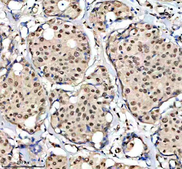
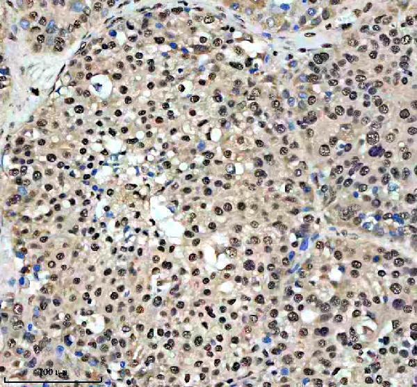
| Sample | Paraffin-embedded human breast cancer tissue; fixative not specified (datasheet A01729-2) |
| Fixation | Image fixative and duration unreported (datasheet A01729-2); verify before use. |
| Sectioning | 4–5 µm sections on charged slides (standard) |
| Deparaffinisation | Xylene, graded ethanol series to water (standard) |
| Antigen retrieval | Heat retrieval: EDTA pH 8.0 (datasheet A01729-2); 20 min, 95–100 °C (standard) |
| Peroxidase block | 3% H2O2, 10 min, room temperature (standard) |
| Blocking | 10% goat serum (datasheet A01729-2) |
| Primary antibody | Rabbit anti-HCFC1, 2-5 μg/ml (datasheet A01729-2) |
| Primary incubation | Overnight at 4 °C (datasheet A01729-2) |
| Detection | HRP-conjugated secondary, DAB chromogen (datasheet A01729-2) |
| Counterstain | Hematoxylin, blue, dehydrate and mount (standard) |
| Expected result | HCFC1-positive staining in ciliated cells (cell body) of nasopharynx (HPA tissue IHC: High). HPA tissue profile: Nuclear and cytoplasmic expression in varying fraction cells in most tissues. No signal in the no-primary control. |
HCFC1 is found in the nucleus and cytoplasm, with no transmembrane segment (UniProt P51610). In paraffin-section IHC, expect staining in a varying fraction of cells across most tissues (HPA tissue IHC: Supported; medium consistency with RNA). Nasopharyngeal ciliated cell bodies provide a high-staining example; bronchial ciliated cells and lung macrophages are medium-staining examples (HPA tissue IHC).
| Nuclear and cytoplasmic staining appears in a varying fraction of cells within a tissue (HPA tissue IHC). | This matches the reported tissue pattern. Assess nuclei and cell bodies separately, and record the fraction stained; uniform staining of every cell is not required by the HPA observation (HPA tissue IHC). |
| Strong signal outlines cell membranes or fills extracellular spaces, with little nuclear or cell-body staining. | Treat this as suspect compartmental staining: HCFC1 is nuclear and cytoplasmic and has no transmembrane segment (UniProt P51610). Review morphology, antibody controls and detection background before interpreting it as HCFC1. |
| Prominent staining occurs in adrenal gland glandular cells or lymph-node germinal-center cells. | These specific cell populations were not detected in HPA tissue IHC. Check whether the signal follows the expected intracellular pattern, and consider cross-reactivity or endogenous detection activity; the HPA finding does not make the entire tissue a negative control. |
| Chromogen appears broadly across tissue, including areas without recognizable cell boundaries. | A diffuse field cannot establish the reported cellular pattern (HPA tissue IHC). In general IHC practice, inspect the no-primary control and detection reagents for background before assigning positive cells or scoring intensity. |
| Nasopharyngeal ciliated cell bodies show no convincing signal. | This conflicts with HPA's high-staining example (HPA tissue IHC). Confirm that the relevant cells are present, then review the catalog antibody's IHC-P conditions and assay controls; one absent signal does not by itself establish lack of HCFC1. |
| Tissue and cell selection | HPA reports high staining in nasopharyngeal ciliated cell bodies; bronchial ciliated cells, lung macrophages and placental trophoblastic cells are medium (HPA tissue IHC). Compare the named cell population, since a tissue-wide average can hide a focal result. |
| Strength of the tissue evidence | The tissue IHC pattern is Supported, with medium consistency between antibody staining and RNA expression (HPA tissue IHC). Treat a surprising result as a reason to review controls and morphology, rather than as a definitive biological exception. |
| Subcellular context | Tissue IHC reports nuclear and cytoplasmic expression (HPA tissue IHC); ICC-IF supports nucleoplasm as the main location (HPA subcellular). UniProt notes that HCFC1R1 overexpression can accumulate HCFC1 in cytoplasm (UniProt P51610), but this does not establish a shift in a given tissue. |
| Processing and epitope coverage | HCFC1 produces associated N- and C-terminal cleavage products (UniProt P51610). The supplied antibody record gives no epitope, so staining cannot identify which chain or cleavage state the catalog antibody recognizes. |
| Isoforms and antibody validation | Four isoforms are listed (UniProt P51610), while HPA078588 is Supported for both IHC and ICC (HPA antibodies). Neither entry establishes equal recognition of all isoforms or transfers an ICC-IF image result to paraffin sections. |
| Situation | Likely cause | Next action |
|---|---|---|
| No signal appears in the expected nasopharyngeal ciliated cells (HPA tissue IHC). | The scored section may lack intact target cells, or the IHC run may have failed; the image alone cannot distinguish these possibilities. | Confirm cell identity and preservation, then review the catalog antibody's IHC-P instructions, reagent performance and a concurrent positive control. General IHC practice: change one workflow variable at a time. |
| Only membrane outlines or extracellular material stain. | That distribution is difficult to reconcile with nuclear and cytoplasmic HCFC1 and its lack of a transmembrane segment (UniProt P51610). | Compare with a no-primary control and examine whether chromogen tracks tissue edges or damaged areas. General IHC practice: resolve detection background before interpreting compartment-specific signal. |
| Unexpected cells stain strongly while expected cells remain faint. | The named HPA-negative populations are cell-specific observations, and unexpected signal may reflect cross-reactivity or endogenous detection activity (HPA tissue IHC; general IHC practice). | Identify the stained cells on the counterstain; compare their compartment and morphology with a known positive example, then inspect no-primary and detection controls. |
| A broad, weak haze obscures nuclei and cell bodies. | Diffuse background prevents a reliable call on the nuclear and cytoplasmic pattern reported by HPA (HPA tissue IHC). | General IHC practice: inspect the no-primary control, blocking and washes, and score only discrete cellular staining after the background is controlled. |
| Cytoplasmic staining exceeds nuclear staining in an otherwise interpretable section. | Cytoplasmic HCFC1 is reported in tissue IHC, and HCFC1R1 overexpression can alter localization (HPA tissue IHC; UniProt P51610). Neither source explains a particular specimen's balance. | Record nuclear and cytoplasmic intensity separately, check a comparable section and controls, and avoid inferring HCFC1R1 status from staining alone. |
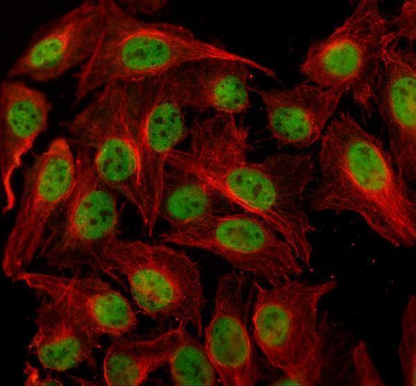
| What should an IF/ICC image show? | HPA identifies nucleoplasm as the supported main ICC-IF location and lists A-549, K-562 and U2OS images (HPA subcellular). | Look for nucleoplasmic signal when interpreting ICC-IF (HPA subcellular). Use the separate IF/ICC guide for that application; the HPA image result alone does not specify an IHC-P protocol. |


Comprehensive Human Protein Atlas IHC scoring per tissue (reliability: Supported — Medium consistency between antibody staining and RNA expression data.). Rows are taken directly from the HPA tissue chart — click any row's HPA link to view the source.
| Tissue | Cell type | Level | Evidence | Source |
|---|---|---|---|---|
| Nasopharynx | Ciliated cells (cell body) | High | Protein (IHC) | HPA → |
| Bronchus | Ciliated cells (cell body) | Medium | Protein (IHC) | HPA → |
| Cerebellum | Purkinje cells | Medium | Protein (IHC) | HPA → |
| Cerebral cortex | Neuronal cells | Medium | Protein (IHC) | HPA → |
| Fallopian tube | Ciliated cells (cell body) | Medium | Protein (IHC) | HPA → |
| Tissue | Cell type | Level | Evidence | Source |
|---|---|---|---|---|
| Adrenal gland | Glandular cells | Not detected | Protein (IHC) | HPA → |
| Appendix | Endocrine cells | Not detected | Protein (IHC) | HPA → |
| Colon | Endocrine cells | Not detected | Protein (IHC) | HPA → |
| Lymph node | Germinal center cells | Not detected | Protein (IHC) | HPA → |
| Soft tissue | Peripheral nerve | Not detected | Protein (IHC) | HPA → |
These questions address HCFC1 staining in paraffin sections with chromogenic IHC detection, with one section on IF/ICC.
A01729-2 has IHC images from human breast and liver cancer sections and mouse and rat liver sections (catalog IHC captions), plus IF data from U2OS cells (catalog IF caption).
A01729-2 is shown in paraffin sections of human breast cancer, human liver cancer, mouse liver and rat liver (catalog IHC captions). The same SKU is shown in U2OS cells by IF (catalog IF caption).
Which to pick: For tissue IHC, choose A01729-2: its images document paraffin sections with EDTA retrieval at pH 8.0 and primary antibody at 2 μg/ml (catalog IHC captions); the fixative is unreported (catalog IHC captions). For IF/ICC, A01729-2 has a U2OS cell image using 5 μg/ml primary antibody (catalog IF caption). For cross-species IHC, A01729-2 has images from human, mouse and rat tissue (catalog IHC captions); its IF reactivity is listed for human samples (catalog application data).