HDAC3 / Histone deacetylase 3 · IHC design guide

Design Immunohistochemistry for HDAC3

Plan chromogenic HDAC3 IHC in paraffin sections using nuclear staining as the main readout (HPA tissue IHC). Compare high staining in cerebellar granular-layer cells with low staining in parathyroid glandular cells (HPA tissue IHC), and start PA1600-1 at 0.5–1 μg/mL (datasheet PA1600-1).

Evidence assembled Oct 2026 · For research use; verify linked source records and product datasheet before use
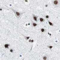
Immunohistochemistry protocol sheet for HDAC3 (IHC for HDAC3): expected localisation Predominantly nuclear (HPA tissue IHC); cytoplasm annotated (UniProt), antibody PA1600-1, validated IHC image, and IHC protocol steps
Printable HDAC3 IHC protocol sheet — expected localisation Predominantly nuclear (HPA tissue IHC); cytoplasm annotated (UniProt), antibody PA1600-1, controls and protocol steps. Open the full HDAC3 IHC guide →

HDAC3 Immunohistochemistry Experimental Design Guide

Expected localisation, validated protocols, controls and antibodies — the at-a-glance facts below, then the full design guide.

Must know before staining
Expected localisation Predominantly nuclear (HPA tissue IHC); cytoplasm annotated (UniProt)
Staining pattern General nuclear staining across tissue cell types (HPA tissue IHC)
Antigen retrieval Citrate pH 6 HIER, heat-mediated (datasheet PA1600-1)
Positive control ⓘ Cerebellum+4 more · see all
Negative control ⓘ None in HPA (detected in all 45 tissues); use no-primary + isotype controls
Important caveats
Reasons your staining may differ from the expected pattern.
Fixation Keep fixation consistent across sections. (standard IHC practice; not target-specific)
Caveat Cytoplasmic localization can complicate nuclear scoring (UniProt)
Regulation CCAR2 favors nuclear enrichment (UniProt)
Isoform / epitope 2 isoforms; confirm epitope coverage (UniProt)
Section 1

Recommended HDAC3 IHC & IF Protocols

The catalog antibody has a datasheet IHC-P protocol (datasheet: PA1600-1). The four published HDAC3 IHC protocols below provide additional tissue examples (PMC7471029; PMC4270766; PMC7670781; PMC6015978).

Recommended immunohistochemistry (IHC-P) protocol parameters
SampleParaffin-embedded human intestinal cancer tissues; fixative not specified (datasheet PA1600-1)
FixationImage fixative and duration unreported (datasheet PA1600-1); verify before use.
Sectioning4–5 µm sections on charged slides (standard)
DeparaffinisationXylene, graded ethanol series to water (standard)
Antigen retrievalHeat retrieval: Citrate pH 6, 20 min (datasheet PA1600-1)
Peroxidase block3% H2O2, 10 min, room temperature (standard)
Blocking10% goat serum (datasheet PA1600-1)
Primary antibodyRabbit anti-HDAC3, 0.5-1μg/ml (datasheet PA1600-1)
Primary incubationOvernight at 4 °C (datasheet PA1600-1)
DetectionStreptavidin-biotin complex (SABC), DAB chromogen (datasheet PA1600-1)
CounterstainHematoxylin, blue, dehydrate and mount (standard)
Expected resultHDAC3-positive staining in cells in granular layer of cerebellum (HPA tissue IHC: High). HPA tissue profile: General nuclear expression. No signal in the no-primary control.
💡Decision noteStart with heat-mediated citrate pH 6 retrieval (datasheet: PA1600-1); one published protocol specifies 121°C for 2 minutes 30 seconds (PMC7471029).
Section 2

What Is the Expected HDAC3 Staining Pattern?

HDAC3 is predominantly nuclear, with cytoplasmic localization also reported (UniProt O15379); it has no transmembrane segment (UniProt O15379 topology). In paraffin sections, expect general nuclear staining across many cell types (HPA tissue IHC: Supported; general nuclear expression). Particularly strong examples are cerebellar granular-layer cells, cortical neurons, epididymal glandular cells, and bladder urothelial cells (HPA tissue IHC: High).

What am I looking at on my slide?
Distinct nuclear staining in the expected cells, with limited background.This fits the general nuclear tissue pattern (HPA tissue IHC: Supported). Compare nuclei within the same section: HPA reports High staining in bladder urothelial cells and Medium staining in adrenal glandular cells, so uniform intensity across tissues is not required (HPA tissue IHC).
Strong, widespread membrane staining or cytoplasm-only staining with unstained nuclei.Reassess specificity and detection conditions: neither pattern matches the general nuclear tissue profile (HPA tissue IHC) or HDAC3's lack of a transmembrane segment (UniProt O15379 topology). Some cytoplasmic signal can still be plausible (UniProt O15379 localization), so do not reject it solely because it is extranuclear.
The strongest signal is in cells expected to stain weakly, or it appears in unexpected cell types.Treat the mismatch as a prompt to investigate cross-reactivity or endogenous detection activity (general IHC practice). For context, HPA reports Low staining in lung alveolar cells and High staining in bladder urothelial cells (HPA tissue IHC); Low is not an absence criterion.
Diffuse color covers nuclei, cytoplasm, and tissue spaces without clear cell boundaries.This is difficult to score as HDAC3 because it lacks the predominant nuclear pattern (HPA tissue IHC; UniProt O15379 localization). Check background controls, blocking, antibody concentration, washing, and chromogen development (general IHC practice) before interpreting compartment or intensity.
No convincing nuclear signal in a known High tissue example.A negative result in cerebellar granular-layer cells or bladder urothelial cells conflicts with the reported High pattern (HPA tissue IHC). Verify tissue preservation, retrieval, antibody and detection reagents, and run a known-positive section (general IHC practice); this finding alone does not prove HDAC3 is absent.
💡Expected HDAC3 appearanceCall positive staining when identifiable nuclei stain in the relevant cells, especially the reported High cell groups (HPA tissue IHC); strong membrane-only color or diffuse cell-free deposit is suspect (UniProt O15379 topology; general IHC practice).
How each factor affects the staining
Tissue and cell choiceUse a reported High group as a positive reference, such as cortical neuronal cells or epididymal glandular cells (HPA tissue IHC: High). A Low group, such as salivary glandular cells, may stain weakly and is not a validated negative control (HPA tissue IHC: Low).
Compartment and topologyPredominantly nuclear staining is the primary IHC expectation (HPA tissue IHC; UniProt O15379 localization). UniProt also lists cytoplasm and cytosol, while reporting no transmembrane segment or signal peptide (UniProt O15379); interpret extranuclear staining in its cellular context.
Antibody evidenceHPA rates the tissue pattern Supported, citing consistency between staining and RNA data (HPA tissue IHC). Its listed antibodies HPA052052, CAB005583, and CAB072336 each have Supported IHC status (HPA antibodies). This supports comparison with the reported pattern, without establishing every new assay condition.
Isoform coverageUniProt lists two HDAC3 isoforms (UniProt O15379). The supplied HPA summaries do not assign their tissue staining to individual isoforms (HPA tissue IHC; HPA antibodies); do not infer isoform identity from chromogenic staining alone.
IF/ICC Q&A: What localization should I expect?Mainly nucleoplasmic, with additional cytosol and Golgi apparatus localization reported in ICC-IF (HPA subcellular). That cell-imaging result can inform interpretation, but it does not establish that Golgi staining must be visible in a paraffin tissue section (HPA subcellular; HPA tissue IHC).
Why is my staining missing, weak or wrong?
SituationLikely causeNext action
High reference tissue stains weakly or shows no nuclear signal.Retrieval, reagent performance, or detection settings may need checking (general IHC practice); HPA reports High staining in cerebellar granular-layer cells (HPA tissue IHC).Review the section and controls, then optimize retrieval and antibody concentration within the assay workflow (general IHC practice). Compare with another reported High cell group (HPA tissue IHC). No HDAC3-specific fixation sensitivity is established by the supplied sources.
Color appears in the no-primary control.Endogenous enzyme activity or nonspecific detection-reagent binding can produce chromogenic signal (general IHC practice).Address endogenous activity with the appropriate blocking step, check the detection reagents, and rerun the no-primary control (general IHC practice). Do not count control-matched color as HDAC3 staining.
Most cells show diffuse, heavy color that obscures nuclei.Excess antibody or chromogen development, insufficient washing, or nonspecific background may obscure compartment assessment (general IHC practice).Review antibody concentration, wash steps, and development time; retain a counterstain that lets you identify nuclei (general IHC practice). Judge the result against the nuclear HPA tissue pattern (HPA tissue IHC).
A Low HPA tissue is being treated as a definitive negative.Low describes observed staining level, not proven absence of HDAC3 (HPA tissue IHC); UniProt describes HDAC3 as widely expressed (UniProt O15379).Use the Low tissue as an intensity comparison, and use a no-primary control to assess detection background (general IHC practice). Include a reported High cell group as the positive reference (HPA tissue IHC).
Staining looks exclusively membranous.That distribution conflicts with the nuclear tissue profile and the absence of a transmembrane segment (HPA tissue IHC; UniProt O15379 topology).Check controls and cellular landmarks, then reassess antibody concentration and detection background (general IHC practice). Seek a clear nuclear pattern in a reported High cell group before scoring the result (HPA tissue IHC).
Cytoplasmic staining accompanies a clear nuclear signal.Extranuclear HDAC3 is plausible: UniProt lists cytoplasm and cytosol, and HPA ICC-IF reports additional cytosol localization (UniProt O15379; HPA subcellular).Record nuclear and cytoplasmic compartments separately and compare the nuclear component with the tissue pattern (HPA tissue IHC). Check background controls before assigning diffuse cytoplasmic color to HDAC3 (general IHC practice).

Sample controls for HDAC3 IHC & IF

🧪Run cerebellum first and score cells in the granular layer, where HDAC3 staining is High (HPA: cerebellum, cells in granular layer, High). HPA detects HDAC3 in all 45 scored tissues, so there is no supported negative tissue; use no-primary and isotype controls, and treat cells without specific nuclear staining on the positive slide as internal background rather than a validated negative cell population (HPA: no negative rows; UniProt O15379: nucleus).
Positive control tissue: Cerebellum (Cells in granular layer, HPA High)
Negative control tissue: None in HPA: HDAC3 is detected in all 45 scored tissues. Use a no-primary (secondary-only) and an isotype control instead.
ICC-IF cell lines (HPA subcellular resource): HPA ICC-IF images show HDAC3 in A-431, U-251MG, U2OS, NIH 3T3, with annotated localisation: Nucleoplasm (supported) (HPA subcellular).
Technical controls: Include a no-primary, secondary-only slide and a concentration-matched rabbit IgG isotype control, matching the rabbit primary used in the selected IHC example (caption: rabbit anti-HDAC3, PA1600-1). Confirm specificity with HDAC3 knockout material or a validated peptide block; quench endogenous peroxidase and check endogenous biotin background because the example uses a biotinylated secondary, streptavidin-biotin complex and DAB (caption: PA1600-1).
⚠️Feasibility: A target-specific fixation window or fixation effect is unreported, and the selected paraffin-section caption does not state its fixative (caption: PA1600-1). The reported IHC procedure uses heat retrieval in citrate buffer at pH 6 for 20 minutes, so verify retrieval when adapting it to local specimens (caption: PA1600-1). The supplied evidence does not establish whether frozen sections or IF are easier; with biotin-based DAB detection, check background from endogenous biotin (caption: PA1600-1).

HPA tissue IHC evidence for HDAC3

Comprehensive Human Protein Atlas IHC scoring per tissue (reliability: Supported — High consistency between antibody staining and RNA expression data.). Rows are taken directly from the HPA tissue chart — click any row's HPA link to view the source.

Positive expression · recommended positive controls

TissueCell typeLevelEvidenceSource
Cerebellum Cells in granular layer High Protein (IHC) HPA →
Cerebral cortex Neuronal cells High Protein (IHC) HPA →
Epididymis Glandular cells High Protein (IHC) HPA →
Urinary bladder Urothelial cells High Protein (IHC) HPA →
Adrenal gland Glandular cells Medium Protein (IHC) HPA →

Undetected expression · recommended negative controls

TissueCell typeLevelEvidenceSource
None in HPA: HDAC3 is detected in all 45 scored tissues. Use a no-primary (secondary-only) and an isotype control instead.
Section 3

Advanced HDAC3 IHC Tips

Troubleshoot HDAC3 staining in paraffin sections by checking retrieval, nuclear localisation, controls, and scoring before interpreting changes in signal.

How should I retrieve HDAC3 in paraffin sections when nuclear staining is weak?
Use heat-mediated citrate buffer at pH 6 for 20 minutes on paraffin sections (datasheet PA1600-1). The selected antibody image used this retrieval before overnight incubation at 4°C with 1 µg/ml primary antibody (caption PA1600-1). If nuclei remain weak, compare a fresh retrieval run with the original run while holding section thickness, antibody concentration, and detection conditions constant (standard IHC practice). Excessive heating can damage tissue morphology and make nuclear scoring unreliable, so inspect the counterstain and tissue architecture alongside signal (standard IHC practice). Include a tissue compartment with established nuclear expression to distinguish failed retrieval from a low-expressing region (HPA: general nuclear expression).
Could fixation explain inconsistent HDAC3 staining across my paraffin blocks?
Target-specific sensitivity of HDAC3 to fixation is unknown from the supplied evidence; the selected paraffin-section caption does not state a fixative (caption PA1600-1). Record each block’s fixative, fixation duration, and processing history before comparing staining intensity across blocks (standard IHC practice). Process matched sections together using citrate pH 6 retrieval for 20 minutes and the same detection run to limit avoidable variation (datasheet PA1600-1; standard IHC practice). Check nuclear morphology and a positive control on every run, because poor preservation or uneven processing can complicate interpretation of a nuclear stain (HPA: general nuclear expression; standard IHC practice). Do not assign a fixation effect to HDAC3 without a controlled comparison.
Should HDAC3 staining be nuclear, or can cytoplasmic signal be credible?
Expect predominantly nuclear staining in tissue sections, consistent with the supported general nuclear IHC profile and nucleoplasmic localisation (HPA: tissue IHC; HPA: subcellular). HDAC3 can also occur in cytoplasm and cytosol, and its distribution can depend on binding partners or cellular context (UniProt O15379 localisation). Score nuclear and cytoplasmic staining separately rather than combining them into one intensity value (standard IHC practice). A diffuse cytoplasmic haze without convincing nuclear signal deserves a background check using a primary-antibody omission control (standard IHC practice). If a condition appears to shift localisation, compare matched regions and confirm that counterstained nuclei and tissue architecture remain intact (standard IHC practice).
Can an epitope or isoform difference explain unexpected HDAC3 staining?
HDAC3 has 2 annotated isoforms and a phosphorylated serine at residue 424 (UniProt O15379 isoforms; UniProt O15379 modified residues). The supplied antibody caption does not identify its epitope, so staining alone cannot establish which isoform it recognises or whether phosphorylation changes binding (caption PA1600-1). Ask for epitope information before interpreting differences between samples as isoform-specific or modification-specific (standard IHC practice). Compare staining with an independently validated antibody recognising a disclosed, distinct epitope if that distinction is essential to the experiment (standard IHC practice). Keep retrieval and detection matched across sections, because altered epitope exposure can mimic a biological change in signal (standard IHC practice).
How can I check an HDAC3 localisation result by immunofluorescence?
Use IF as a localisation check alongside chromogenic IHC, with separate nuclear and cytoplasmic assessments (standard IF practice; HPA: subcellular). For cerebral cortex, multiplex HDAC3 with a neuronal cell marker so the expected cell population can be identified (HPA: high staining in cerebral cortex neuronal cells; standard IF practice). Choose a fluorophore away from prominent tissue autofluorescence and include single-colour and no-primary controls when evaluating overlap (standard IF practice). HDAC3 lacks a transmembrane segment and has nuclear and cytosolic locations, so permeabilise sufficiently to admit antibody to intracellular epitopes while preserving structure (UniProt O15379 topology and localisation; standard IF practice). HPA reports mainly nucleoplasmic signal with additional cytosolic signal, which provides a pattern to compare against (HPA: subcellular).
What should I check when HDAC3 DAB staining appears diffuse or widespread?
First inspect a section processed without primary antibody to assess detection-system and tissue-derived colour (standard IHC practice). The selected image used a biotinylated secondary, avidin–biotin detection, and DAB, so controls should follow that detection sequence (caption PA1600-1; standard IHC practice). Confirm that peroxidase blocking, serum blocking, washes, and DAB development were consistent across slides (standard IHC practice). The selected section received 10% goat serum blocking and 1 µg/ml primary antibody overnight at 4°C (caption PA1600-1). Widespread nuclear signal is plausible for a widely expressed protein, but uniform colour over nuclei and surrounding tissue needs control-based assessment (UniProt O15379 tissue specificity; standard IHC practice).
How should I quantify HDAC3 staining across tissue sections? ⚠ ANSWER MARKED FOR VERIFICATION
Define the cell population and tissue region before scoring, then report nuclear HDAC3 separately from cytoplasmic signal (HPA: general nuclear expression; standard IHC practice). For a nuclear H-score, assign intensity grades 0–3, multiply each grade by its percentage of cells, and sum to a 0–300 score (standard IHC practice). Alternatively report the percentage of positive nuclei, with the positivity threshold fixed before comparing groups (standard IHC practice). Normalise positive-cell counts to all evaluable nuclei in the same compartment, or report counts per mm² of viable tissue (standard IHC practice). Exclude folds, damaged edges, and necrotic regions consistently, and keep imaging and threshold settings matched (standard IHC practice).
How do I distinguish genuine HDAC3 positivity from staining artefacts?
A credible result should show interpretable cellular staining, usually nuclear, in intact tissue rather than colour confined to damaged margins (HPA: general nuclear expression; standard IHC practice). Compare the cell population with the expected pattern: HPA reports high staining in cerebellar granular-layer cells and cerebral-cortex neuronal cells (HPA: tissue IHC). Cytoplasmic staining can be biologically plausible, but isolated diffuse cytoplasmic colour warrants a detection-background check (UniProt O15379 localisation; standard IHC practice). Exclude edge effects, folds, and necrotic areas before calling a region positive, and inspect the primary-omission control for endogenous-enzyme or detection colour (standard IHC practice). Interpret group differences only after matching retrieval, detection, and scoring conditions (standard IHC practice).
Boster reagents

Best HDAC3 / Histone deacetylase 3 IHC Antibodies

PA1600-1 has pictured IHC in human and rat paraffin sections and mouse frozen sections, plus IF/ICC in U20S and Hela cells (PA1600-1 image captions).

Real IHC data IHC analysis of HDAC3 using anti-HDAC3 antibody (PA1600-1). HDAC3 was detected in paraffin-embedded section of human intestinal cancer tissues. Heat mediated antigen retrieval was performed in citrate buffer (pH6, epitope retrieval solution) for 20 mins. The tissue section was blocked with 10% goat serum. The tissue section was then incubated with 1μg/ml rabbit anti-HDAC3 Antibody (PA1600-1) overnight at 4°C. Biotinylated goat anti-rabbit IgG was used as secondary antibody and incubated for 30 minutes at 37°C. The tissue section was developed using Strepavidin-Biotin-Complex (SABC)(Catalog # SA1022) with DAB as the chromogen.
Anti-Histone deacetylase 3 HDAC3 Antibody ®
Cat # PA1600-1

The PA1600-1 card shows IHC in paraffin sections of human intestinal and lung cancer tissues and rat intestine (PA1600-1 IHC captions). It also shows IHC in frozen mouse brain sections and IF/ICC in U20S and Hela cells (PA1600-1 IHC/IF captions).

Which to pick: Choose PA1600-1 for paraffin-section tissue IHC; its own human intestinal cancer caption documents citrate retrieval at pH 6 for 20 minutes and 1 μg/ml primary antibody overnight at 4°C, while the fixative is unreported (PA1600-1 IHC caption). For IF/ICC, PA1600-1 has cell-image evidence at 2 μg/ml; M00839 lists IF/ICC but has no IF image in the payload (PA1600-1 IF captions; M00839 catalog entry). For work across species, PA1600-1 has pictured human and rat paraffin-section IHC and mouse frozen-section IHC; both SKUs list human, mouse and rat reactivity, and M00839 is monoclonal whereas PA1600-1 has no clone listed (PA1600-1 IHC captions; catalog entries).

Each figure is that product's own IHC / IF validation image from its datasheet.

References

  1. UniProt Consortium. UniProt entry O15379 (HDAC3_HUMAN, Histone deacetylase 3).
  2. Human Protein Atlas. HDAC3 tissue IHC expression (reliability: Supported).
  3. Human Protein Atlas. HDAC3 subcellular location (ICC-IF): Mainly localized to the nucleoplasm. In addition localized to the Golgi apparatus and cytosol..
  4. Human Protein Atlas. HDAC3 antibody validation summary (3 antibodies).
  5. Aberrant HDAC3 expression correlates with brain metastasis in breast cancer patients. Thoracic cancer 2020 — PMC7471029.
  6. Ginsenoside Rg3 inhibits melanoma cell proliferation through down-regulation of histone deacetylase 3 (HDAC3) and increase of p53 acetylation. PloS one 2014 — PMC4270766.
  7. HDAC3 deteriorates colorectal cancer progression via microRNA-296-3p/TGIF1/TGFβ axis. Journal of experimental & clinical cancer research : CR 2020 — PMC7670781.
  8. Histone Deacetylase-3 Modification of MicroRNA-31 Promotes Cell Proliferation and Aerobic Glycolysis in Breast Cancer and Is Predictive of Poor Prognosis. Journal of breast cancer 2018 — PMC6015978.
  9. PubMed PMID:9464271 — UniProt-cited evidence.
  10. PubMed PMID:9346952 — UniProt-cited evidence.
  11. PubMed PMID:9501169 — UniProt-cited evidence.