This website uses cookies to ensure you get the best experience on our website.


- Table of Contents
Plan paraffin IHC for HDAC6 using the catalog antibody. Expect cytoplasmic staining in most cell types, with prominent signal in renal tubules and cells in seminiferous ducts (HPA tissue IHC); interpret nuclear signal in light of HDAC6 shuttling (UniProt).

Expected localisation, validated protocols, controls and antibodies — the at-a-glance facts below, then the full design guide.
| Expected localisation | Cytoplasmic in tissue (HPA tissue IHC); can shuttle to nucleus (UniProt) | |
| Staining pattern | Cytoplasmic in most cells; strongest in renal tubules and seminiferous ducts (HPA tissue IHC) | |
| Antigen retrieval | EDTA pH 8.0 HIER, heat-mediated (datasheet PB9628) | |
| Positive control | Breast+4 more · see all | |
| Negative control | Adipose tissue+4 more · see all |
| Fixation | Keep fixation consistent across sections (standard IHC practice; not target-specific) | |
| Caveat | Antibody staining and RNA expression have low consistency (HPA tissue IHC) | |
| Regulation | Intensity regulation is unreported (UniProt) | |
| Isoform / epitope | 2 isoforms; no extracellular epitope expected; epitope map unknown (UniProt) |
The catalog antibody’s IHC-P protocol (datasheet PB9628) is accompanied by four published HDAC6 IHC protocols (PMC12127455; PMC13256419; PMC7762972; PMC7751721).
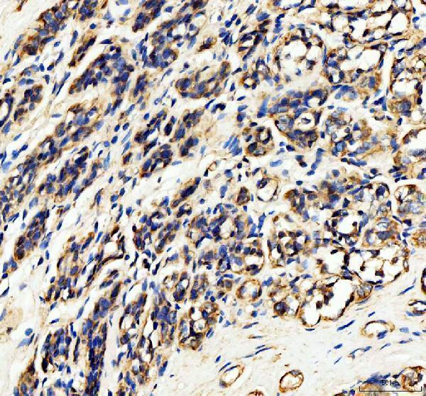
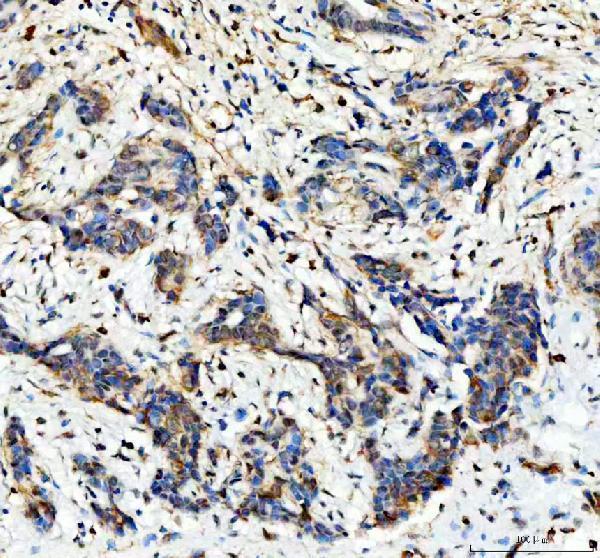
| Sample | Paraffin-embedded human breast cancer tissue; fixative not specified (datasheet PB9628) |
| Fixation | Image fixative and duration unreported (datasheet PB9628); verify before use. |
| Sectioning | 4–5 µm sections on charged slides (standard) |
| Deparaffinisation | Xylene, graded ethanol series to water (standard) |
| Antigen retrieval | Heat retrieval: EDTA pH 8.0 (datasheet PB9628); 20 min, 95–100 °C (standard) |
| Peroxidase block | 3% H2O2, 10 min, room temperature (standard) |
| Blocking | 10% goat serum (datasheet PB9628) |
| Primary antibody | Rabbit anti-HDAC6, 2-5 μg/ml (datasheet PB9628) |
| Primary incubation | Overnight at 4 °C (datasheet PB9628) |
| Detection | HRP-conjugated secondary, DAB chromogen (datasheet PB9628) |
| Counterstain | Hematoxylin, blue, dehydrate and mount (standard) |
| Expected result | HDAC6-positive staining in glandular cells of breast (HPA tissue IHC: High). HPA tissue profile: Cytoplasmic expression in most cell types, most abundant in renal tubules and cells in seminiferous ducts. No signal in the no-primary control. |
HDAC6 should stain mainly the cytoplasm, particularly in breast glandular cells, skin keratinocytes and testicular pachytene spermatocytes, where HPA reports high staining (HPA tissue IHC; UniProt Q9UBN7 localization). It has no transmembrane segment, so a membrane-restricted pattern is unexpected (UniProt Q9UBN7 topology). HPA rates its tissue IHC evidence Supported while reporting low consistency between antibody staining and RNA expression (HPA tissue IHC).
| Cytoplasmic staining is clear in breast glandular cells, skin keratinocytes or pachytene spermatocytes, with weaker staining in some other cell types. | This fits the reported high staining in those cells and cytoplasmic expression across most cell types (HPA tissue IHC). Score the named cell population and its cytoplasmic compartment; a positive area elsewhere in the section does not establish that the intended cells are positive (general IHC practice). |
| Staining is predominantly nuclear, or traces cell membranes while cytoplasm is blank. | A membrane-restricted pattern conflicts with the reported mainly cytoplasmic localization and lack of a transmembrane segment (UniProt Q9UBN7 localization and topology). Predominantly nuclear staining warrants checking the controls and morphology: HDAC6 can shuttle into nuclei under particular conditions, so an isolated nuclear signal is not conclusive evidence of an artefact (UniProt Q9UBN7 localization; general IHC practice). |
| Strong signal appears in adipocytes or skeletal muscle myocytes, while an expected positive cell population is also present. | HPA reports HDAC6 as not detected in adipocytes and skeletal muscle myocytes (HPA tissue IHC). Review that unexpected staining for antibody cross-reactivity or endogenous detection activity, using appropriate controls; a negative HPA observation alone cannot prove either cause in this specimen (general IHC practice). |
| Chromogen is spread across stroma, lumen or much of the section without a readable cellular boundary. | Treat this as diffuse background until cell-associated staining can be distinguished from it (general IHC practice). Excess detection activity, incomplete blocking or overly concentrated antibody are general IHC possibilities; the supplied HPA and UniProt records do not identify which applies to this section. |
| A section containing a reported high-staining cell population has no interpretable cytoplasmic signal. | A blank positive control makes a biological negative call unreliable (general IHC practice). Confirm that the expected cells are present, then check the antibody, retrieval and detection workflow against its validated IHC-P instructions (general IHC practice). HPA's high staining is a reference pattern, not a guarantee for every specimen (HPA tissue IHC). |
| Cell population selected for comparison | HPA reports high staining in breast glandular cells, skin keratinocytes and pachytene spermatocytes; it reports adipocytes and skeletal muscle myocytes as not detected (HPA tissue IHC). Compare like cell types when judging intensity. The HPA profile also describes cytoplasmic expression in most cell types, so a whole-tissue positive/negative label can obscure cellular variation (HPA tissue IHC). |
| Compartment and cell state | HDAC6 is mainly cytoplasmic and microtubule associated, but UniProt also reports nuclear shuttling under particular conditions (UniProt Q9UBN7 localization). Nuclear signal therefore needs its cellular context and controls. HPA ICC-IF supports cytosol, centrosome and basal body localization; its additional nucleoplasm assignment is uncertain (HPA subcellular ICC-IF). |
| Strength of reference evidence | HPA calls tissue IHC reliability Supported and notes low consistency between staining and RNA expression (HPA tissue IHC). Its listed antibodies HPA003714, HPA026321 and CAB004236 each have Supported IHC status (HPA antibody validation). Use those observations as pattern benchmarks, while keeping antibody-specific control results central to an interpretation (general IHC practice). |
| Isoforms and antibody recognition | UniProt lists two HDAC6 isoforms and one annotated protein chain spanning residues 1–1215 (UniProt Q9UBN7 isoforms and processing). The supplied records do not map the catalog antibody's epitope to either isoform. Do not assign a compartment difference or a missing signal to isoform selectivity without antibody-specific evidence. |
| What should IF/ICC show? | Mainly cytosol, centrosome and basal body signal is supported; nucleoplasm is an additional uncertain location (HPA subcellular ICC-IF). That is an IF/ICC localization reference, not an IHC-P protocol or a requirement to resolve those small structures in a chromogenic tissue section (general IHC practice). |
| Situation | Likely cause | Next action |
|---|---|---|
| Expected high-staining cells are blank. | The expected cells may be absent from the section, or the IHC-P staining or detection workflow may have failed (general IHC practice; HPA tissue IHC for expected cells). | Identify the cells on the counterstained section, run a known-positive section alongside it, and verify each step against the IHC-validated antibody's instructions (general IHC practice). Interpret the sample only after the control gives readable cytoplasmic signal. |
| Nuclear signal dominates the positive cells. | This departs from the mainly cytoplasmic baseline, although condition-dependent nuclear HDAC6 is documented (UniProt Q9UBN7 localization). The image alone cannot distinguish a biological shift from staining artefact. | Check nuclear boundaries and matched positive and detection controls, then compare cytoplasmic and nuclear staining in the same cell type (general IHC practice). Record nuclear localization as a separate observation rather than counting every stained nucleus as a routine positive. |
| Adipocytes or skeletal muscle myocytes stain strongly. | Those cell types are reported as not detected by HPA; cross-reactivity or endogenous detection activity are possibilities, not established diagnoses (HPA tissue IHC; general IHC practice). | Check cell identity and morphology, inspect a no-primary control for detection background, and compare the unexpected signal with a reported high-staining cell population on matched sections (general IHC practice; HPA tissue IHC). |
| Brown staining covers extracellular areas and masks cell borders. | Diffuse deposit prevents reliable assignment of stain to cells (general IHC practice). The supplied HDAC6 records do not identify a target-specific source for that background. | Review the no-primary control, blocking and washes, then adjust antibody or detection conditions according to the validated IHC-P workflow (general IHC practice). Reassess localization only when cell boundaries are visible. |
| Only membrane outlines are scored as positive. | A membrane-restricted call conflicts with HDAC6's reported mainly cytoplasmic localization and lack of a transmembrane segment (UniProt Q9UBN7 localization and topology). | Recheck whether cytoplasm contains distinct signal and whether outlines also appear in a detection control (general IHC practice). If staining remains confined to borders, flag the result for specificity review instead of using it as the expected HDAC6 pattern. |
| Two specimens show different proportions of positive cells. | HPA reports variation by cell type, including high, medium, low and not-detected observations; its IHC profile also has low consistency with RNA expression (HPA tissue IHC). A difference alone cannot establish technical failure or biological change. | Compare the same identified cell population, compartment and scoring threshold, and confirm that both runs have usable positive controls (general IHC practice). Report the observed difference with its cell-type context and HPA evidence limit. |
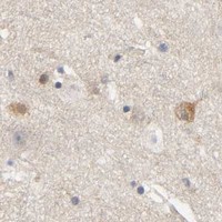
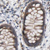


Comprehensive Human Protein Atlas IHC scoring per tissue (reliability: Supported — Low consistency between antibody staining and RNA expression data.). Rows are taken directly from the HPA tissue chart — click any row's HPA link to view the source.
| Tissue | Cell type | Level | Evidence | Source |
|---|---|---|---|---|
| Breast | Glandular cells | High | Protein (IHC) | HPA → |
| Skin | Keratinocytes | High | Protein (IHC) | HPA → |
| Testis | Pachytene spermatocytes | High | Protein (IHC) | HPA → |
| Adrenal gland | Glandular cells | Medium | Protein (IHC) | HPA → |
| Appendix | Glandular cells | Medium | Protein (IHC) | HPA → |
| Tissue | Cell type | Level | Evidence | Source |
|---|---|---|---|---|
| Adipose tissue | Adipocytes | Not detected | Protein (IHC) | HPA → |
| Bronchus | Basal cells | Not detected | Protein (IHC) | HPA → |
| Hippocampus | Glial cells | Not detected | Protein (IHC) | HPA → |
| Skeletal muscle | Myocytes | Not detected | Protein (IHC) | HPA → |
| Smooth muscle | Smooth muscle cells | Not detected | Protein (IHC) | HPA → |
Troubleshoot chromogenic HDAC6 IHC in paraffin sections by checking retrieval, staining pattern, controls and compartment-specific scoring.
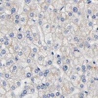
PB9628 has a real IHC figure from a human breast cancer paraffin section (PB9628 image caption). Human IF/ICC is listed for M00595, without an IF figure (M00595 catalog).
PB9628 is the rendered SKU; its IHC figure shows a human breast cancer paraffin section (PB9628 image caption). It is listed for IHC in Human, Mouse, and Rat, although the supplied IHC figure shows human tissue only (PB9628 catalog; PB9628 image caption).
Which to pick: Choose PB9628 for paraffin-section IHC: its own figure documents EDTA retrieval at pH 8.0, 2 μg/ml primary antibody, and DAB detection; the fixative is unreported (PB9628 image caption). For IF/ICC, M00595 is the rabbit monoclonal option listed for human samples, with no supplied IF figure (M00595 catalog). For cross-species work, PB9628 lists Human, Mouse, and Rat reactivity, but its supplied IHC figure documents human tissue only (PB9628 catalog; PB9628 image caption).